Lecture: 4 Heart muscle: the heart as a pump and function of the heart valves Rhythmical excitation of the heart
1/142
There's no tags or description
Looks like no tags are added yet.
Name | Mastery | Learn | Test | Matching | Spaced | Call with Kai |
|---|
No analytics yet
Send a link to your students to track their progress
143 Terms
1. atrial muscle (LA, RA)
2. ventricular muscle (LV, RV)
3. specialized excitatory and conductive muscle fibers (SA node, AV node, bundle of his, purkine fibers)
What are three major types of cardiac muscle?

atrial and ventricular types of muscles
What types of cardiac muscle contract?
they contain few contractile fibrils
The specialized excitatory and conductive fibers of the heart, however, contract only feebly because
Lub
What is the first heart sound?
closure of the AV valves
The first heart sound is caused by what?
Dup
What is the second heart sound?
to closure of semilunar valves
What is the second heart sound caused by?
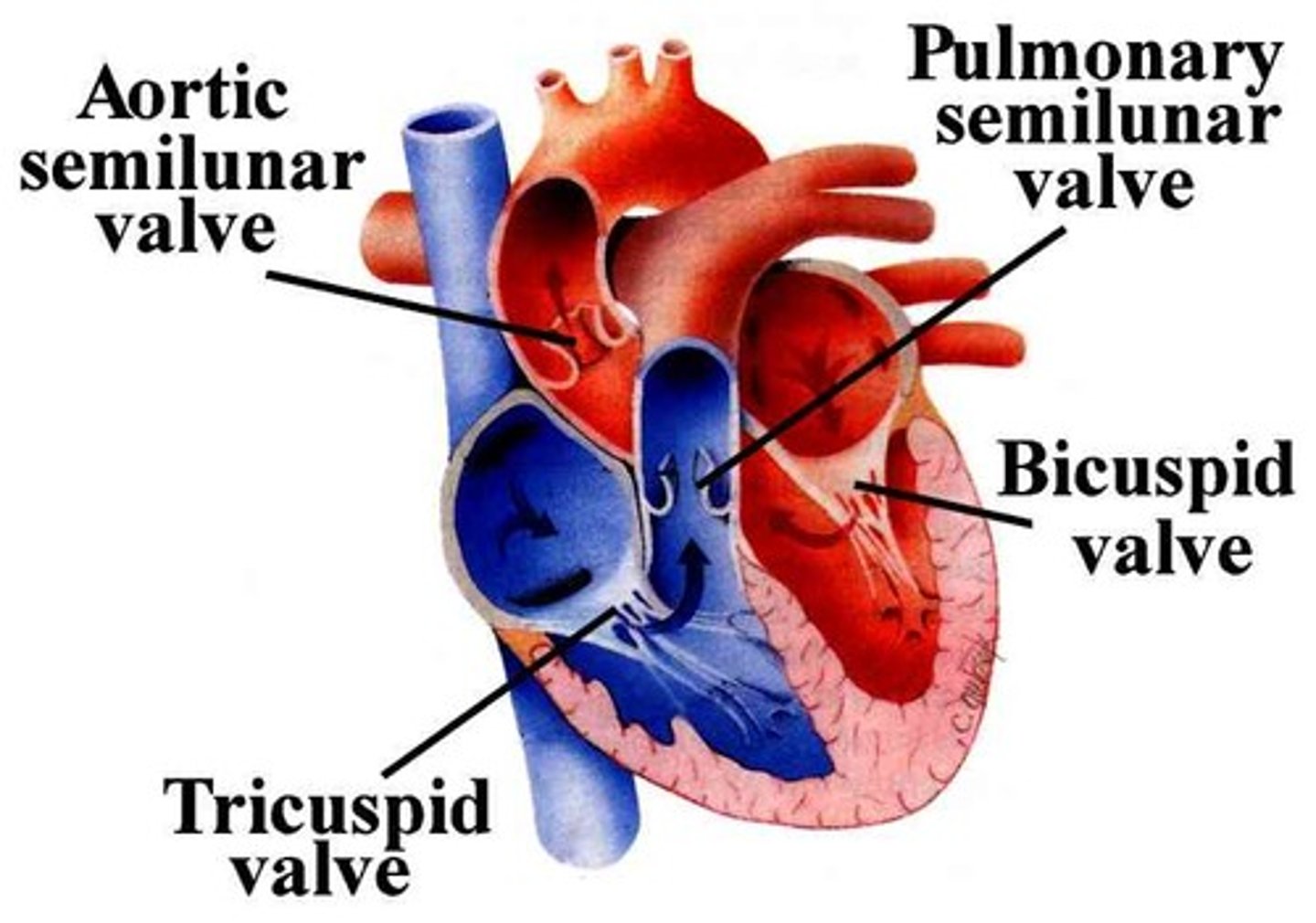
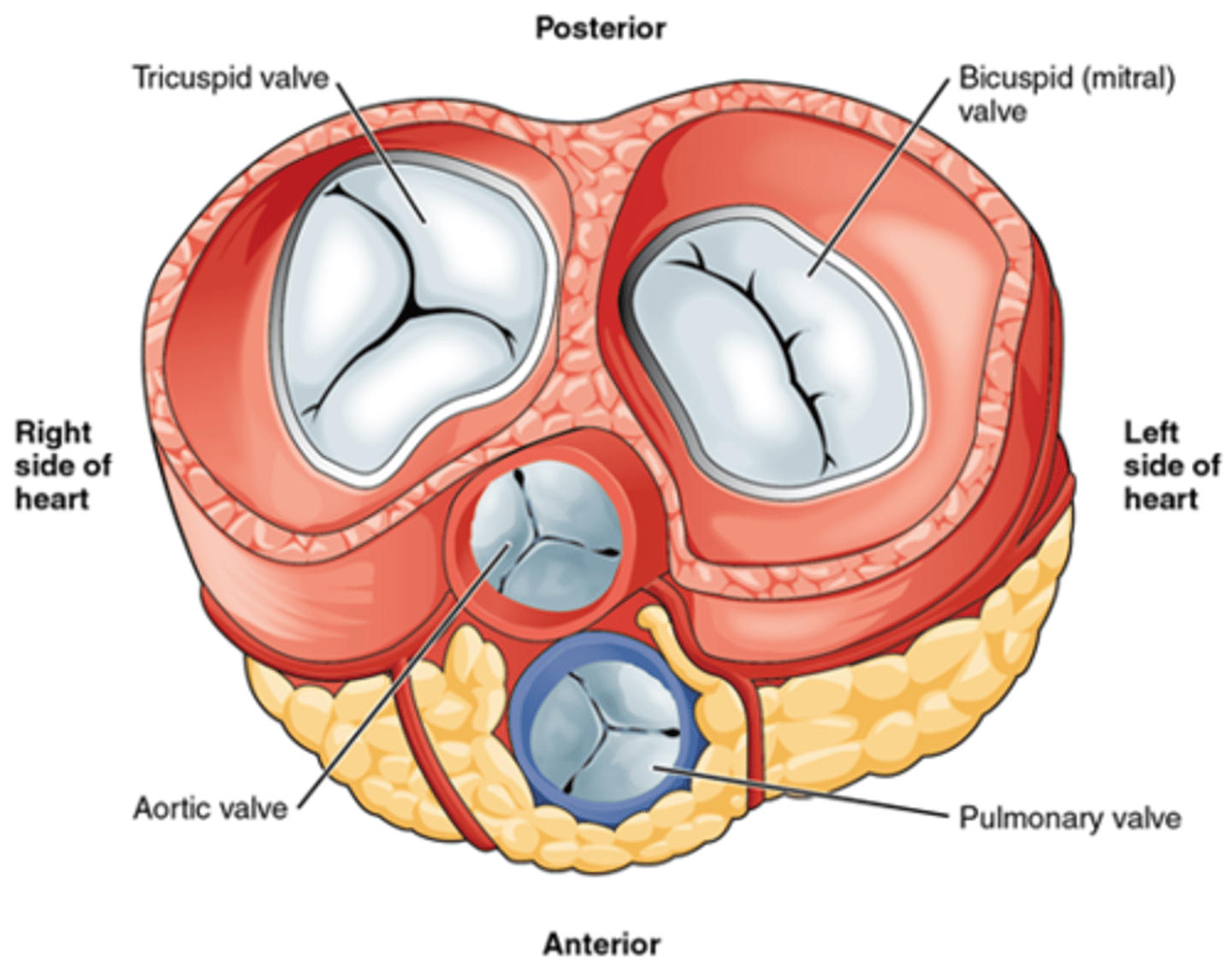
Cuspid valves
What are the AV valves also known as?

semi-lunar valves
What are the valves between ventricles and great arteries are also known as
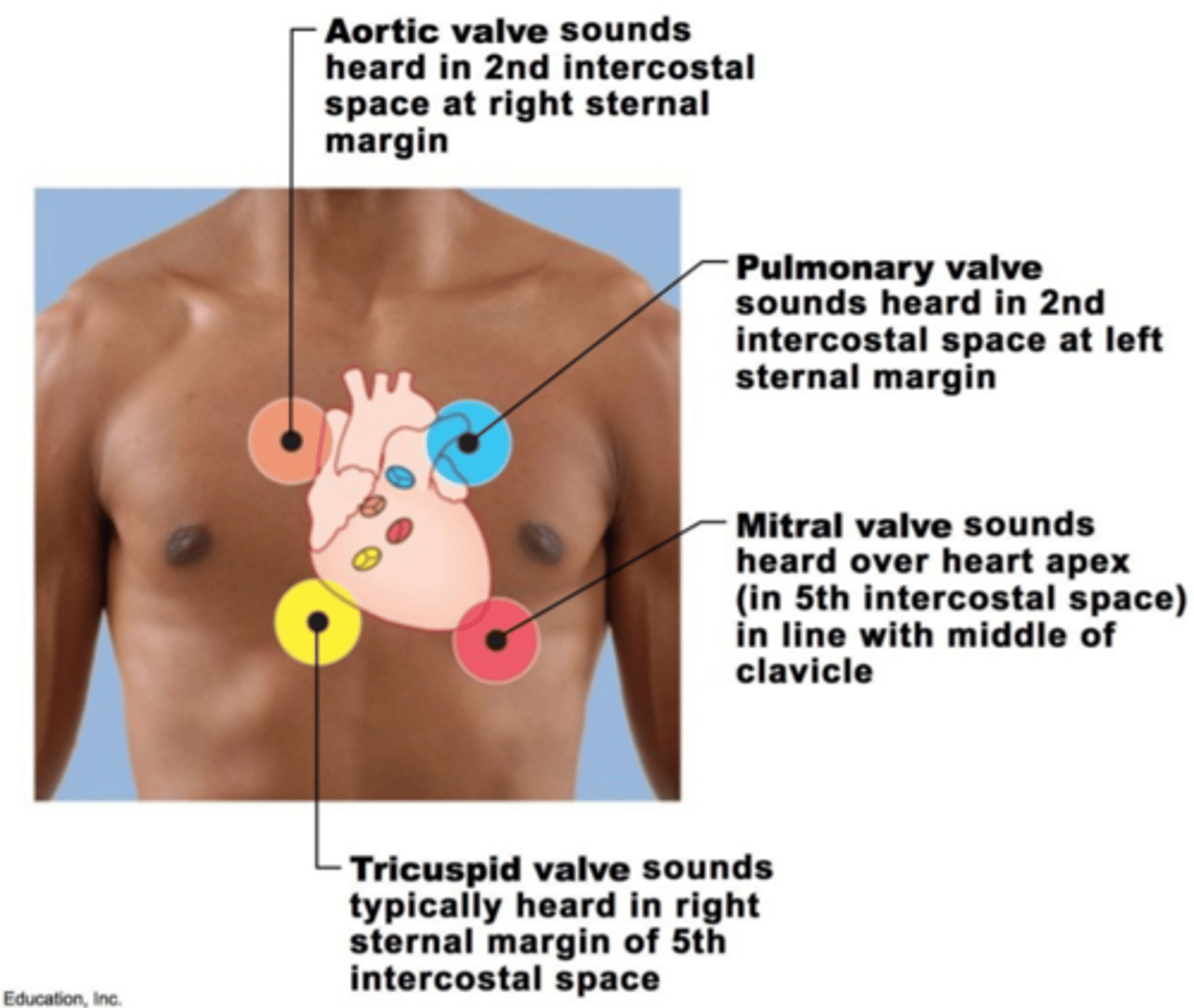
2nd intercostal space at right sternal margin
Where are the sounds of the aortic semilunar valve heard in?
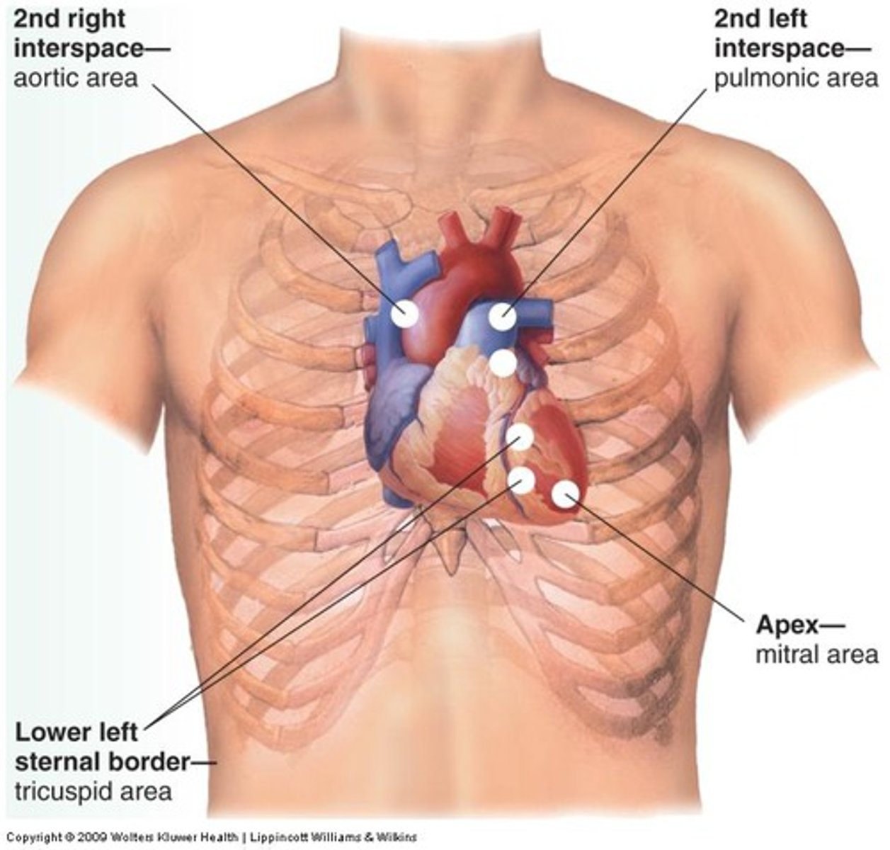
2nd intercostal space at left sternal margin
Where are the sounds of the pulmonary semilunar valves heard in?
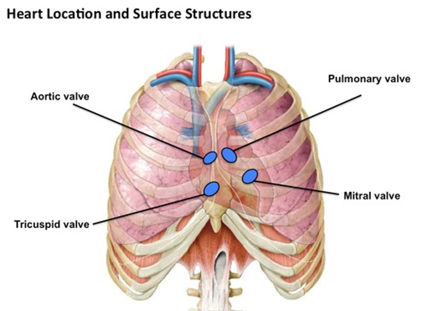
heard over the heart apex, in 5th intercostal space in line with middle of clavicle.
Where are the sounds of the mitral valve heard over and what space? (D)
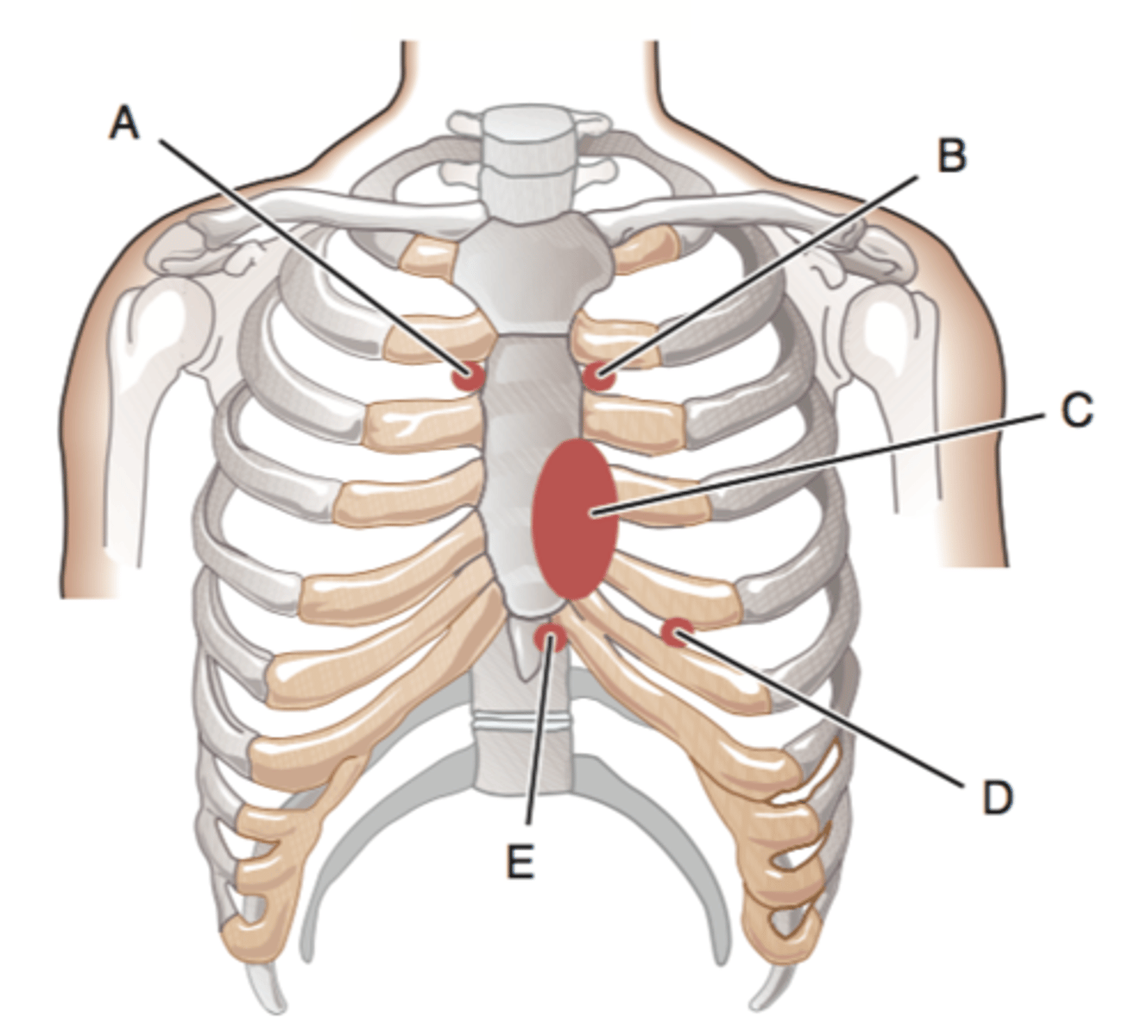
right sternal margin of 5th intercostal space; variations include over sternum or over lefter sternal margin in 5th intercostal space.
where are the sounds of the tricuspid valve heard in?
commissures
the divisions between the cusps are known as
The leaflets of the valves are connective tissue covered with endothelial tissue
The leaflets of the valves are what type of tissue? What are they covered with?
chordae tendinae
The A-V valves have their ventricular surface anchored to the ventricular wall by way of the
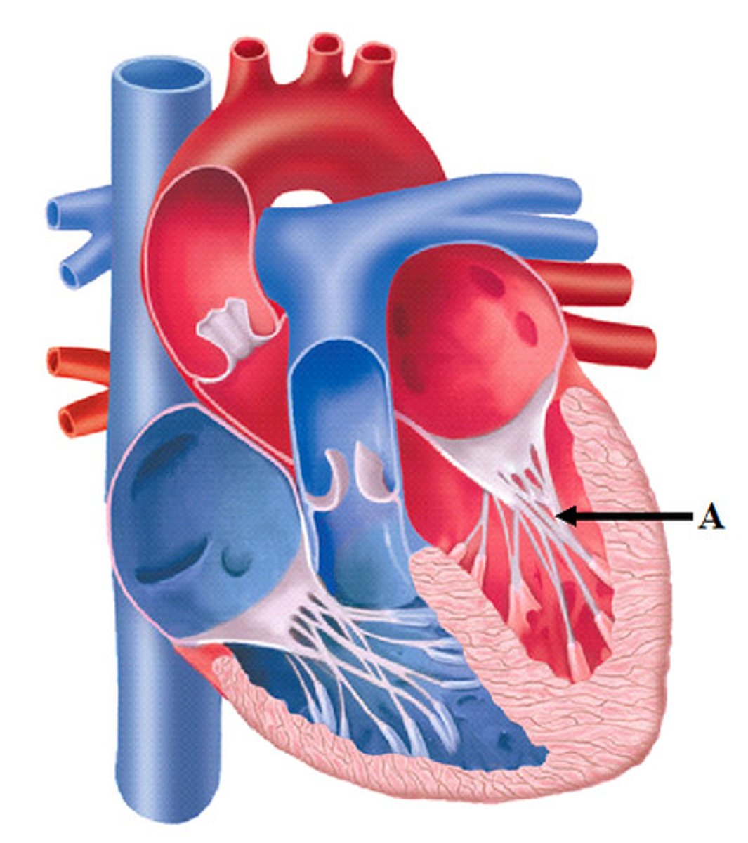
myocardial tissue
elastic cords inserted into the papillary muscles composed of what type of tissue?
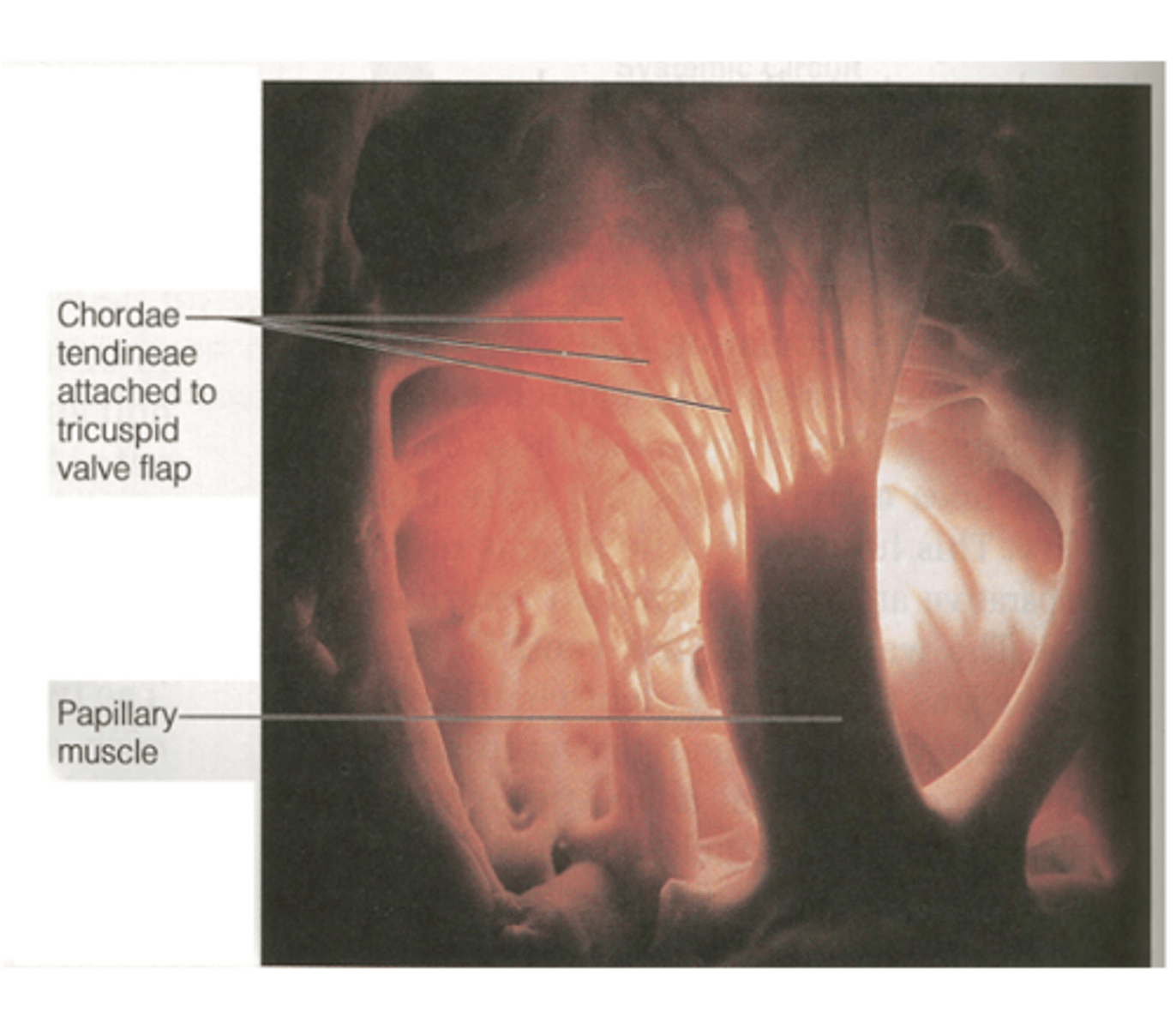
their proper closure during ventricle contraction preventing blood reflux.
elastic cords composed of myocardial tissue contracts simultaneously with the ventricles assuring what?
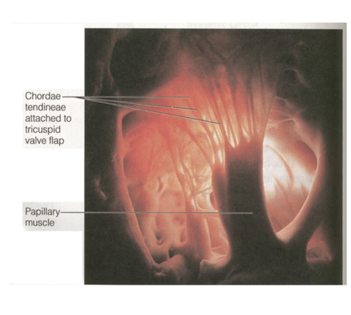
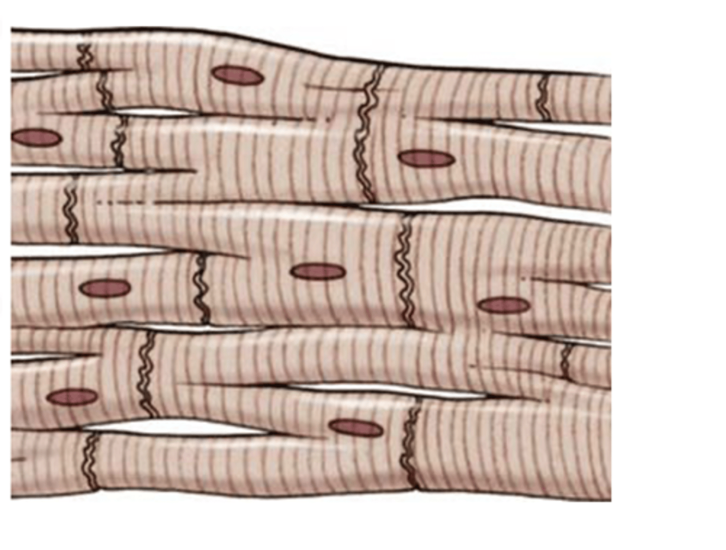
cell membranes that separate individual cardiac muscle cells from one another
What are intercalated discs in cardiac muscle?
permeable “communicating” junctions (gap junctions) that allow rapid diffusion of ions.
At each intercalated disc the cell membranes fuse with one another in such a way that they form
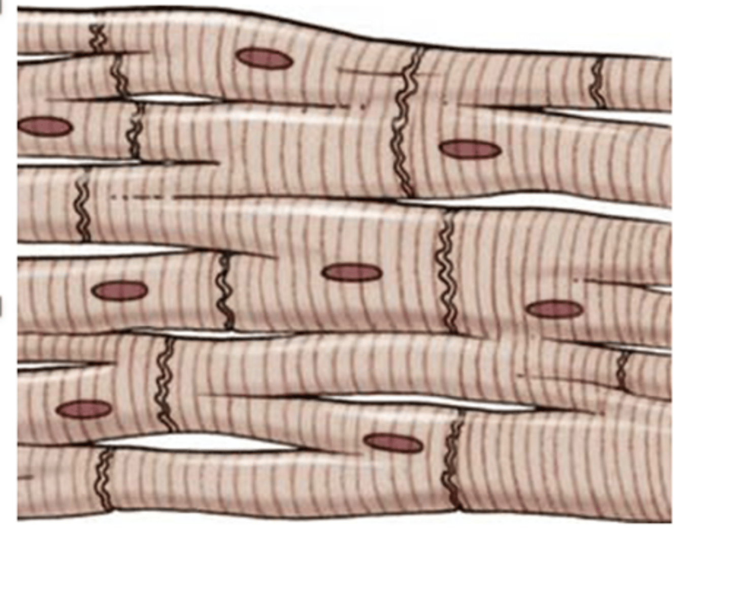
latticework interconnections.
In cardiac cells the action potential spreads to all of them, from cell to cell throughout the
the volume of blood ejected by each ventricle per unit of time.
Cardiac output (CO) is defined as
heart rate x stroke volume
CO (cardiac output) =
5-6 L/min
Cardiac output at rest is normally what?
SV is affected by the
1. venous return
2. peripheral resistance
3. Autonomic Nervous System
What is stoke volume (SV) affected by? (3)
high proportionally
if the HR (heart rate) is high 90/min, the CO (cardiac output) is?
remains the same because the Stroke volume (SV) low in proportion to the increase in the HR due to the fact that ventricles have less time to fill during diastole.
If the HR is high from 90 to 140/min, what is the cardiac output?
brain 13%
cardiac muscle 4%
kidneys 20 - 25%
What is the cardiac output percentage of the brain, cardiac muscle, and kidneys?
left ventricle (LV), normally 70-80 mL/per systole
Heart rate (HR) is normally 80 beats/min and the Stroke volume (SV) is usually the same for both ventricles but CO usually refers to the
chronotropic influences on the spontaneous electrical activity of SA nodal cells
What is the heart rate controlled by?
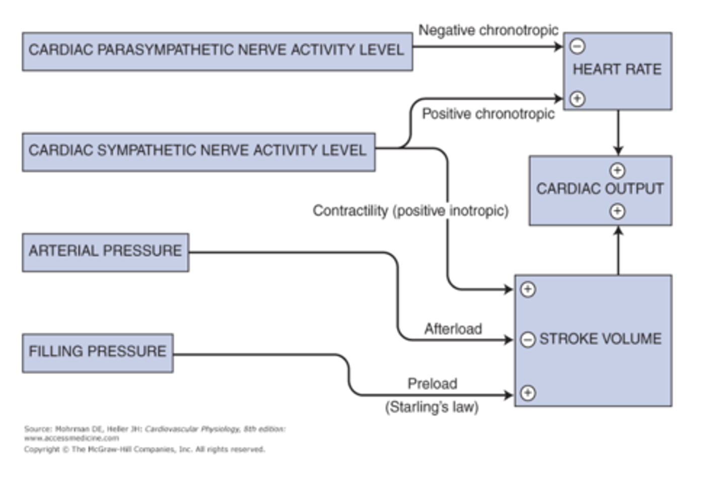
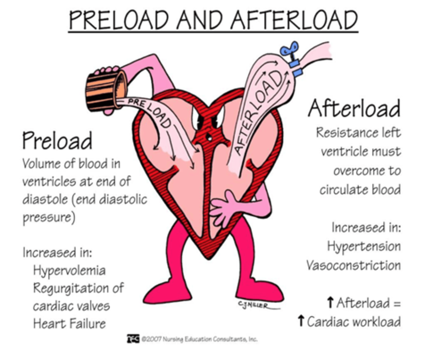
SV is controlled by influences on the contractile performance of the ventricular cardiac muscle, in particular, its degree of shortening in the after loaded situation.
What is SV (stroke volume) controlled by?
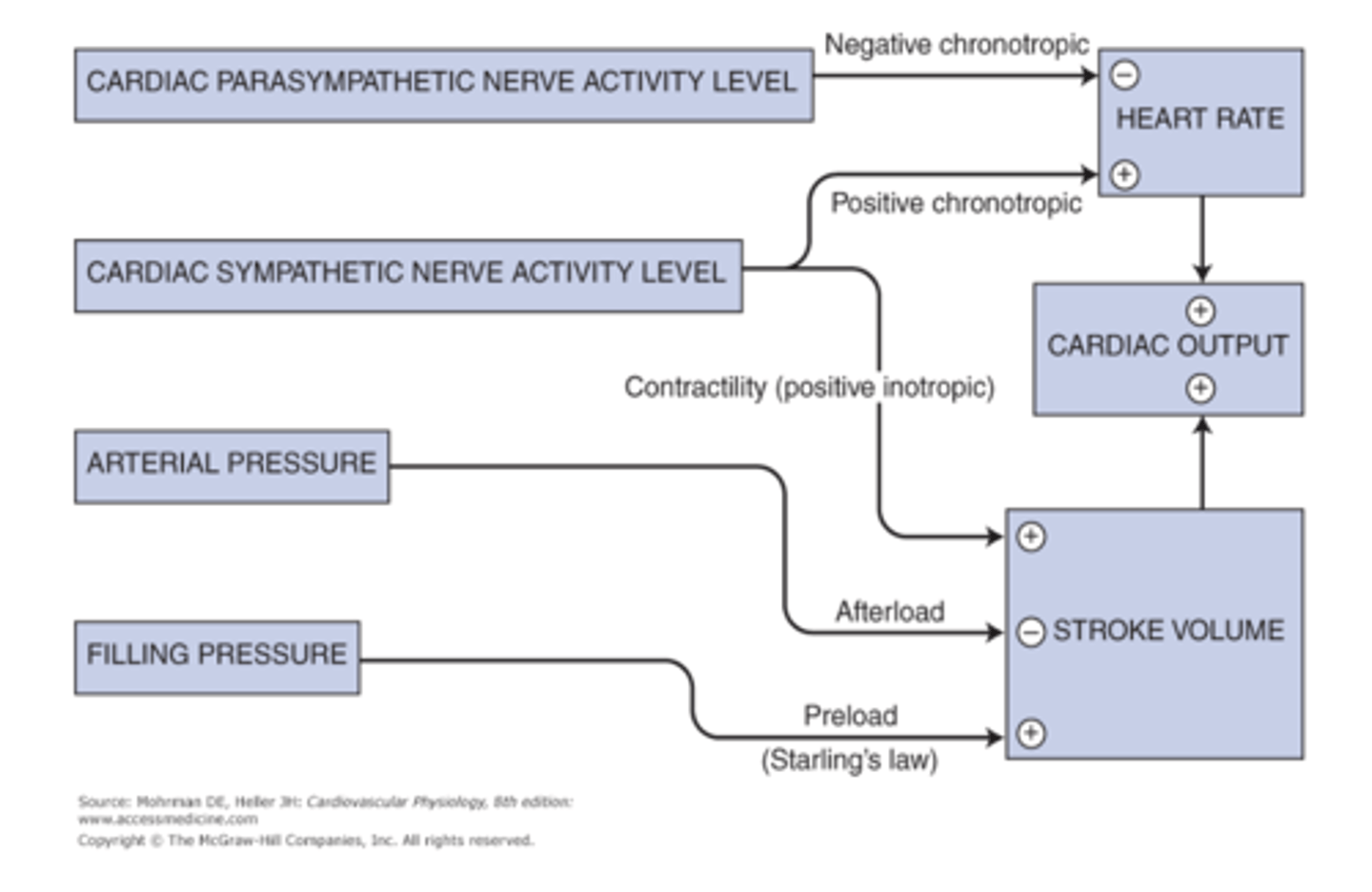
the force of the contraction
If all other factors remain constant, the SV (stroke volume) is determined by
This is possible because of the nature of striated and cardiac muscle, which has a force of contraction proportional to the degree to which it was stretched.
The heart will automatically eject all the blood that enters it over a broad range of volume. This is possible because of?
1. Contraction
2. Elasticity (elastic recoil)
The myocardial wall has two important properties during systole what are they?
SV (stroke volume) and therefore in the CO (cardiac output)
According to the Starling's law, any change in the venous return (increased preload) has a direct effect on the
SV; CO
Increase in the venous return causes an increase in the strength of the contraction which causes an increase in the ______which increases the ________.
Frank- Startling mechanism
the stretched ventricular walls recoil back to their resting position with greater force.This phenomenon is called the

length of the Ventricular Muscle Fibers, which implies that all the extra blood resulting from increased venous return is pumped out.
The strength of contraction increases in proportion to the
The quantity of blood in the ventricle just prior to their contraction determines the initial length of the ventricular muscle fibers
What is as End-diastolic volume?
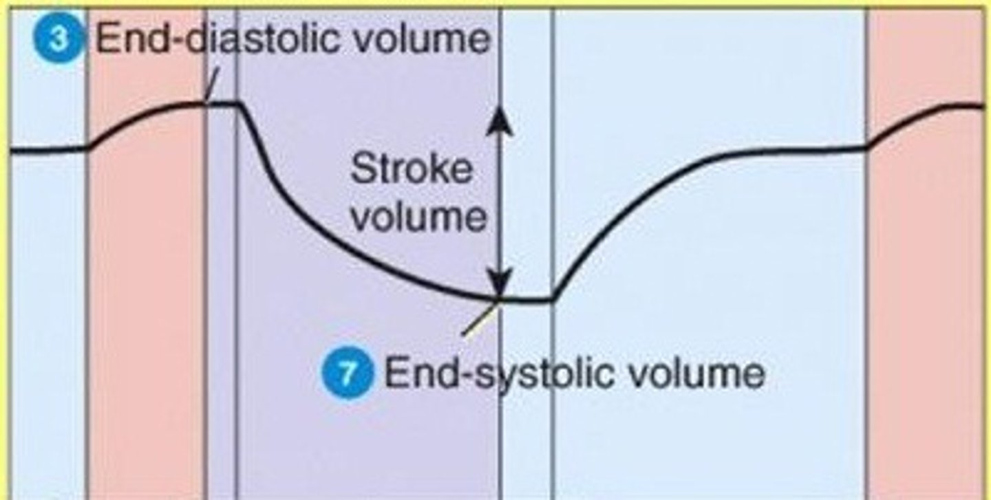
the degree of tension of the muscle when it begins to contract.
What is pre load?
SV; end-diastolic volume; SV
Increased blood pressure, increases the resistance which decreases the ______ which causes an increase in the _______which increases the strength of myocardial contraction which increases the _______.
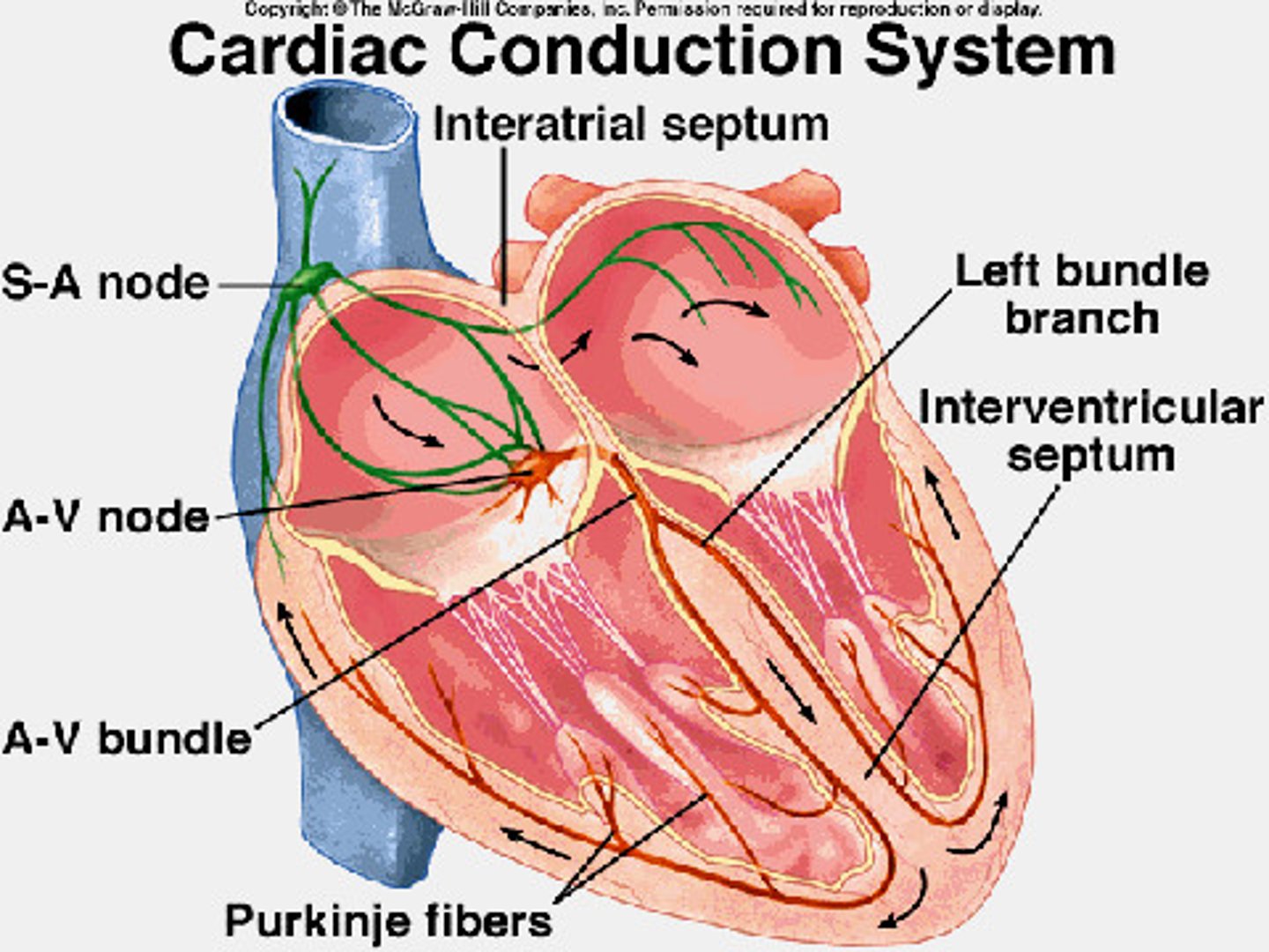
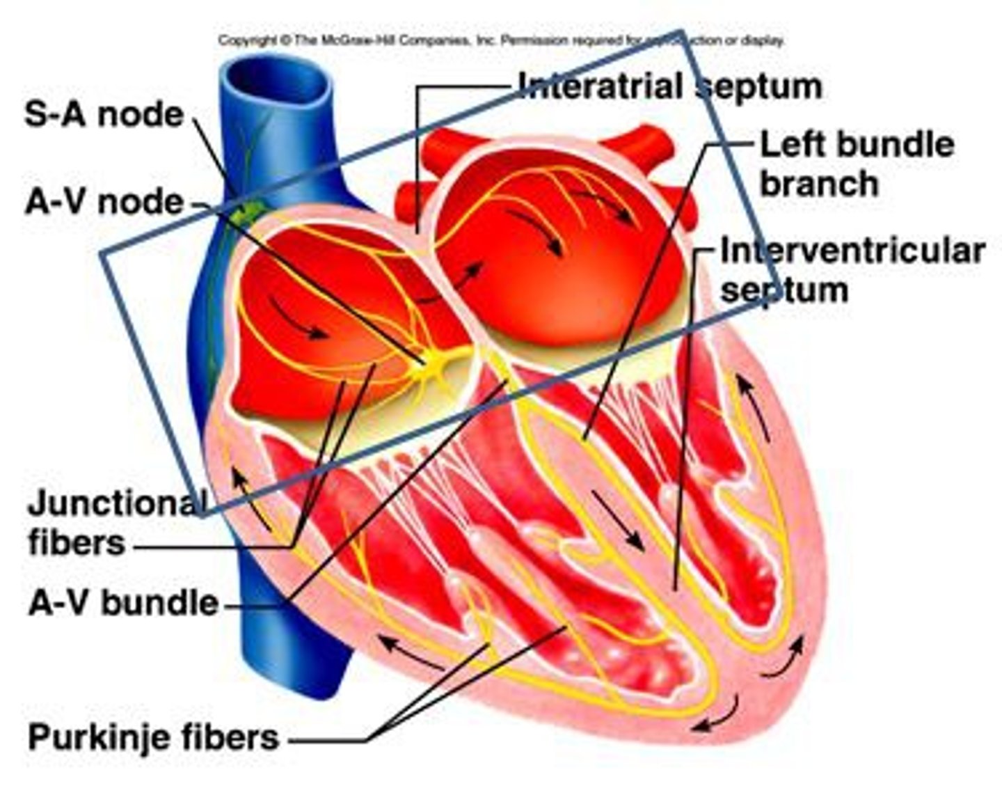
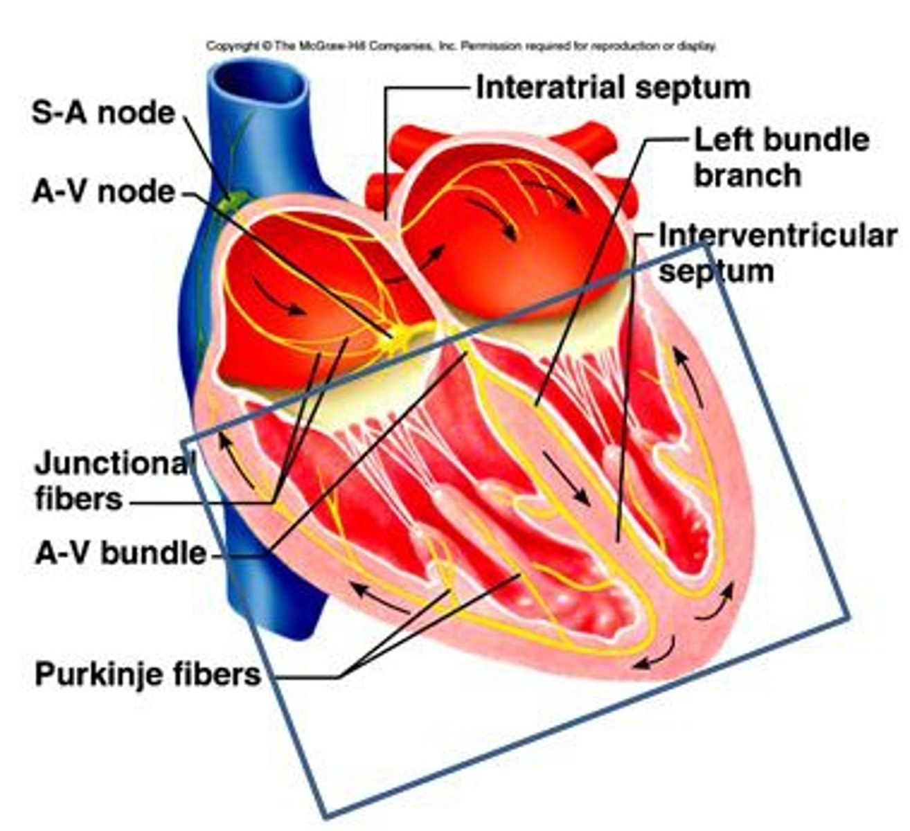
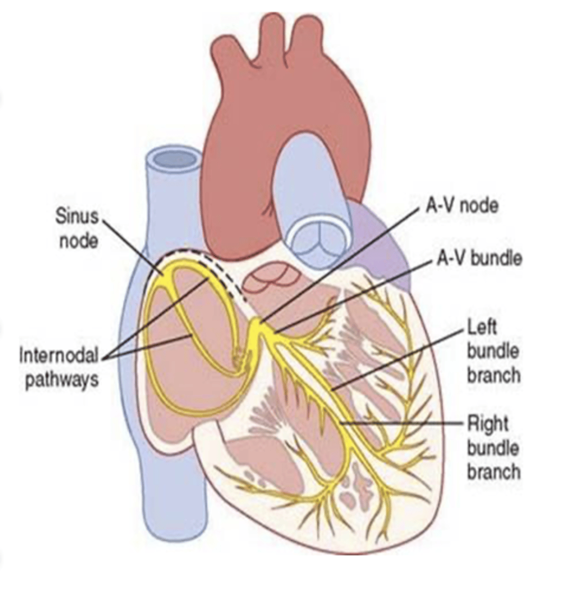
(1) generating rhythmical electrical impulses to cause rhythmical contraction of the heart muscle and
(2) conducting these impulses rapidly through the heart.
the heart has a special system for what (2)?
1/6
when the Rhythmical Excitation of the Heart functions normally, the atria contract about _____ of a second ahead of ventricular contraction, allowing filling of the ventricles before they pump the blood through the lungs and peripheral circulation.
most effective pressure generation in the ventricular chambers.
The Rhythmical Excitation of the Heart allows all portions of the ventricles to contract almost simultaneously, which is essential for
the atrial syncytium and ventricular syncytium
The heart actually is composed of two syncytium's. What are they?
constitutes the walls of the two atria
What does the atrial syncytium constitute?
constitutes the walls of the two ventricles.
What does the ventricular syncytium constitute?
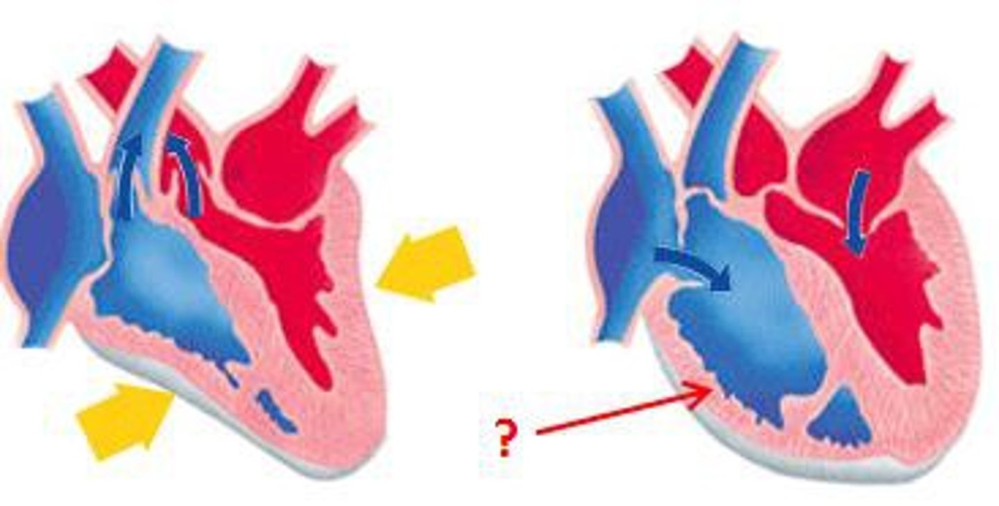
The atria are separated from the ventricles by fibrous tissue that surrounds the atrioventricular (A-V) valvular openings between the atria and ventricles.
What are the atria separated from the ventricles by?
The cardiac cycle is divided in diastole (relaxing) and systole (contract)
What is the cardiac cycle divided into?
defined as the cardiac events that occur from the beginning of one cycle to the beginning of the next.
What is the cardiac cycle defined as?
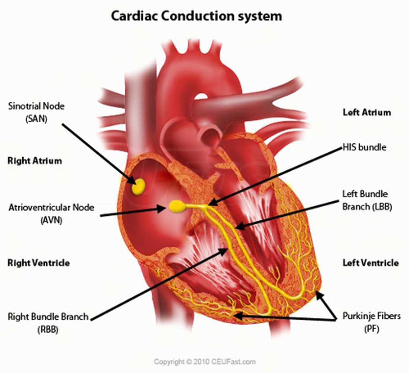
Each cycle is initiated by spontaneous generation of an AP (action potential) in the sinoatrial node (S-A node).
What is each cardiac cycle initiated by?
The action potential travels through both atria and reaches the atrioventricular (A-V) node, bundle of his, bundle branches, Purkinje fibers and finally all the cardiac fibers.
what does the action potential of the cardiac cycle travel though and reach?
Some cardiac fibers have the capability of self-excitation, a process that can cause automatic rhythmical discharge and contraction
What is automatic rhythmicity?
Sinus node
What node has ordinary controls the rate of beat of the entire heart (automatic rhythmicity)
allowing the atria to contract a little before than the ventricle, thus contributing to fill the ventricle
There's a delay in the impulse going from atria to ventricle, allowing for what to happen?
not contraction but impulse generation and transmission.
The conductive system is composed of specialized muscle tissue which function is what?
1 second
There're 4 components in the cardiac cycle and since the heart rate (HR) is usually 70 beats/min, all 4 components are accomplished in less than
pressure gradient across them.
The opening and closing of valves are passive processes determined by the
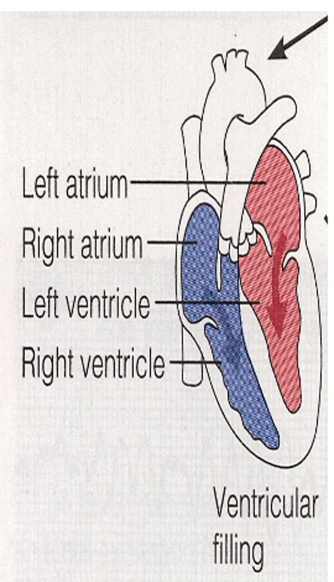
Technically, the cycle begins at the end of systole when all valves are closed and both atria are being filled with blood returning to the heart cause pressure within the atria till is higher than in the relaxed ventricle. Leading to opening of A-V valves starting ventricular filling.
When does the cardiac cycle technically begin?
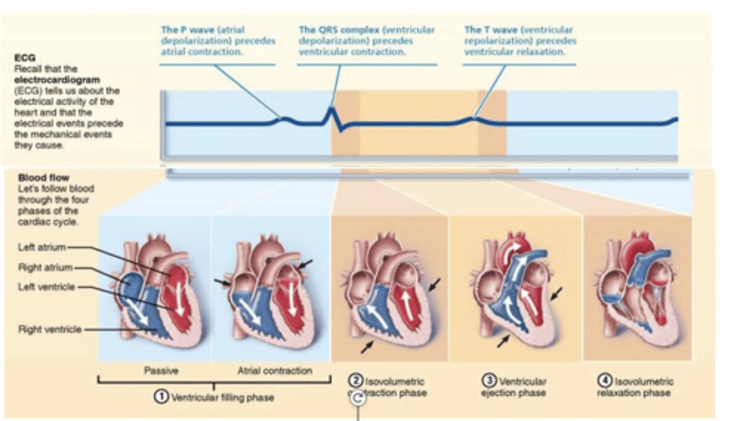
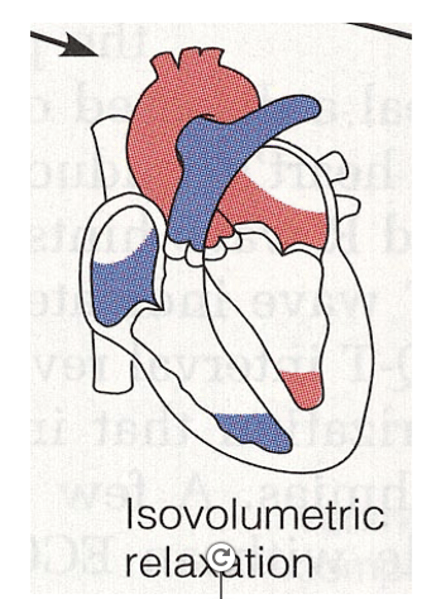
A = Diastole
B = Systole
C = isovolumetric contraction
D = ejection
E = isovolumetric relaxation
The cardiac cycle phases in the chart are what
A = _____
B = _____
B is divided into
C = _____
D = _____
E = ______
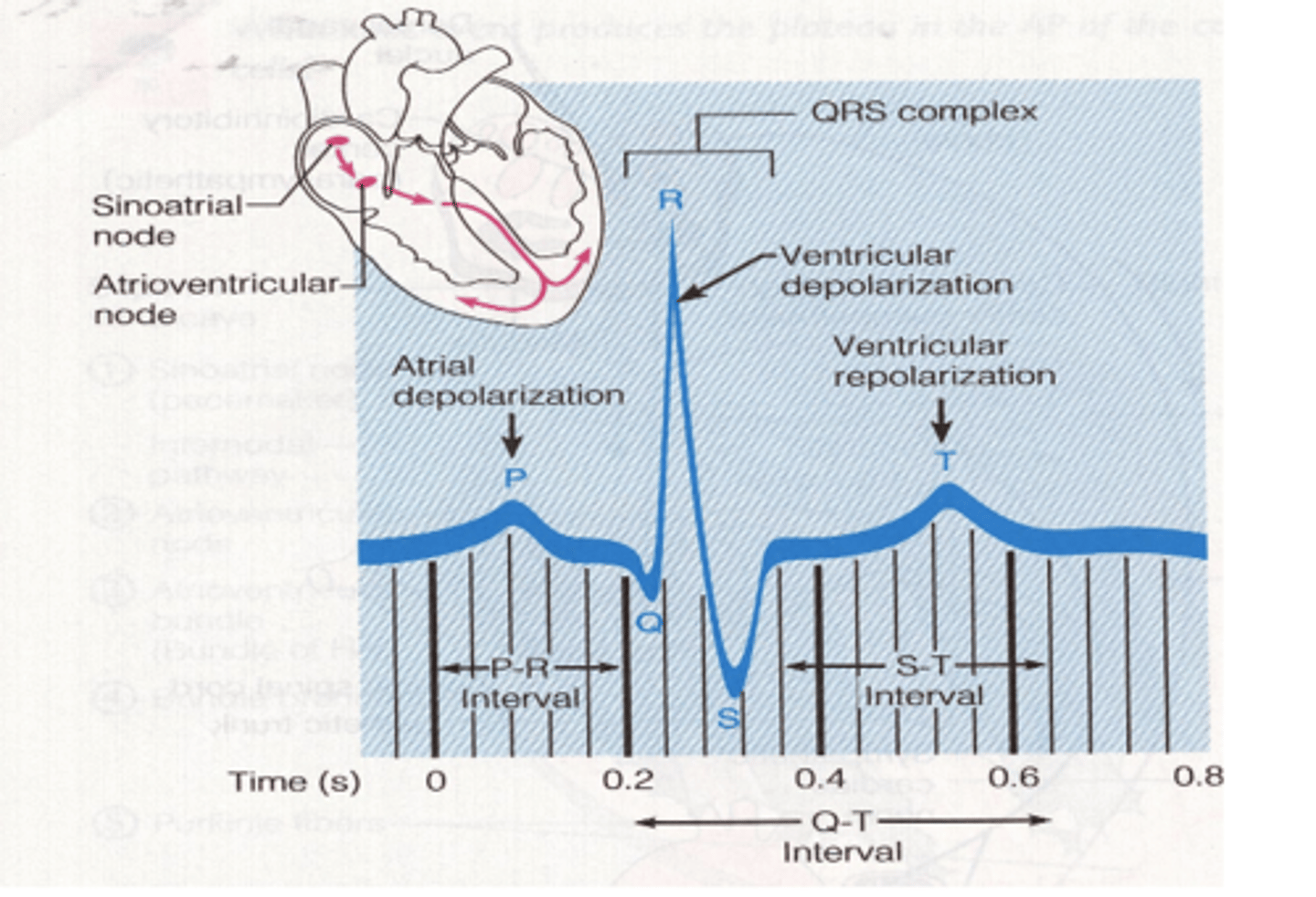
P wave represents atrial depolarization. It occurs just before atrial contraction
What does the P wave represent in a ECG (EKG)?
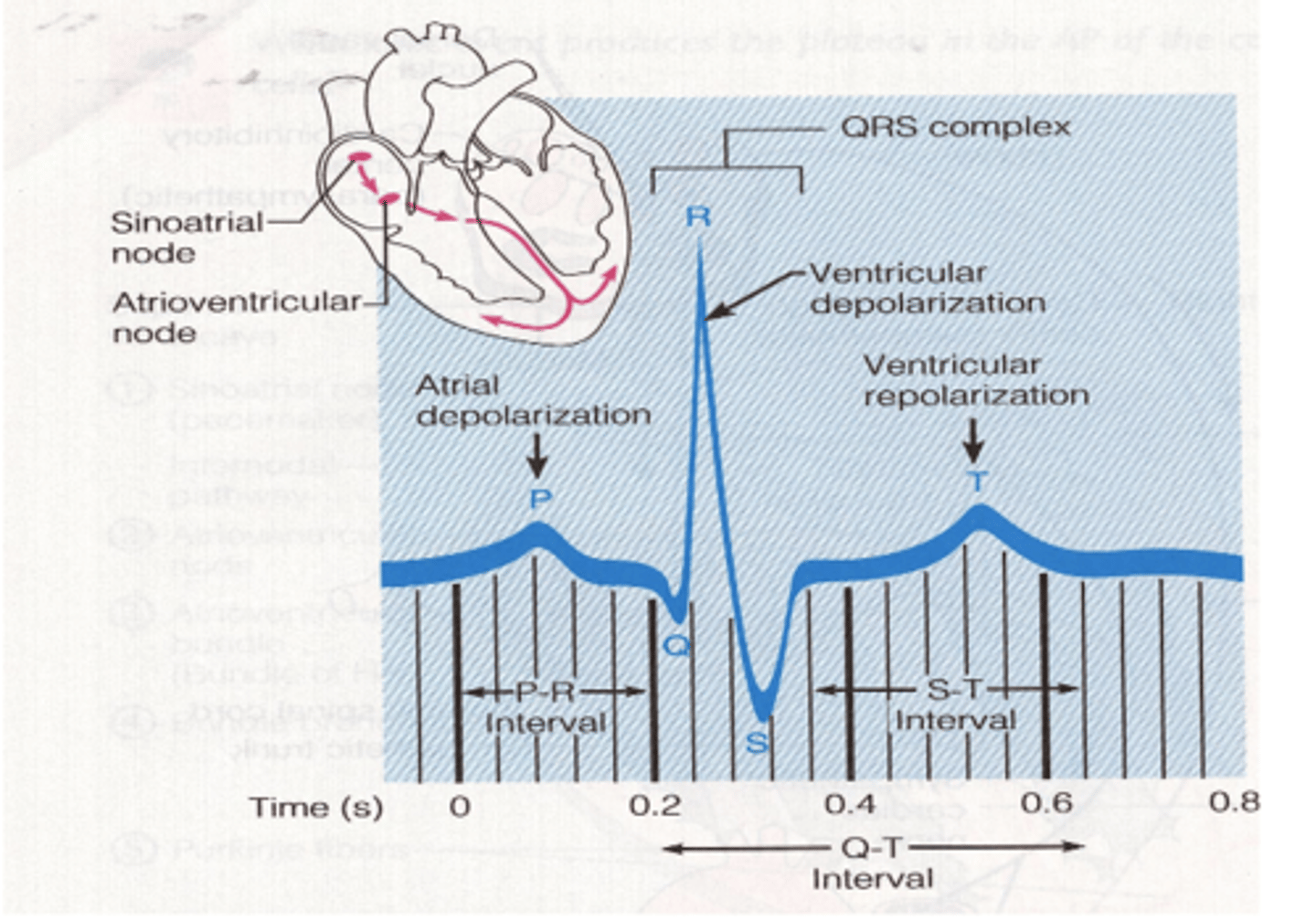
QRS complex represents ventricular contraction
What does the QRS complex represent in a ECG (EKG)?
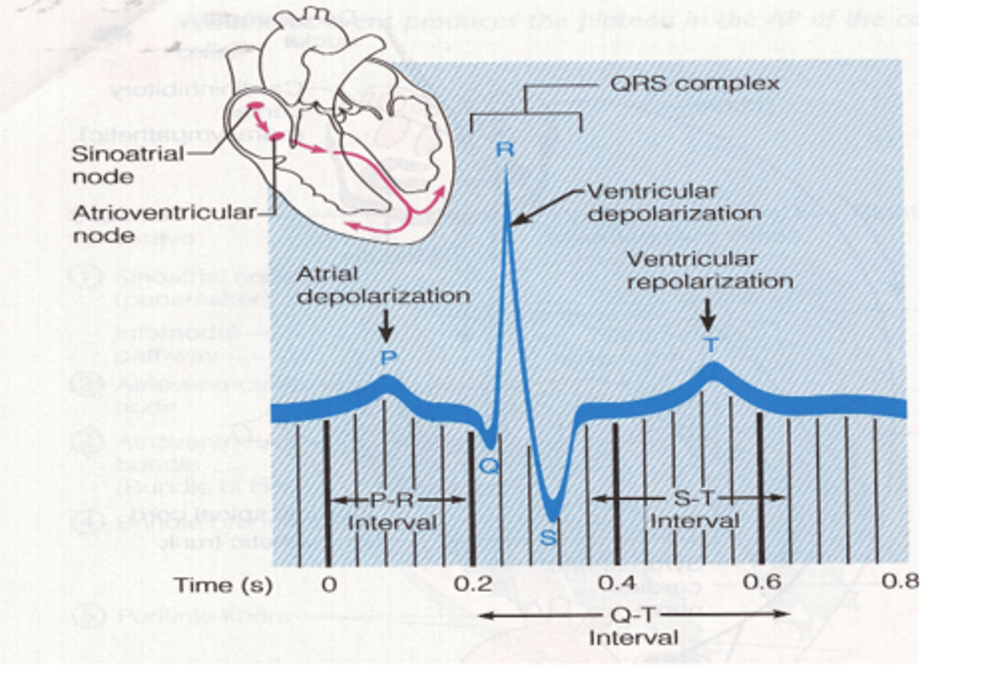
T wave represents repolarization of ventricle.
What does the T wave represent in a ECG (EKG)?
There are 8 phases total. 5 phases during diastole.
The cardiac cycle can also be described in terms of the phases of ventricular pumping. How many are there?
Fall in IV pressure and closing of aortic valve.
What is protodiastole?
Ventricle is closed; muscle relaxing
but not lengthening.
What happens during isovolumetric relaxation?
Rapid filling:
Immediately after the opening
of AV valves.
What happens during ventricular filling?
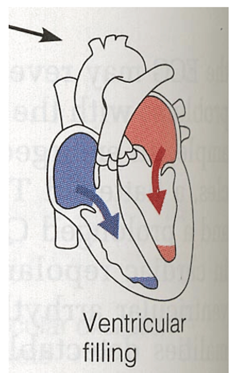
almost no blood flow is flowing from the atria.
What happens during Diastasis?
increase ventricular filling
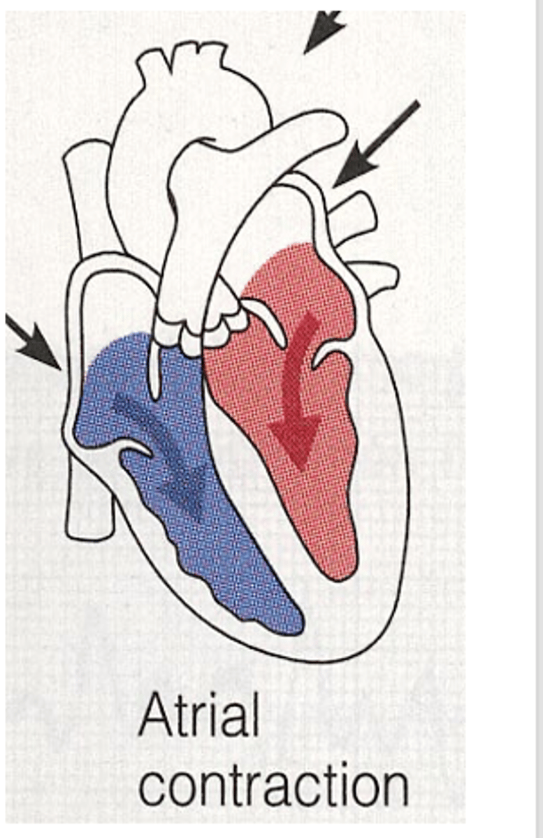
What happens during atrial contraction?
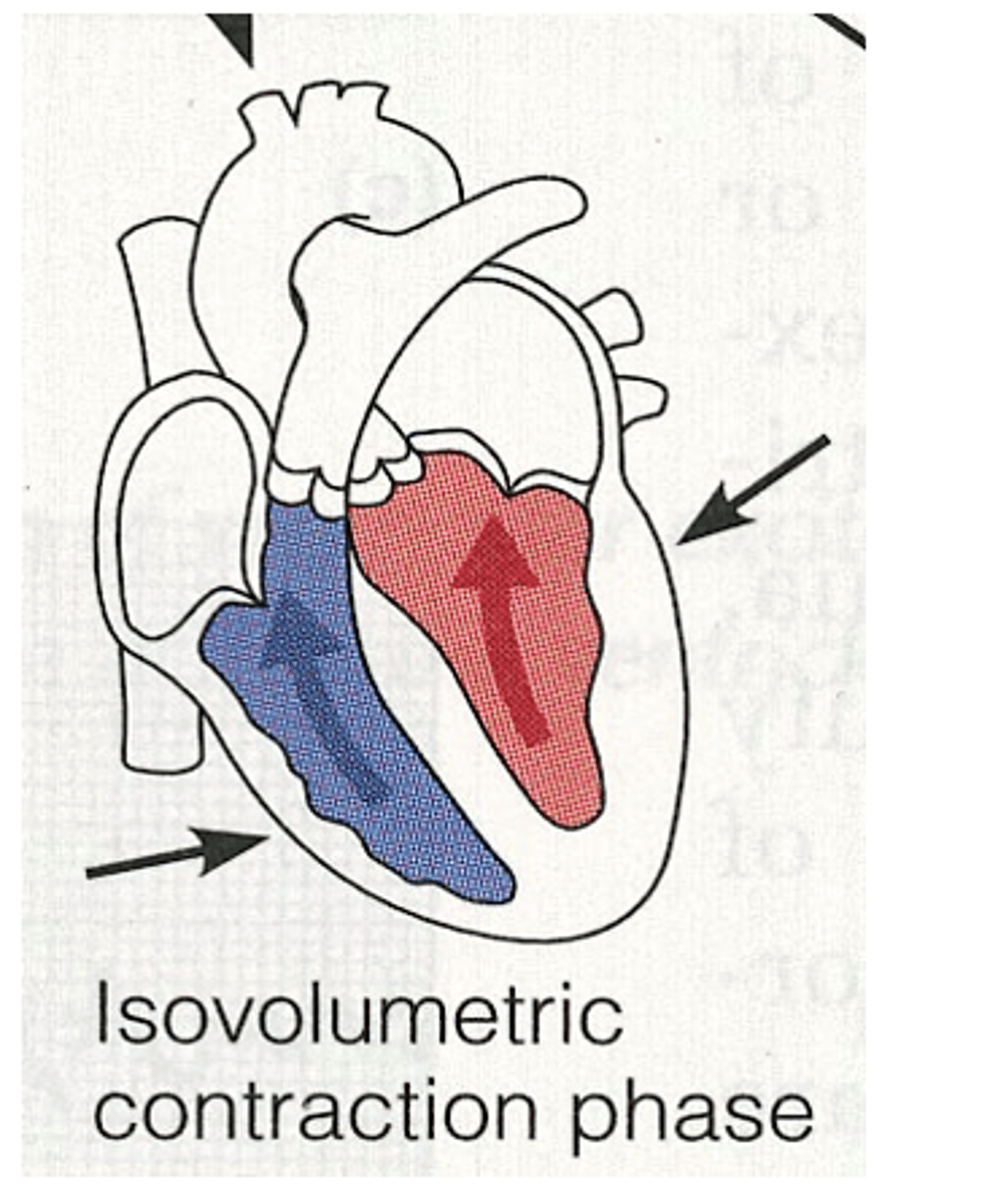
1. Isometric contraction
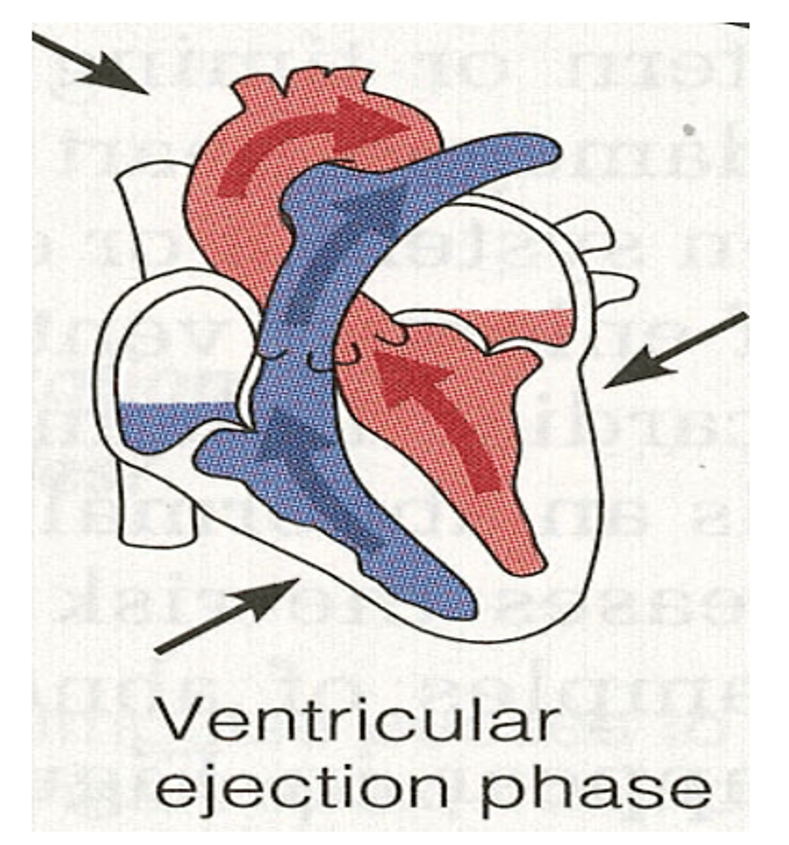
2. Rapid ejection
3. Reduced ejection
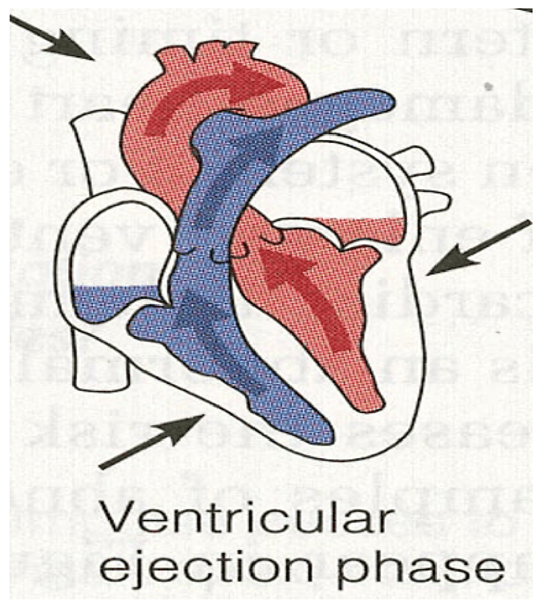
What are the three phases during systole?
Just after the beginning of ventricular systole. But before opening of semi-lunar valves.
During systole when does isometric contraction occur?
Semi-lunar valve. Open and blood is
pushed into the great artery.
During systole when does rapid ejection occur?
Ventricle remain contracted but little blood is pumped out.
During systole when does reduced ejection occur?
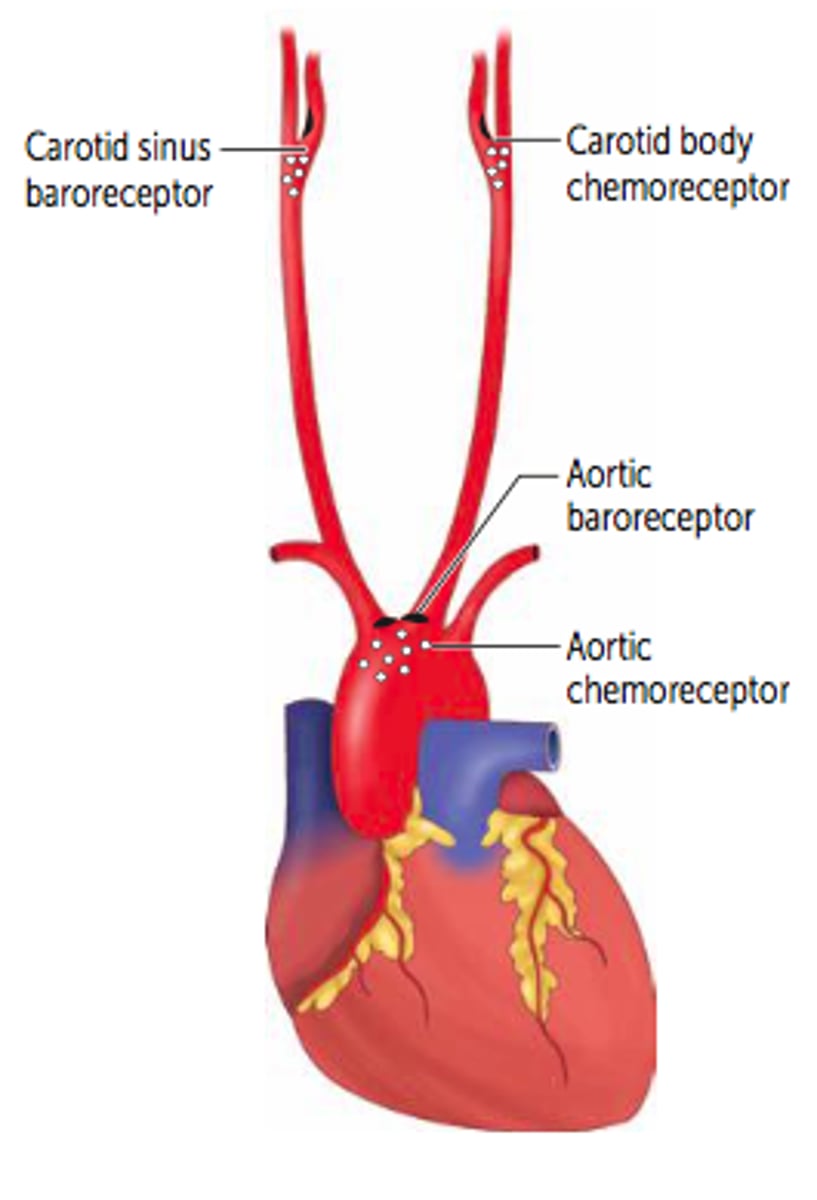
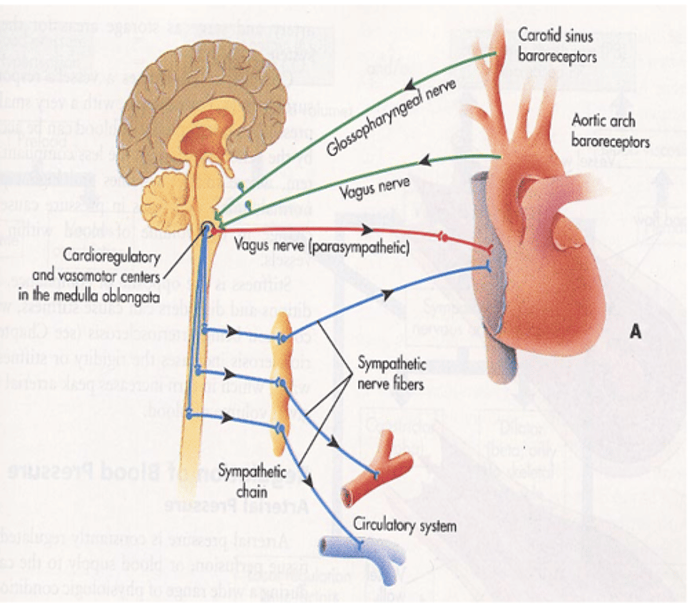
Sensors that respond to distension of arteries wall as a result of increase in pressure.
What are baroreceptors?
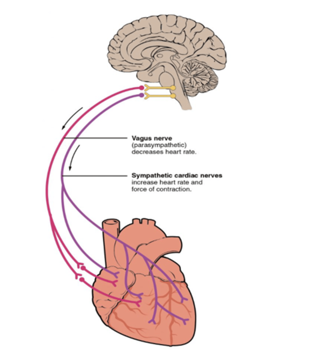
sending impulses to the cardiac center (medulla oblongata) specifically the cardioinhibitory center which is activated triggering parasympathetic stimulation
What do baroreceptors send impulses to when there is responce to distension of arteries wall as a result of increase in pressure.
to reduce the heart rate. This, along with increasing vasodilation of vessels, acts to reduce the arterial pressure.
If an increase in arterial pressure is detected by baroreceptors the parasympathetic pathway is activated to
to increase the heart rate and the contractility of the heart. This, along with increasing vasoconstriction of vessels, acts to increase the arterial pressure.
If a decrease in arterial pressure is detected by baroreceptors the sympathetic pathway is activated to
baroreceptors not stimulated
- no parasympathetic discharge
- Sympathetic acts through cardioaccelerator center.
if blood pressure decreases are baroreceptors stimulated?
located in the aortic arch and innervated by the vagus nerve
where is the Aortic sinus located and what is it innervated by?
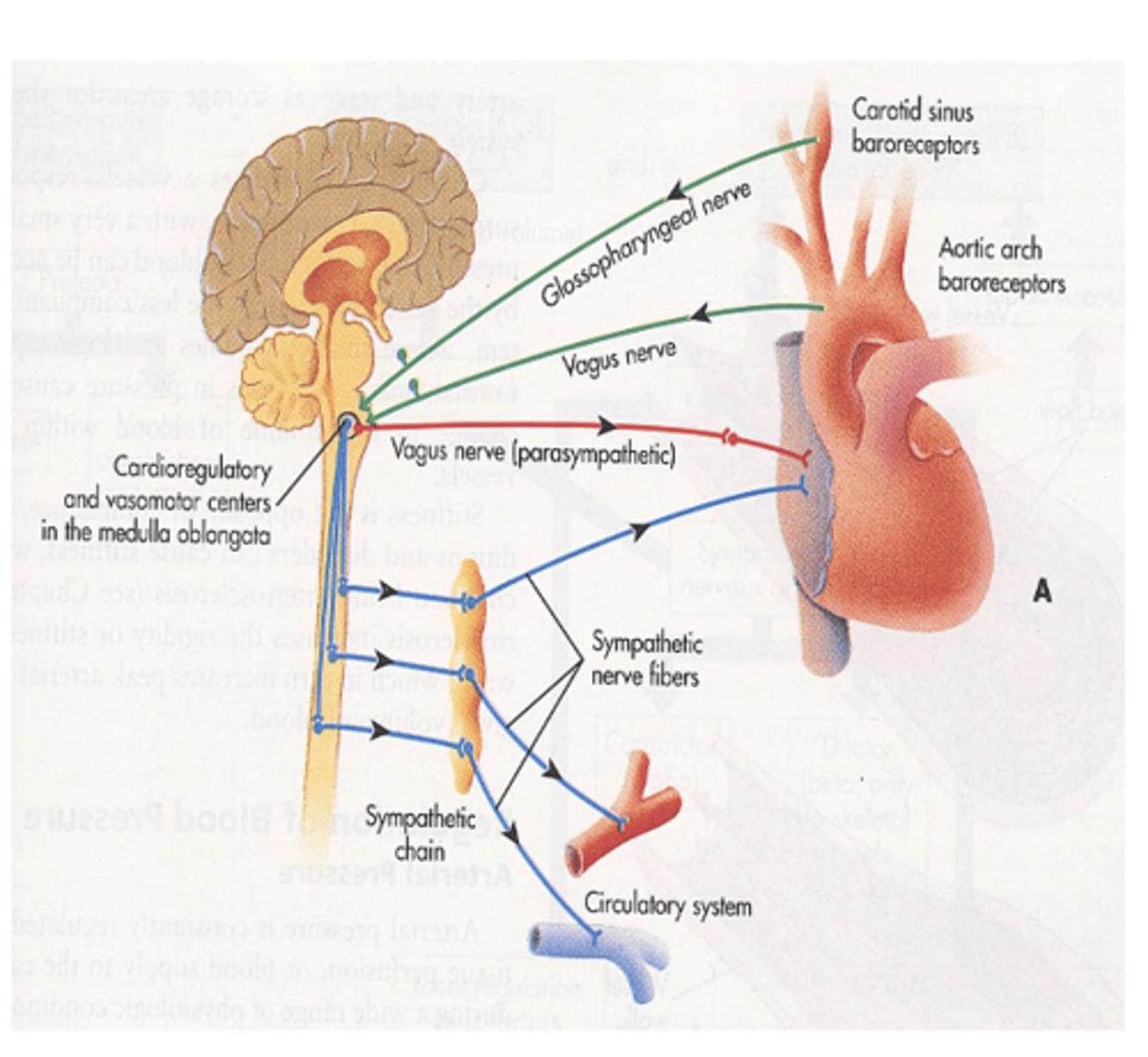
located at the bifurcation of each common carotid innervated by the carotid sinus nerves or Hering’s nerve
Where is the carotid sinus located and innervated by?
O2 and CO2 in the blood (po2 and pco2).
What do chemoreceptors respond to changes in?
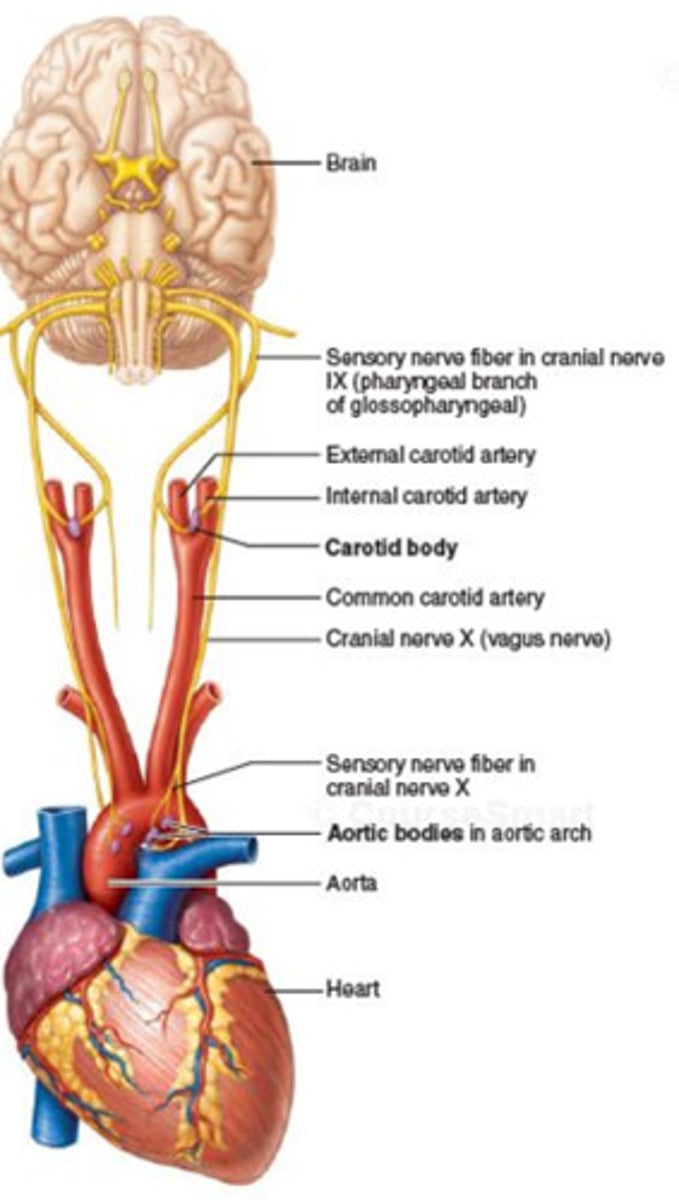
There're located near the aortic and carotid sinuses and known as Carotid and aortic bodies.
Where are chemoreceptors located near? What are they known as? (2)
Medulla oblongata.
Hypoxemia causes Increase CO but it is thought to be the result of direct stimulation of the
Decreased or unchanged
If hypoxemia is limited to the carotid and aortic bodies,
The heart action is
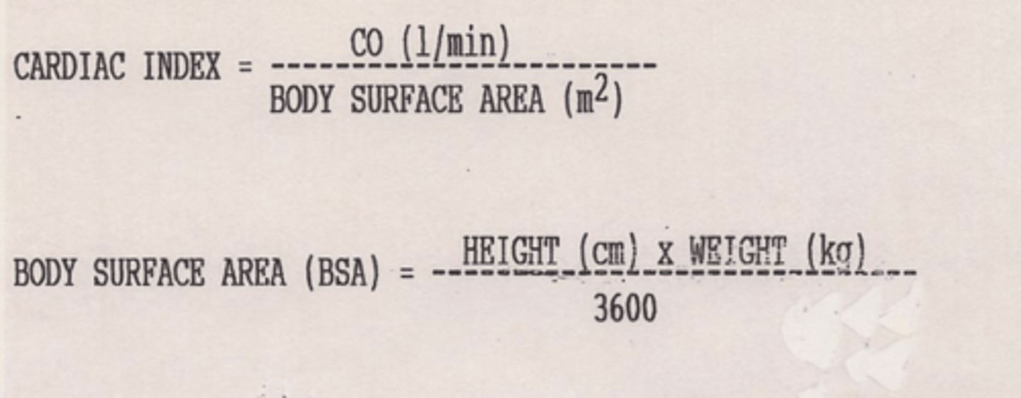
This value adjusts the cardiac output to the individual person's body size
by representing blood flow relative to a square meter of body surface area.
What is cardiac index?
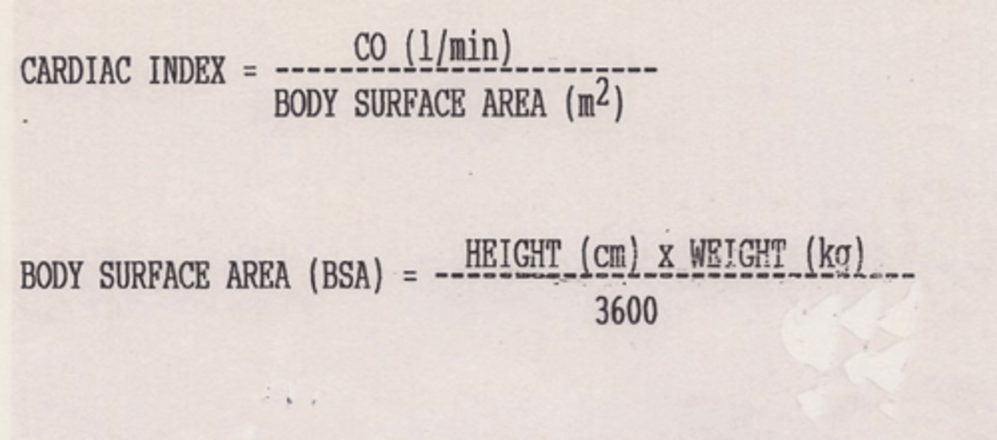
2.5-4.2 L/min/m2
What is the normal adult cardiac index range?
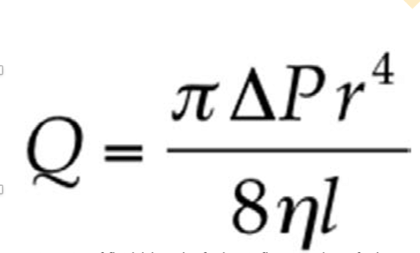
1
The resistance is expressed in PRU (peripheral resistance unit).
If Delta p = 1 mmHg and
Blood flow:
Q = 1 ml/sec then _____ PRU
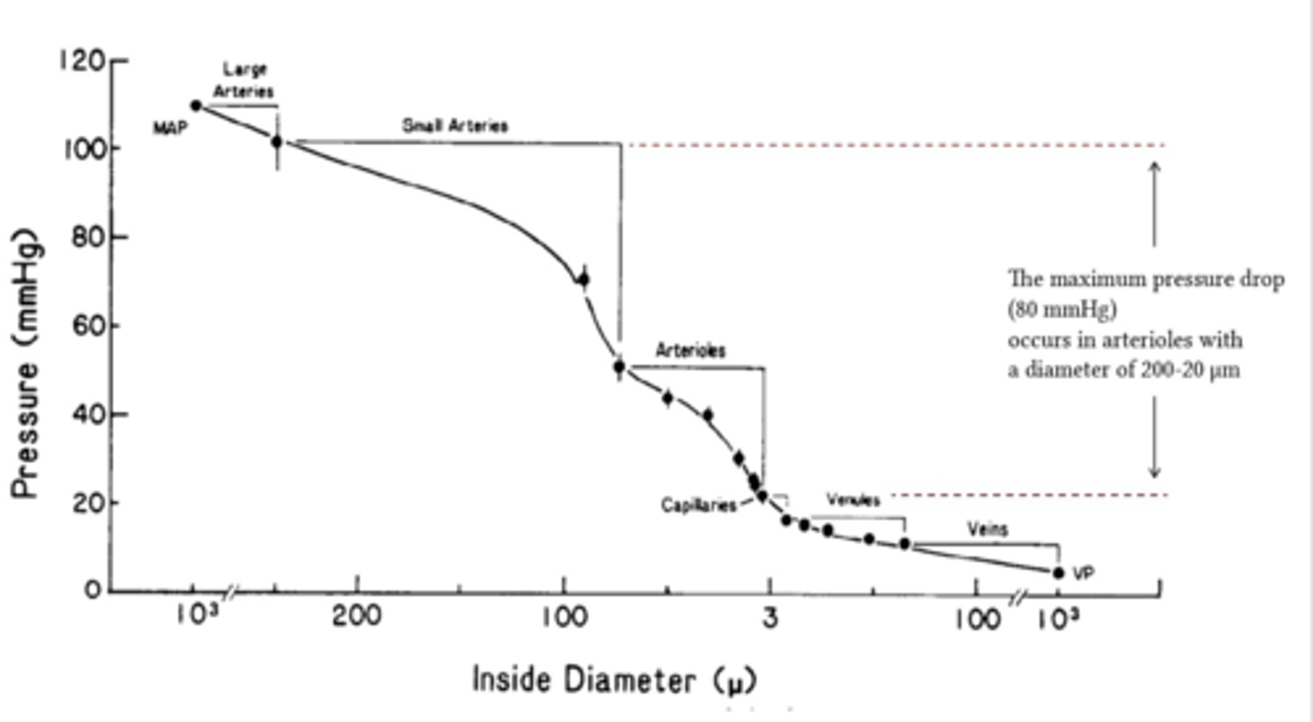
the diameter of the blood vessel (which is equal to twice the radius) plays by far the greatest role of all factors.
the rate of blood flow is directly proportional to the 4th power of the radius.
In Poiseuille's law what plays by far the greatest role of all factors?
1. Generating rhythmical impulses to trigger contraction of the heart muscle
2. Conducting these impulses quickly throughout the heart.
What does the heart has a specialized system for? (2)
an ion will move down its concentration gradient
What is chemical potential?
an ion will move away from ions/molecules of like charge.
What is electrical potential?
the electrical potential difference (voltage) between the inside and the outside of a cell.
What is the transmembrane potential (TMP)?
more +ve
When there is a net movement of +ve ions into a cell, the TMP becomes
-ve
when there is a net movement of +ve ions out of a cell, TMP becomes more
help maintain ionic concentration gradients and charge differentials between the inside and outside of the cardiomyocytes
What do ion channels help maintain?
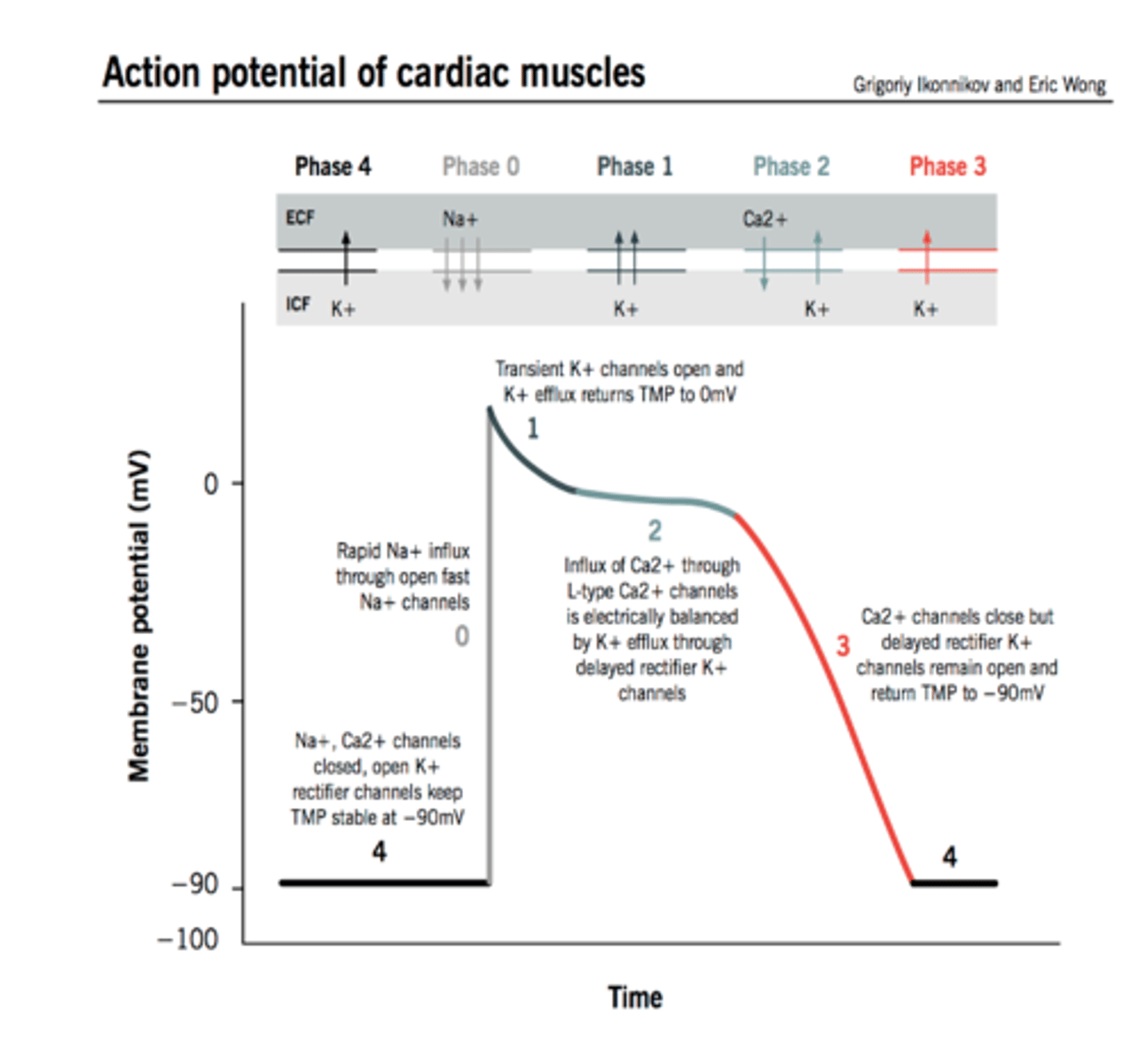
Phase 4: The resting phase
Phase 0: Depolarization
Phase 2: Early repolarization
Phase 3: Repolarization
The action potential in typical cardiomyocytes is composed of 5 phases (0-4), beginning and ending with phase 4. What are the 4 phases?
Phase 4: _____
Phase 0: _____
Phase 2: _____
Phase 3: _____
Naturally leaky to Na+
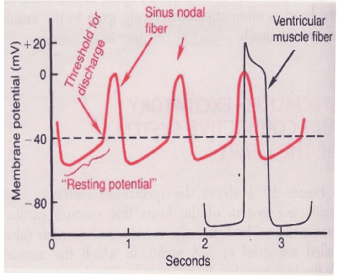
The potential of the S-A fibers between discharges has a negativity of only 55-60 millivolts, compared to 85-90 mv for the ventricular muscle fibers, since the S-A fibers are
1. Fast sodium channels
2. Slow calcium channels
3. Potassium channels
There're 3 types of membrane ion channels playing an important role in causing the voltage changes of the action potential what are they?
because it has the fastest depolarizing rate, also known as the intrinsic rate (60-100 impulses/min).
Why does the S-A node act like the pacemaker of the heart?
60-100 imp/min
What is the S-A nodes intrinsic rate?