(2.5-2.8) Trachea, Bronchi, cavum thoracis, Lungs
1/13
Earn XP
Description and Tags
include from page 53 trachea, bronchi, cavum thoracis, lungs chapter page. 61.
Name | Mastery | Learn | Test | Matching | Spaced | Call with Kai |
|---|
No analytics yet
Send a link to your students to track their progress
14 Terms
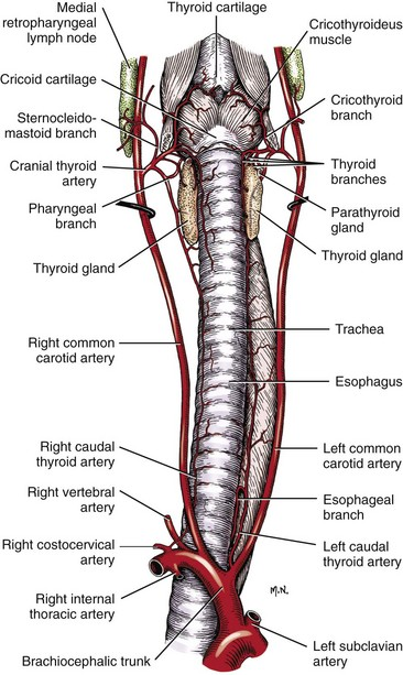
Where is the start and end of trachea? what is the two main parts?
Transports air from larynx to main bronchi.
pars cervicalis
ventrally to esophagus + m. longus colli on neck.
thyroid gland: caudally to larynx on both sides of first tracheal cartilages.
trachea → goes to thoracic cavity by thoracic inlet, ventrally to m. longus colli.
pars thoracica
bw. right + left lung in mediastinum.
accompanied by esopagus + cranial vena cava dorsally.
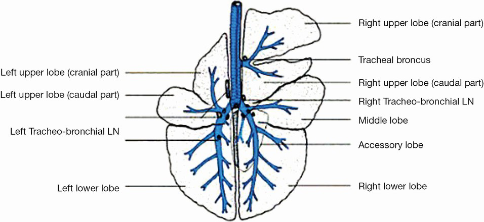
Trachea → divides into bronchi principales dexter et sinister dorsally to the base of heart. This division is at the level of 4th to 6th intercostal space → bifurcatio tracheae.
In ru + su: bronchus trachealis → arises in short distance to bifurcation and to the right. Gets air into lobus cranialis dexter.
The wall of trachea consist of which 4 layers?
tunica mucosa (covred with resp. epithelium)
tela submucosa (contains gll. tracheales)
cartilagines tracheales (incomplete rings formed from the hyaline cartilage)
number + shape - differ acc. to species.
connected by ligg. anularia.
paries menbranaceus (loose CT, interconnects the rings)
m. trachealis (bw. CT + mucous membrane, only in car + cun tis muscle is on the external side of the rings)
tunica adentitia (connects trachea with surrounding tissues, in pars thoracica, trachea is covered by serous membrane)
eq: flattened
bo + ov: free ends of tracheal cartilages are in contact by their internal surface which forms the dorsally located crest → carina tracheae.
cap: shape of tracheal cartilages are similar to letter U.
su: free ends interleaved one over the other, in cross-section, they are circular.
car: circular in cross section, free ends of tracheal cartilages are connected by thickened adventitia.
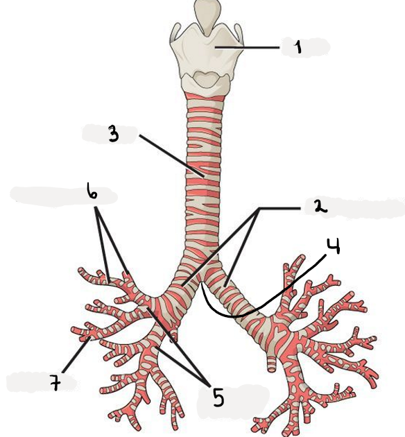
Structures of bronchii.
larynx
trachea
bronchus trachealis (third branch in ru + su, cranially)
bronchus principales dexter et sinister
bifurcatio trachea (start of arbor bronchialis (the tree)
bronchi lobares (when bronchus principales divides, these enters the lung)
bronchi segmentales (from lobares)
bronchuli (arise from segmentales, less than 1 mm.)
These divide into 2 bronchuli respiratorii, which then have branches called ductuli alveolares, which are surrounded by alveoli pulmonis (small cavities).
The ductuli ends in sacculi alveolares (surrounded completely by alveoli pulmonis, in these, gas exchange happens)
These Bronchii makes up the arbor bronchalis (formation of the bronchial tree)
structures in the pictures.
Cavum thoracis: consisting of the bony base of thorax made up of thoracic vertebrae, ribs and sternum
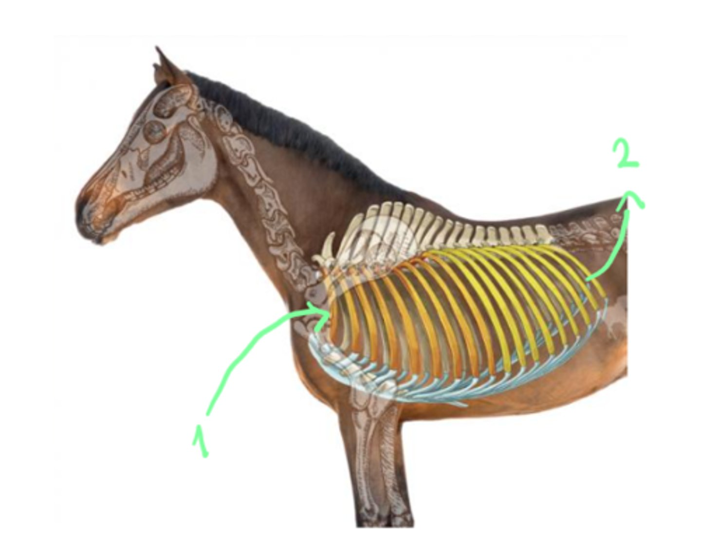
the cavum thoracis opens by means of apertura thoracis cranialis and closes by apertura thoracis caudalis.
thoracic inlet: by first thoracic vertebra, first pair of ribs + manubrium sterni. m. longus colli, esophageus, trachea, arteries + veins goes here.
thoracic outlet: by last thoracic vertebra, last pair of rib, costal arch + xiphoid process.
diaphragm separates the thoracic and abdominal cavity.
The internal surface of ribs, intercostal muscles, sternum, transverse thoracic muscle and diaphragm are covered by the?
fascia endothoracica
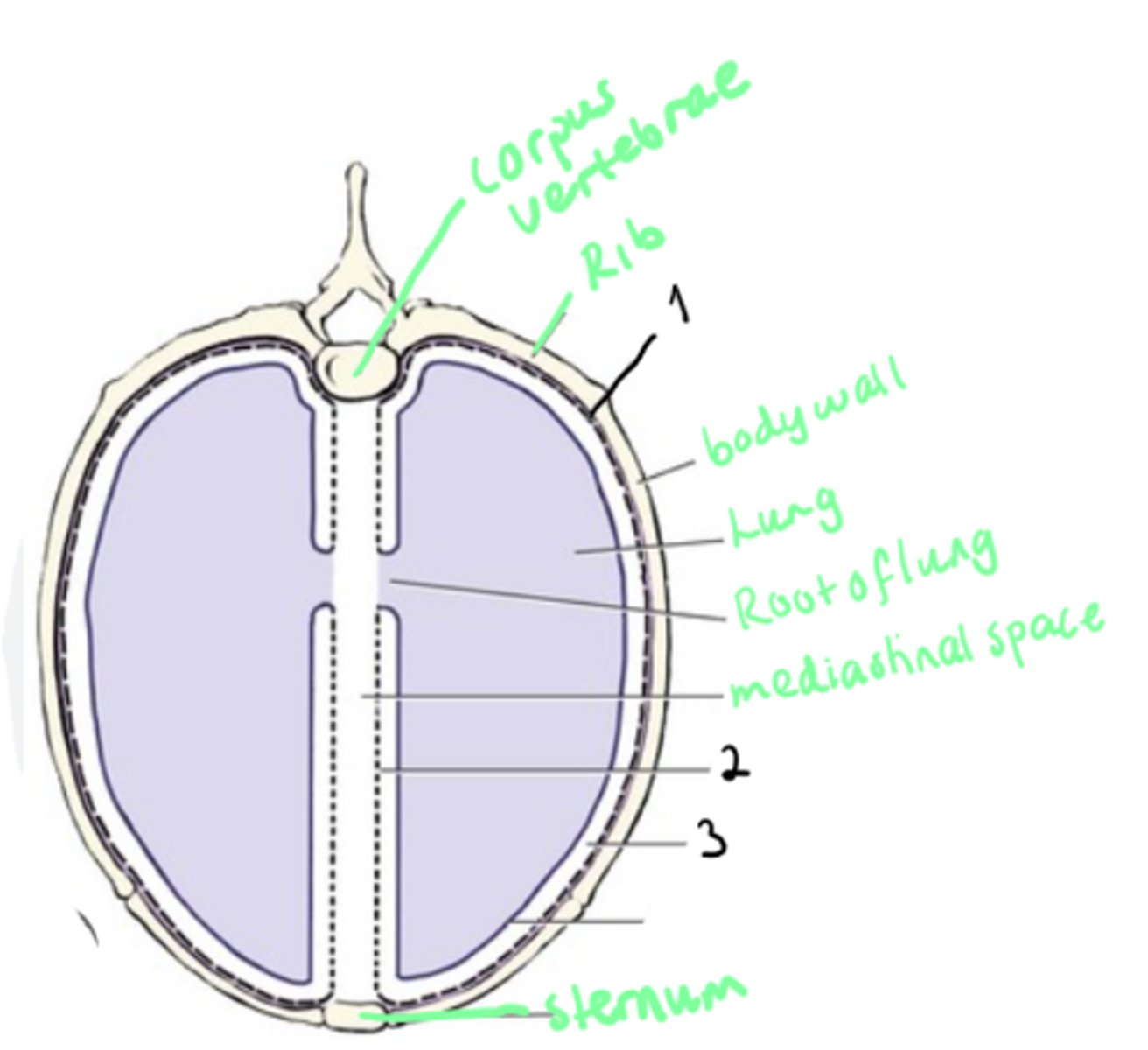
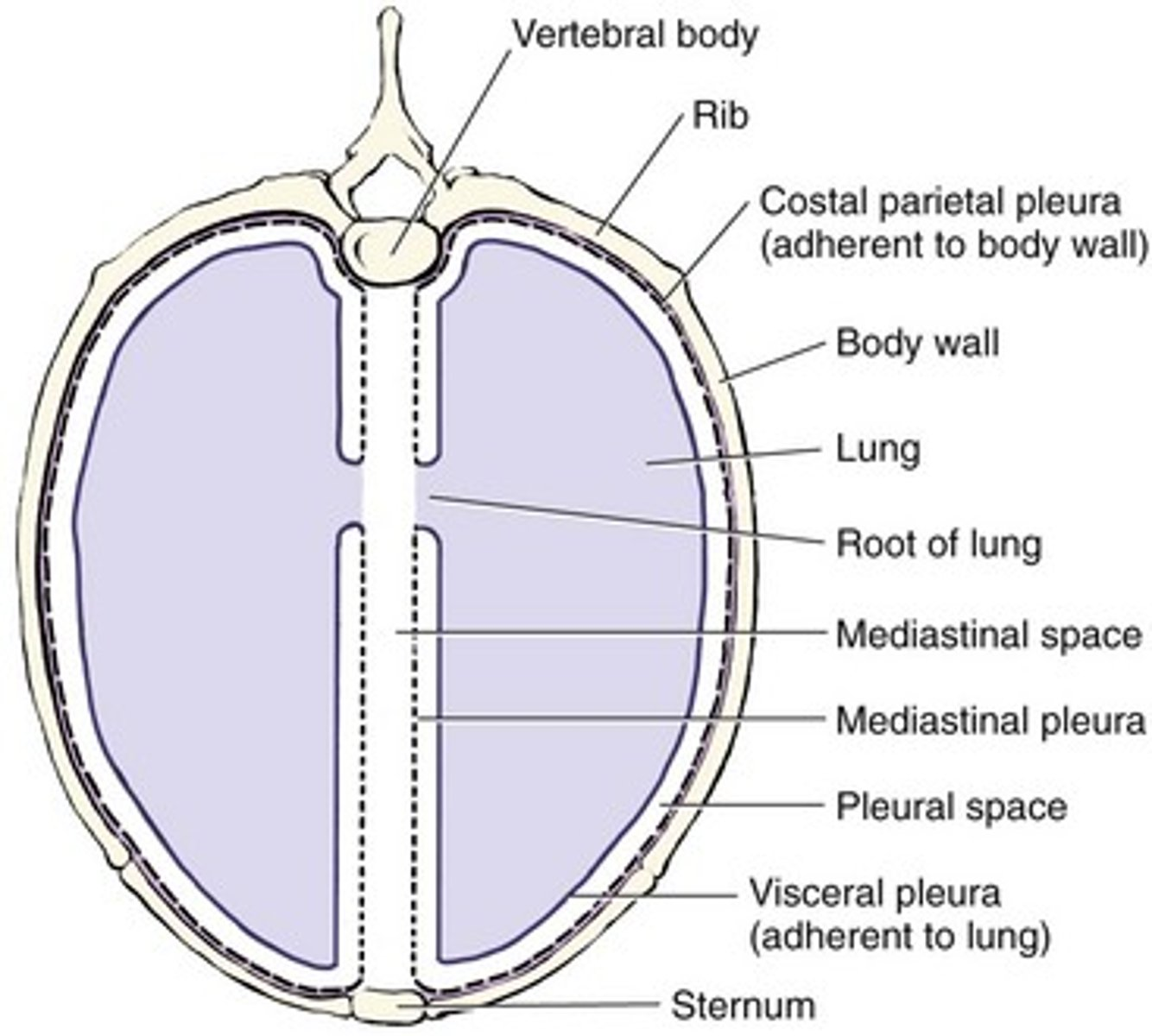
What is the pleura? Name the structures.
Pleura: covers walls of thoracic cavity + organs inside of it. Serous mebrane forming two pleural sacs which enclose the cavum pleura inside with corresponding lung.
consisting of the Pleura parietalis (part of pleura covering the walls)
pleura parietalis costalis (subdivision of p. parietalis, covers ribs)
Pleura mediastinalis (forms walls of mediastinum, subdivision of p. parietalis)
lig. pulmonale (connects this to to pleura pulmonalis)
cavum pleurae (fissure like space between the pleura parietalis and pleura pulmonis)
Car: right + left cavum pleurae protrude the level of first ribs as the cupulae pleurae.
small ru: cupula pleurae is formed only at right side.
others: Pleura diaphragmatica (another subdivision, lying on the diaphragm) and pleura pericardiaca (another subdivision, closing the pericard)
Lungs are covered by the Pleura Pulmonalis
What are the pleural recesses? (There are 3 types)
Recessus Pleurales → allows for movements of the walls and lungs during inspiration and expiration. Located bw. speicfic parts of the pleura parietalis:
recessus costodiaphragmaticus (bw. pleura costalis and diaphragmatica)
recessus costomediastinalis (bw. pleura costalis and mediastinalis)
recessus lumbodiaphragmaticus (in small ru, su + car, formed by pleura going to vertebral end of last rib to first lumbar vertebra)
Mediastinum consists of which 3 parts? 2 new parts also.
Mediastinum: narrow space bw. medial surfaces of left and right lung, covered by pleura mediastinalis.
mediastinum craniale (cranially to heart)
incl. m. longus colli, trachea, esophageus, arteries + veins.
mediastinum medium (contains the heart)
heart with vessels, esophageus, bifurcatio trachea, vagal nerves, bronchial lymph nodes.
mediastinum caudale (caudally to heart)
esophageus, aorta, lymph nodes, dorsal + ventral vagal trunks, left phrenic nerve.
But also:
mediastinum ventrale (between pericardium and sternum)
mediastinum dorsale (bounded by bodies of the corresponding thoracic vertebrae + pericardium)
In recessus mediastini is located the?
the accessory lobe of right lung.
walls of recessus mediastini: pericardium, mediastinum caudale, plica vena cavae + diaphragm.
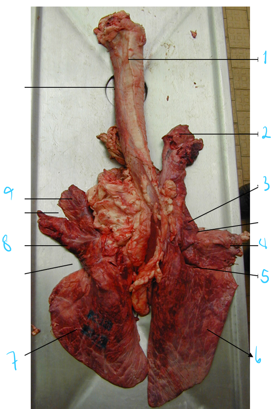
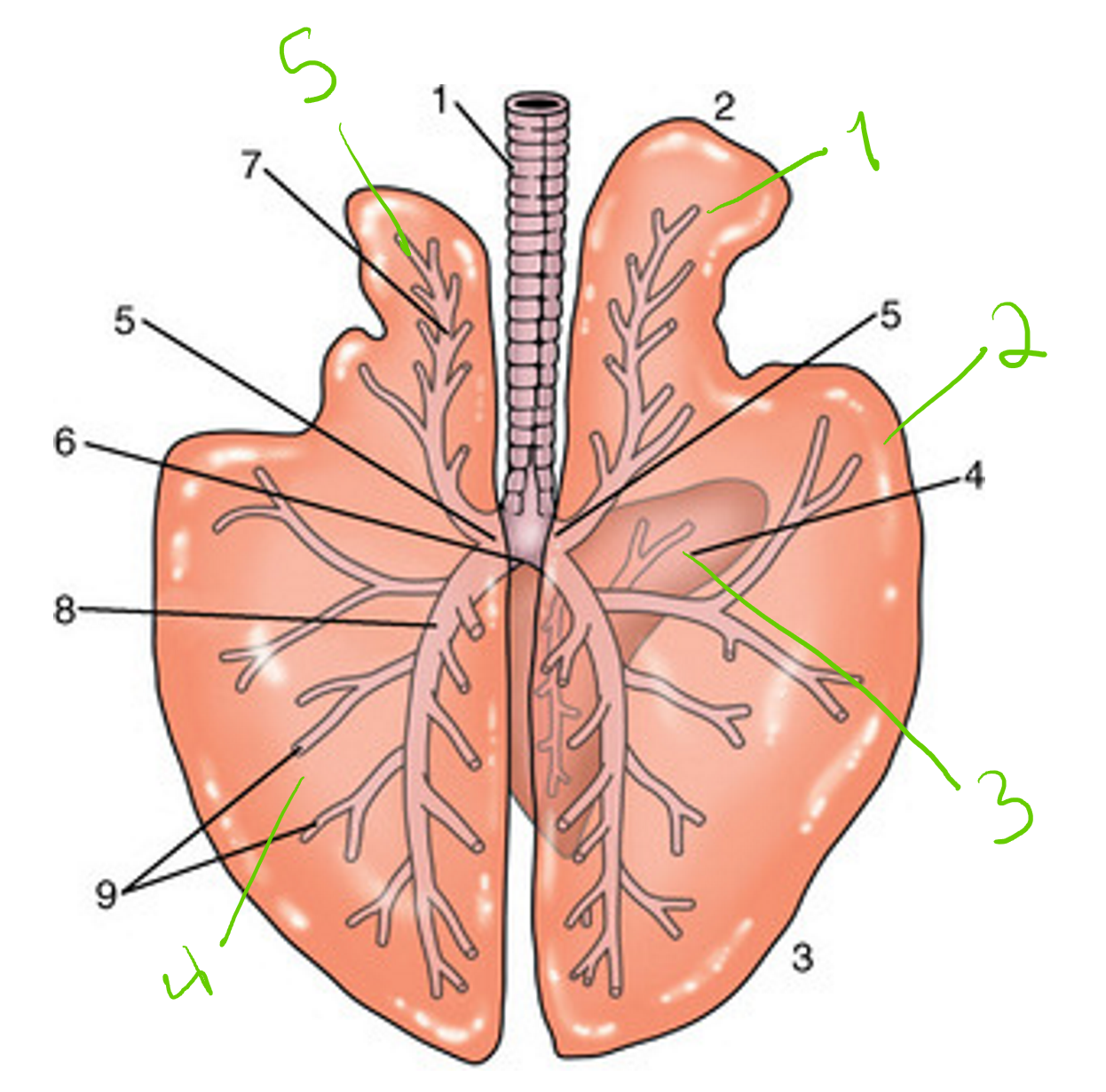
identify these of RU lung.
Seen on facies costalis.
trachea
pars cranialis of lobus cranialis dexter
pars caudalis of lobus cranialis dexter
lobus medius
lobus accessorius under (on right lung)
lobus caudalis dexter
lobus caudalis sinister
pars caudalis of lobus cranialis sinister
pars cranialis of lobus cranialis sinister
Margo medialis (inner side, in middle, we also have hilus pulmonalis here)
margo acutus (the thin outer border)
Apex pulmonis + basis pulmonis
incisura cardiaca pulmonis dextri et sinistri (bw. the lobes)
fissura interlobularis cranialis (bw. lobus medius + lobus cranialis dexter)
fissura interlobularis caudalis (bw. lobus medius + lobus caudalis dexter)
Ru: left: divided lobus cranialis + undivided lobus caudalis, while right: lobus cranialis is divided, medius and accessorius, caudalis undivided.
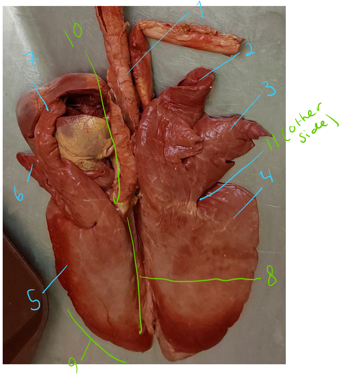
identify parts of this lung of pig/car. What structures separates the lobes?
trachea
lobus cranialis dexter (lateralis)
lobus medius
fissura interlobaris caudalis (bw. lobus medius and caudalis dexter)
lobus caudalis dexter
lobus caudalis sinister
pars caudalis of lobus cranialis sinister
incisura cardiaca pulmonis sinistri (the one to divide the lobus cranialis sinister)
pars cranialis of lobus cranialis sinister
facies medialis (inside) and margo dorsalis
margo acutus
hilus pulmonis here on f. medialis
lobus accessorius (seen on opposite side)
fissura interlobaris cranialis (separates the lobus caudalis et cranialis, also separates lobus cranialis dexter from lobus medius)
In car: notches + fissures bw. lobes are deeper. Apex is rounded in ca, pointed in fe.
seen on f. costalis (opposite is f. diaphragmatica). Seven lobes in total.
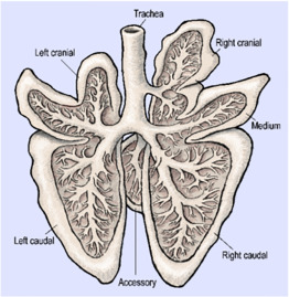
identify these structures, lungs of horse. What basic facies/parts are the lungs divided into?
lobus cranialis dexter
lobus caudalis dexter
lobus accessorius (to right lung, f. medialis)
lobus caudalis sinister
lobus cranialis sinister
Incisura cardiaca pulmonis sinistri → separates lobus caudalis et cranialis sinistri.
incisura cardiaca pulmonis dextri (bw. lobus caudalis et cranialis dextri)
We can also describe, hilus pulmonalis on f. medialis (towards mediastinum), margo dorsalis, margo acutus (consist of margo basalis (caudoventr.) and margo ventralis), basis et apex pulmonis. We see the facies costalis here (against ribs).
pars vertebralis (dorsally located, in contact with vertebral bodies of thoracic vertebrae)
pars mediastinalis (faces mediastinum)
facies interlobares (directed to the fissures between the lobes)
what forms the radix pulmonis?
On medial surface of lung, you have hilus pulmonis, which is where the main bronchus, pulmonary and bronchial blood vessels, lymphatic vessels and nerves pass through this and goes to th lungs from the mediastinum. All these forms the radix pulmonis.
Explain briefly the blood supply of lungs. Divided into functional and nutritional.
Lungs → by parenchyma and interstiital tissue framework. This is mainly of collagenous + elastic fibers. Lobes are divided into small irregular units + surrounded by interstitial tissue → lobuli pulmonis
functional: a small blood circulation, used to oxygenate the blood in the lungs. Arises from right ventricle of heart by truncus pulmonalis → De-O2 blood into lungs. → oxygenation of alveoli pulmonis → arterial blood leaves by 2-4 pulmonary veins. → left atrium of heart.
nutritional: done by bronchoesophageal artery from aorta thoracica → oxygenation → the deoxygenated blood is transported from lungs by bronchial veins.