Lecture 5 - Flow Cytometry (FINISH OFF)
1/43
There's no tags or description
Looks like no tags are added yet.
Name | Mastery | Learn | Test | Matching | Spaced | Call with Kai |
|---|
No analytics yet
Send a link to your students to track their progress
44 Terms
What is flow cytometry?
Technique which analyses the characteristics of cells pass singly in a fluid suspension through a beam of light
What information can flow cytometry tell you?
Only flow cytometry
Cell shape
Cell size
Using fluorescent tags
Cell components
What are the two scatters of flow cytometry?
Forward scatter
(90 degree) side scatter
What can forward scatter tell you?
Usually a measure of cell size
What can side scatter tell you?
Usually a measure of internal complexity (e.g. granules)
Lymphocytes are ___ FSC and ___ SSC.
Fill in the blank.
Lymphocytes are low FSC and low SSC.
Smaller cells with few granules (internal complexity)
Monocytes are ___ FSC and ___ SSC.
Fill in the blank.
Lymphocytes are high FSC and low SSC.
Larger cells with few granules (internal complexity)
Cells with a high FSC are smaller or larger?
Larger cells have a high FSC (e.g. monocytes)
Cells with a high SSC are more or less granular?
Granular cells have a high SSC (e.g. neutrophils)
What are the two spectrums that fluorescent molecules have?
Absorption/excitation spectrum
Emission spectrum
What is the absorption/excitation spectrum?
The range of wavelengths of light which are best at exciting the fluorescent molecule
Example: at 480nm some of the fluorophore molecules will be excited however some won’t
When 550nm is used, it excites more fluorophore molecules than any other wavelength
What is the emission spectrum?
The range of wavelengths emitted by fluorophore molecules following excitation
What is the purpose of dichroic filters in flow cytometry?
Filter specific wavelengths of light out from the emission
Example: a dichroic filter may allow red, yellow and green light to pass but reflect blue light
This allows the emission produced by flow cytometry to be separated to different Photomultiplier tubes (PMT) allowing detection of specific markers
What are band pass filters?
Filters only allow a narrow range of light through
Example: A 630/20nm BandPass filter will only let 620-640nm light through
What is the purpose of dichroic filters and BandPass filters in flow cytometry?
Allow specific fluorescent molecules to be detected independently of each other (dichroic filter) AND control signal-to-noise ratio by restricting what wavelengths of light can hit the PMT (BandPass filter)
How can DNA content be measured using flow cytometry?
DNA can be stained with a fluorescent dye which intercalates with DNA
As each dye molecule will bind so many nucleotides, you can calculate the amount of detected DNA
This allows G0/G1 phase, S phase, and G2/M phase cells to be identified based on the amount of DNA detected
If a G2 phase inhibitor is given what will happen to the graph of DNA content?
As G1 phase inhibitor is given, cells in G1/S phase will not be effected and will continue through the cycle
However G2 phase cells will begin accumulating and the number of G1/S phase cells will decrease
What is DNA index and how is it calculated?
Measure of amount of DNA in a cell compared to a normal cell
Mass of G0/G1 DNA peak of tumour / mass of G0/G1 DNA peak of a 2N standard
This produces the DNA index
A DNA index of 1.5 means what?
The cell has hyperdipoidy
More DNA than normal
Why is diagnosing a cancer as hyper/hypodiploid important?
Key in cancer treatment as hyper and hypodiploidy cancer can have different outcomes.
What is an example of the differences between hypodiploidy and hyperdiploidy in cancer outcome?
In Acute Lymphoblastic Leukaemia (ALL)
Hyperdiploidy is associated with a good prognosis
Hypodiploidy is associated with a poor prognosis
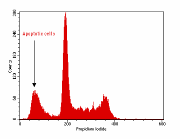
How can apoptosis be identified by flow cytometry?
During apoptosis, calcium and magnesium-dependent nucleases are activated which degrade DNA and small fragments of DNA can leave the cell
This will result in the cell taking up less stain due to having lost DNA
On a graph, this produces a new peak to the left of the G1 peak
Called the sub G1 peak
FINISH OFF