Lecture 3 - Passive Stay
1/10
There's no tags or description
Looks like no tags are added yet.
Name | Mastery | Learn | Test | Matching | Spaced | Call with Kai |
|---|
No analytics yet
Send a link to your students to track their progress
11 Terms
explain the importance of the passive stay apparatus
horses spend ~80% of their time standing, even during light sleep
they typically stand on both front limbs plus one hind limb
horses have special connective tissue system that allows them to conserve muscle energy while standing → avoids muscles fatiguing
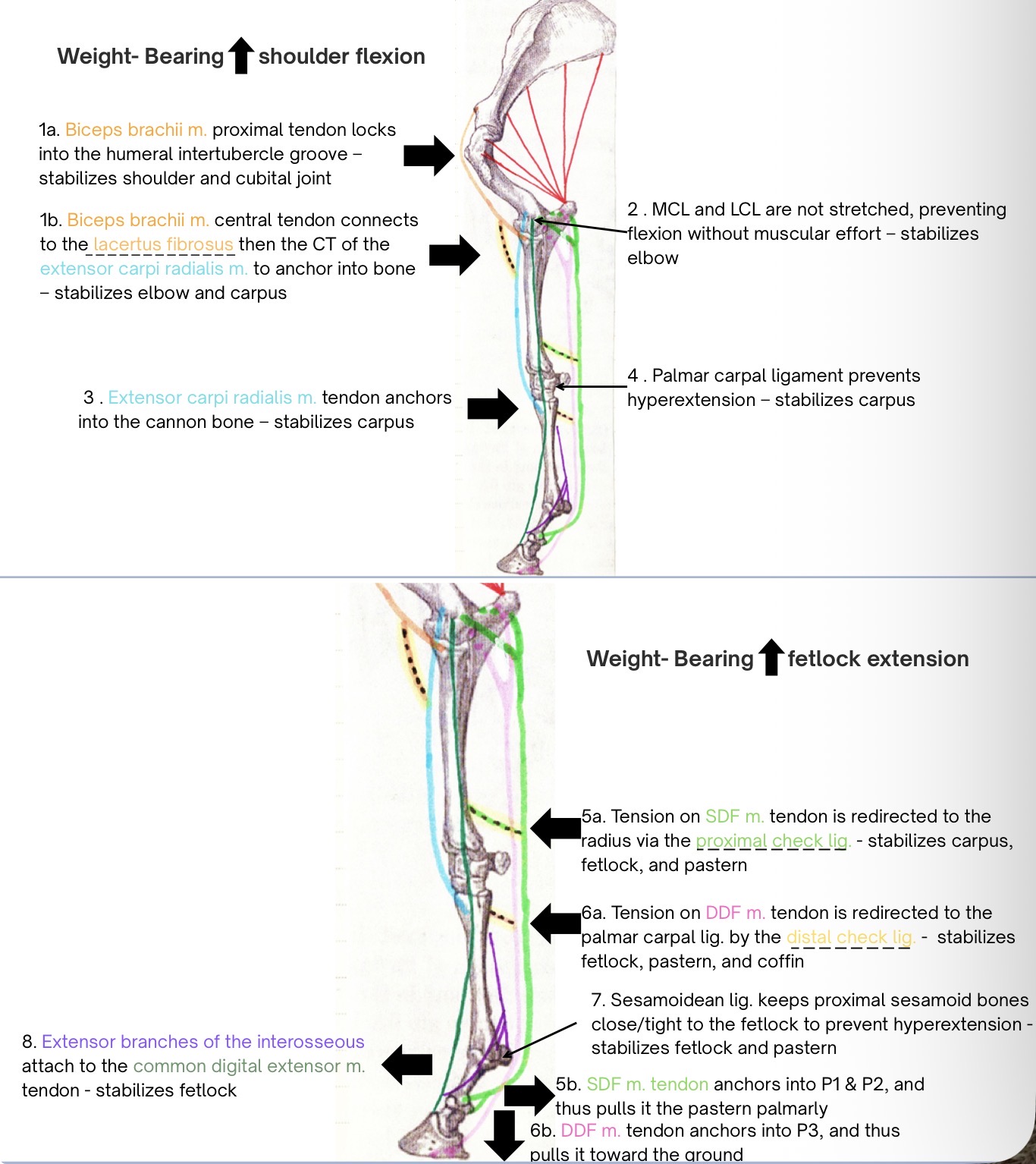
in the thoracic limb, this is called the passive forelimb stay apparatus, which either prevents flexion or hyperextension of specific joints to prevent collapse of the limb
the forelimb passive stay apparatus includes structures spanning from the distal scapula to the distal phalanx
parts of passive stay
limb support
shoulder support
cubital support
carpal support
fetlock support
pastern and coffin joint support
limb support - supportive mechanisms, muscles that can relax, and the connective tissue structures that provide support
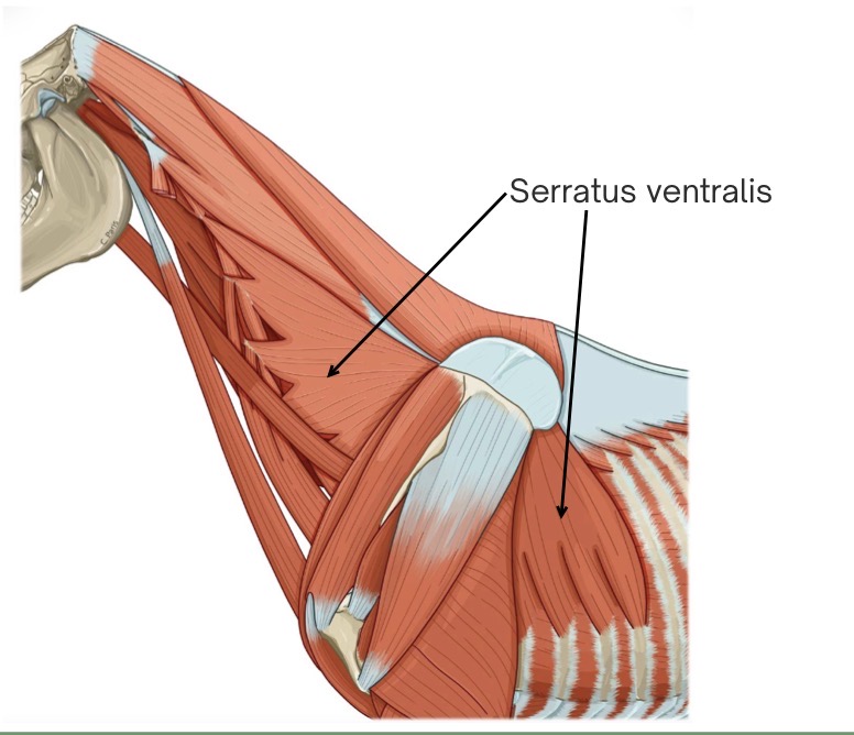
the serratus ventralis m. acts like a sling to attach the forelimb to the body
the dorsoscapular ligament connects the trunk (via thoracolumbar fascia) to the medial surface of the scapula
reinforces the serratus ventralis m.
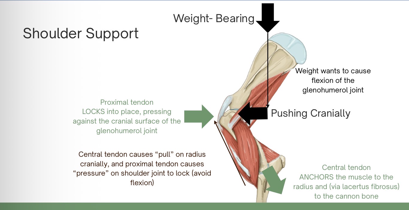
shoulder support - supportive mechanisms, muscles that can relax, and the connective tissue structures that provide support
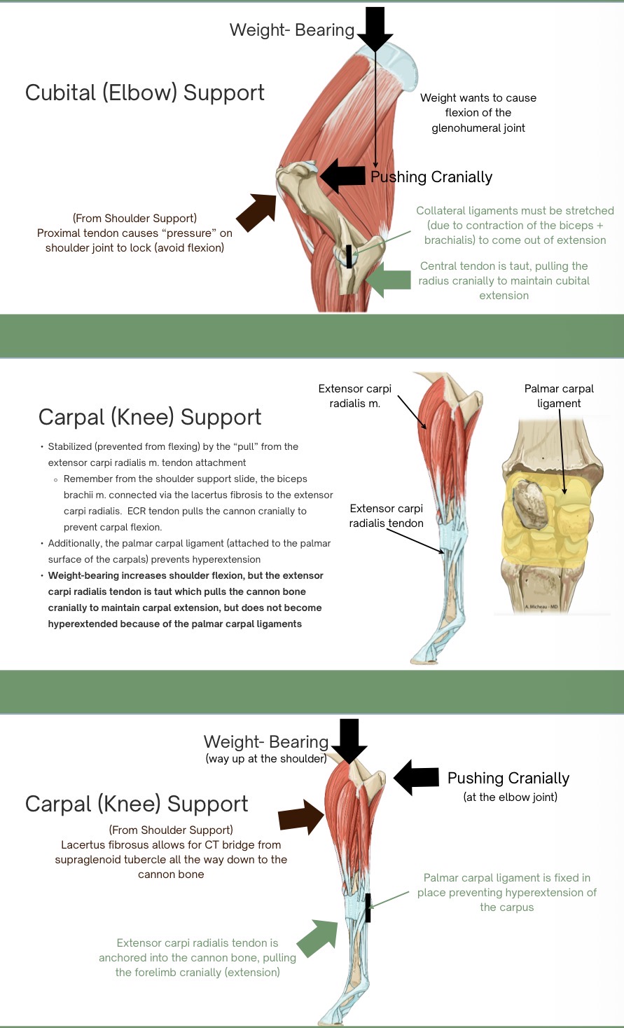
the biceps brachii m. attaches to the supraglenoid tubercle of the scapula via its proximal tendon
the proximal tendon fits the shape of the intermediate tubercle and intertubercle groove of the humerus
the tendon is continuous with a central tendon that runs within the biceps brachii m. and attaches distally into the connective tissue of the extensor carpi radialis m. via the lacertus fibrosus
the extensor carpi radialis m. attaches distally to the metacarpal tuberosity of the cannon bone
weight-bearing increases shoulder flexion, which locks the proximal tendon of the biceps under the intermediate tubercle of the humerus
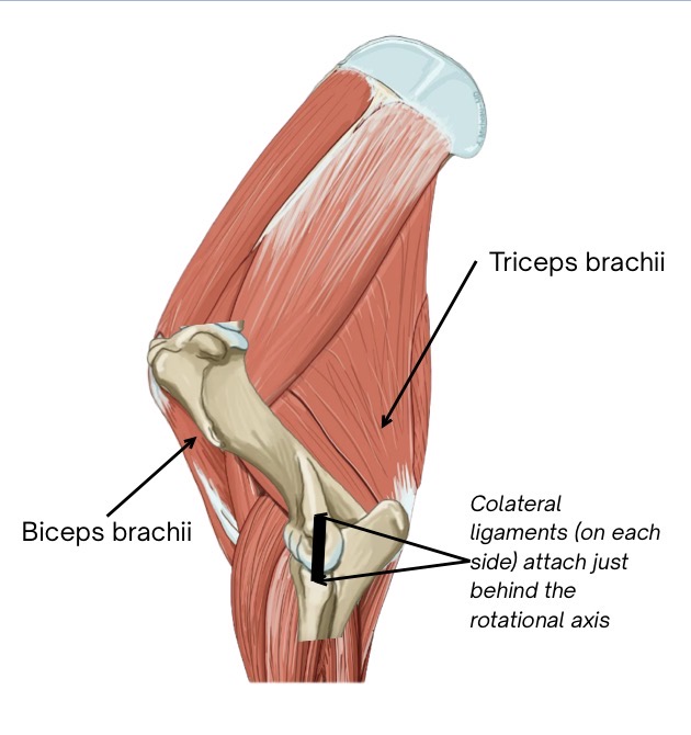
cubital support - supportive mechanisms, muscles that can relax, and the connective tissue structures that provide support
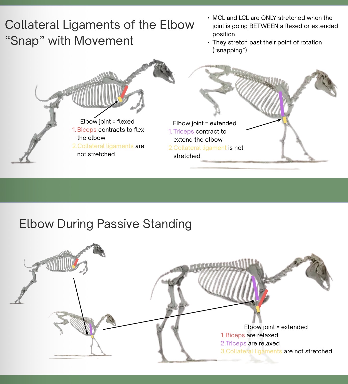
MCL and LCL of cubital joint attach caudal to axis of joint rotation (and the humeral condyles are elongated) therefore it takes effort to “snap” the joint between extension and flexion
the collateral ligg. also prevent medial and lateral movement of the joint
joint is fully extended when passively standing
if the triceps m. were to contract, the cubital joint snaps into extension, but then the triceps is able to relax once extended
in order for the joint to come out of extension, the biceps and brachialis m. contract to flex the joint
weight bearing increases shoulder flexion → the biceps can relax but the central tendon is still taut, so the radius is pilled cranially to maintain cubital extension
carpal support - supportive mechanisms, muscles that can relax, and the connective tissue structures that provide support
fetlock support - supportive mechanisms, muscles that can relax, and the connective tissue structures that provide support
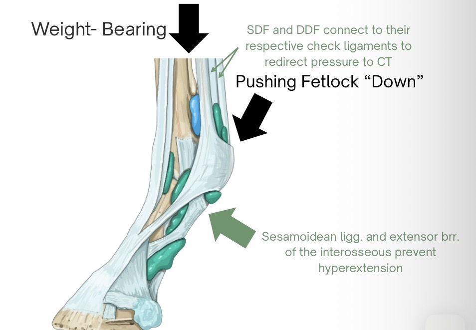
the fetlock joint needs to be stabilized to prevent it from sinking towards the ground
tension in the distal SDF tendon is redirected to the radius via the proximal check ligg. and tension in the distal DDF tendon is redirected to the palmar carpal ligg. via the distal check ligg.
this allows muscles to relax, as the joints are being supported by CT alone (CT doesn’t fatigue)
proximal sesamoidean ligg. press the proximal sesamoid bones against the palmar aspect of the fetlock joint
extensor branches of the interosseuous m. (suspensory lig.) connect to the common digital extensor m. tendon to prevent hyperextension
weight-bearing increases fetlock extension, which puts tension on the SDF and DDF tendons to prevent hyperextension, along with some suspensory ligg. and proximal sesamoidean ligg. to help
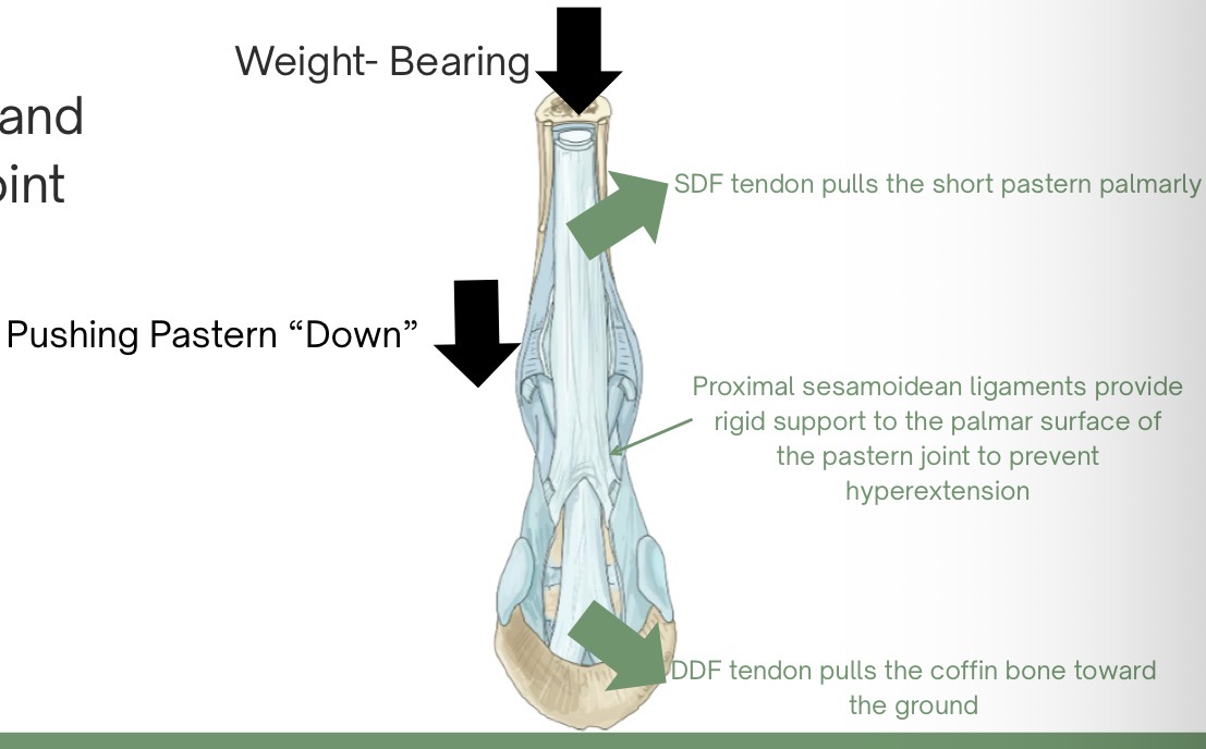
pastern and coffin joint support - supportive mechanisms, muscles that can relax, and the connective tissue structures that provide support
proximal sesamoidena ligg. support the palmar surface of the joint, preventing hyperextension
the DDF tendon pulls P3 (coffin) toward the ground, while SDF tendon pulls P1 (long pastern) and P2 (short pastern) palmarly, further preventing the buckling of the pastern
weight-bearing increases fetlock and coffin joint extension, which puts tension on the SDF and DDF tendons, and the proximal sesamoidean ligg.
explain the cubital joint and collateral ligament “snapping” mechanism
MCL and LCL are only stretched when the joint is going between a flexed or extended position
they stretch past their point of rotation (“snapping”)
list which joints are capable of flexion or hyperextension due to weight-bearing (that the passive stay apparatus works to avoid)
shoulder - avoid flexion
cubital - avoid flexion
carpal (knee) - avoid hyperextension
fetlock - avoid hyperextension
pastern and coffin - avoid hyperextension
identify and draw the passive stay apparatus components on a given diagram or picture