Micro LAB review 1
1/50
There's no tags or description
Looks like no tags are added yet.
Name | Mastery | Learn | Test | Matching | Spaced | Call with Kai |
|---|
No analytics yet
Send a link to your students to track their progress
51 Terms
What is the magnification and total magnification of the oil immersion objective?
100X, 1000X (with 10X eyepiece)
What is the magnification and total magnification of the high dry objective?
40X, 400X (with 10X eyepiece)
What is the magnification and total magnification of the low dry objective?
10X, 100X (with 10X eyepiece)
What is the magnification and total magnification of the scanning objective?
4X, 40X (with 10X eyepiece)
What is resolution?
Ability to see 2 objects as 2 separate objects
What is parfocal?
When focused on low power, automatically focused on high power
What are two reasons to heat fix a bacterial slide?
Kill bacteria; make them stick to the glass slide
Why does negative staining give a more accurate measure of cell size?
No heat fixing, so no cell distortion
What is the purpose of simple stain (methylene blue)?
Tells you shape and arrangement of bacteria
What is the difference between simple and differential staining?
Simple shows only shape; differential shows shape and distinguishes between two different species
What is the gram stain?
Differential stain; distinguishes between G+ and G-
What are the steps of the gram stain procedure?
Prepare smear, air dry, heat fix 2. Apply primary stain crystal violet for 2 minutes (don’t let it dry) 3. Rinse with distilled water 4. Apply gram’s iodine (mordant) until smear is fully covered, leave for 2 minutes (forms insoluble complex with crystal violet in G+ cell walls and G- outer membrane) 5. Rinse with distilled water 6. Apply gram’s decolorizer (95% ethanol) while tilted until stain runs off (~30 seconds); don’t over-apply 7. Rinse with distilled water 8. Apply counterstain safranin for 2 minutes 9. Rinse with distilled water, blot dry, and examine
What are the steps of the acid fast-Ziehl method?
Prepare smear 2. Saturate with carbolfuchsin stain 3. Leave slide on hot plate at 80°C 4. Wash with acid alcohol 5. Saturate with methylene blue
What is the acid fast-Ziehl method used for?
Differential stain that distinguishes acid-fast bacteria (e.g. Mycobacterium) from non-acid-fast bacteria based on mycolic acid in the cell wall
What is the primary stain, mordant, decolorizer, counter stain, and heat fix status for negative/capsule staining?
Primary stain: acid fuchsin (stains bacteria); mordant: none; decolorizer: none; counter stain: congo red (stains slide); heat fix: no
What is the primary stain, mordant, decolorizer, counter stain, and heat fix status for endospore staining?
Primary stain: malachite green; mordant: heat; decolorizer: none; counter stain: safranin; heat fix: yes
What is the primary stain, mordant, decolorizer, counter stain, and heat fix status for acid fast staining?
Primary stain: carbol fuchsin; mordant: heat; decolorizer: acid alcohol; counter stain: methylene blue; heat fix: yes
What is the primary stain, mordant, decolorizer, counter stain, and heat fix status for gram staining?
Primary stain: crystal violet; mordant: iodine; decolorizer: ethanol; counter stain: safranin; heat fix: yes
What is the primary stain, mordant, decolorizer, counter stain, and heat fix status for simple staining?
Primary stain: methylene blue; mordant: none; decolorizer: none; counter stain: none; heat fix: yes
What does a mordant do?
Makes the primary stain adhere to cells
What is a stain that is negatively charged (acidic)?
Congo red, nigrosine
What color are Gram + cells?
Purple (crystal violet); example: Bacillus, Staph aureus
What are the steps of the endospore stain procedure?
Prepare thin smear (avoid too many bacteria); heat fix 2. Cover smear with cut blotting paper, saturate with malachite green 3. Place slide on hot plate at 90°C for 10-15 minutes, adding more malachite green as needed 4. Remove slide carefully with slide holder 5. Remove and dispose of blotting paper 6. Rinse front and back of slide with distilled water (no decolorization in this technique) 7. Apply safranin for 1 minute, rinse, blot, and examine
What color are gram – cells, an example, and why they differ from gram +?
Pink (safranin); example: Salmonella, Escherichia coli; difference due to amount of lipid outside cell wall and thickness of cell wall
What genus of bacteria are acid fast, why, and what colors result for acid fast vs non-acid fast?
Genus: Mycobacterium; acid fast due to mycolic acid in cell wall; acid fast color: pink (carbol fuchsin); non-acid fast (methylene blue)
What two genera are endospore formers, what color are spores vs vegetative cells after staining, and how do you completely get rid of spore-forming bacteria?
Genera: Clostridium, Bacillus; spores: green (malachite green); vegetative cells: pink (safranin); to get rid of spores: autoclave (121°C, 15 psi, 15-20 min)
After negative capsule staining, what colors are the capsule, vegetative cells, and background?
Capsule: clear; vegetative cells: pink (acid fuchsin); background: blue (congo red + acid)
What is Staphylococcus medium?
Selective medium containing 7.5% salt; selects for halophiles (Staph aureus)
What is Thayer Martin medium?
Enriched medium with blood + antibiotics; selects for Neisseria gonorrhoeae
What is MacConkey agar and why is it selective and differential?
Selective: contains crystal violet and bile salts which select for G- and inhibit G+. Differential: contains lactose and neutral red dye; lactose fermenters (e.g. E. coli) turn red, non-fermenters (e.g. Salmonella, Serratia marcescens) stay uncolored
What is blood agar and what do alpha, beta, and gamma hemolysis look like?
Distinguishes between alpha, beta, and gamma hemolysis. Alpha: colonies and surrounding agar turn brown/green (partial hemolysis). Beta: clear colonies (complete hemolysis). Gamma: agar remains red (no hemolysis)
What is Mannitol Salt Agar and how do you read results?
Nutrient agar with 7.5% NaCl (selects for halophiles), enriched with mannitol. Staph aureus and Staph epidermidis (halophiles) grow; E. coli does not grow. If colonies and media turn yellow (pH indicator): Staph aureus. If colonies grow but mannitol not fermented: Staph epidermidis
What type of medium is Mannitol Salt Agar, its purpose, and how do you interpret results?
Type: selective and differential. Purpose: selects for Staphylococci (grow at high salt concentrations); differentiates Staph aureus from other Staphylococci. Interpretation: Staph aureus is yellow (ferments mannitol); other staphylococci are white
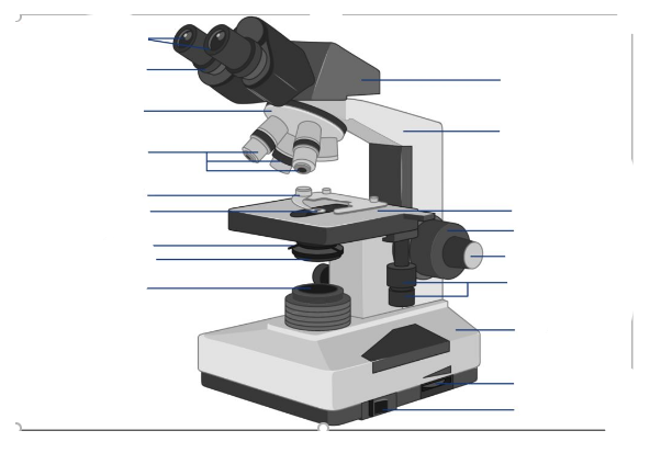
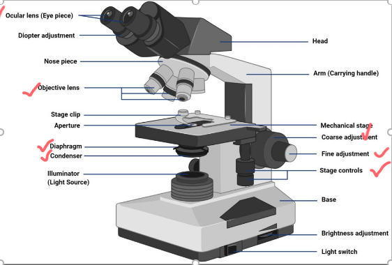
What is the condenser and what does it do?
Third lens system, located under the stage; collects and directs light from the light source to the slide. Substage adjustment knobs move it up and down; normally kept within about 2 turns from the stage
What is the iris diaphragm and how else is light controlled?
Controls the amount of light reaching the slide by opening or closing. Light can also be controlled by the rheostat, which changes the electrical current coming into the microscope
What is the purpose of aseptic technique?
To prevent contamination of materials; all materials must be clean and sterile before use
What are key safety practices for aseptic technique lab setup?
Secure long hair, wear closed-toe shoes (lab coat optional); minimize materials on floor/bench to prevent accidents and cross-contamination; wash hands before/after, wear gloves; wipe down tables with alcohol before/after; prepare clean slides by washing with soap and water, dry with Kim wipe
How do you sterilize the inoculating loop?
Hold like a pencil in dominant hand; hold wire parallel to burner and place into flame from base to tip; allow wire to become visibly hot before removing; sterilize before and after each use/transfer; keep hands away from flame; when not in use, place in test tube rack with loop up and away from you
How do you safely retrieve microbes from broth cultures?
Hold cap in pinky finger of dominant hand (don't set it down); hold tubes at an angle to minimize airborne contamination; pass open end of tube through flame 2-3 times; dip sterile loop into culture and transfer a drop onto slide/agar/broth; replace cap immediately after transfer
How do you prepare a slide after transferring a sample?
Spread the sample in a thin, even layer on the slide; flame the wire after spreading
What materials are needed for the simple stain procedure?
Stain pan, wire loop/slides, methylene blue, bacterial culture, Meker-Fisher burner, distilled water
What are the steps of the simple stain procedure?
Flame loop 2. Make smear, air dry, and heat fix 3. Place slide on stain pan 4. Cover smear with methylene blue for 2 minutes 5. Rinse briefly with distilled water 6. Blot-dry water from slide 7. Examine
What materials are needed for negative staining?
Slides, nigrosine, wire loop, bacterial culture
What are the steps of the capsule stain (congo red + acid fuchsin) procedure?
1. Clean slide; apply one small drop of congo red to slide 2. Inoculate congo red with one loop of bacteria from broth culture 3. Spread mixture into thin film using wire loop 4. Allow smear to air dry completely 5. Immerse slide in coplin jar of acid fuchsin for 30 seconds 6. Remove slide, drain excess stain with blotting paper (do not rinse, not heat fixed) 7. Examine at 100X, 400X, and 1000X
What are the steps of the negative staining procedure?
Clean slide 2. Add 1 drop of nigrosine on end of slide, flame loop 3. Transfer 1 loopful of bacteria into stain from surface of agar culture, flame the loop 4. Using a second clean slide, spread mixture of nigrosine and bacteria into a thin film covering the entire slide 5. Don't rinse the slide with water 6. Let stain air dry completely 7. Examine using all three lenses
Endospore Stain: What Happens If You Skip a Step?
Malachite Green (Primary Stain)
Endospore: colorless - false negative (no green stain enters the spore)
Vegetative cell: red/pink - correct (safranin still stains the cell)Heat (Steam)
Endospore: red/pink - false negative (malachite green cannot penetrate the tough spore coat)
Vegetative cell: red/pink - correctWater Decolorizer
Endospore: green - correct
Vegetative cell: green - false positive (vegetative cells should lose malachite green)Safranin (Counterstain)
Endospore: green - correct
Vegetative cell: colorless - incorrect (vegetative cells should be red/pink)
Gram Stain: What Happens If You Skip a Step?
Crystal Violet (Primary Stain)
Gram+: pink - false gram-negative (no crystal violet applied, so gram+ cells can't appear purple)
Gram-: pink - correct (gram- cells are normally pink after safranin)Gram's Iodine (Mordant)
Gram+: pink - false gram-negative (without iodine, crystal violet isn't trapped and washes out during decolorization)
Gram-: pink - correct (gram- cells lose crystal violet and stain pink)Decolorizer
Gram+: purple - correct (gram+ cells should retain crystal violet)
Gram-: purple - false gram-positive (without decolorization, gram- cells never lose the purple stain)Safranin (Counterstain)
Gram+: purple - correct (gram+ cells remain purple)
Gram-: colorless - incorrect (gram- cells lose crystal violet but receive no pink counterstain)
Acid-Fast Stain: What Happens If You Skip a Step?
Carbolfuchsin
Acid-fast: blue - false negative (no red stain available)
Non-acid-fast: blue - correctHeat
Acid-fast: blue - false negative (carbolfuchsin can't penetrate waxy wall)
Non-acid-fast: blue - correctAcid-Alcohol
Acid-fast: red - correct
Non-acid-fast: red - false positive (should be blue)Methylene Blue
Acid-fast: red - correct
Non-acid-fast: colorless - incorrect (should be blue)
Negative/Capsule Stain: What Happens If You Skip a Step?
Congo Red
Capsule: not visible - incorrect
Cell: red/pink - correct
Background: light/unstained - incorrect
Why wrong: without a dark background, the clear capsule blends into the slide and cannot be distinguishedAcid Fuchsin
Capsule: not visible - incorrect
Cell: colorless - incorrect
Background: dark - correct
Why wrong: without a stained cell, there is nothing for the capsule halo to surround, making the capsule difficult or impossible to identify
Simple Stain (Methylene Blue): What Happens If You Skip a Step?
Primary Stain
Cell: colorless - incorrect
Background: light - correctWater Rinse
Cell: blue - correct
Background: blue - background too darkBlot Dry
Cell: blue - correct
Background: light - correct (but slide may be difficult to view due to excess water)