Exam 4 Sensation & Perception
1/59
There's no tags or description
Looks like no tags are added yet.
Name | Mastery | Learn | Test | Matching | Spaced | Call with Kai |
|---|
No analytics yet
Send a link to your students to track their progress
60 Terms
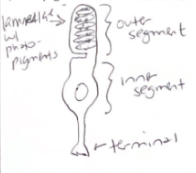
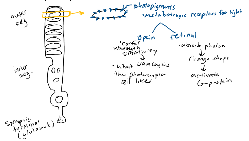
Draw and diagram the structure of a rod or cone
outer segment
inner segment
synaptic terminal
Label the outer and inner segments of a rod/cone (?)
outer segment (contains lamella, which contains photopigments)
inner segment (contains nucleus)
synaptic terminal (releases glutamate to cells, bipolar & horizontal)
In the structure of a rod/cone: Where are the lamellae? Where are the photopigments? Where is the nucleus?
layer of membrane in outer segment containing photopigments; inner segment contains nucleus
(In relation to rods/cones) What are photopigments? What are the two main components? Where are they made & where are they stored?
the actual receptors for light (metabotropic); retinal & opsin; made in inner segment & stored in lamellae (outer segment)
What makes different photoreceptors sensitive to different wavelengths of light?
structure of opsin molecule (long = red, medium = green, short = blue)
What is going on in the dark… What is the dark current?
in dark, photoreceptor cell is depolarized and releases glutamate, which inhibits bipolar cell; sodium entering cell in the dark
What are the steps in phototransduction? (?)
rhodopsin absorbs photon →
photon causes retinal to change shape (cis to all-trans), so no longer fits in binding site w opsin →
retinal is released and binds w & activates G-protein (transducin) →
G-protein activates a 2nd messenger →
Photoreceptor is hyperpolarized (inhibited) → disinhibited & depolarized which activates RGC
How does hyperpolarization of the photoreceptor lead to an action potential in the optic nerve (ie, what happens at each successive layer of nerve cells in the retina?)?
photoreceptor depolarizes in dark (cuz sodium/cation channel is open) & is releasing glutamate that inhibits bipolar cell
light striking a photoreceptor produces a hyperpolarization = photoreceptor releases less glutamate (& depolarization of bipolar cell… bcuz NT normally hyperpolarizes bipolar cell and it’s been reduced)
Depolarization (of bipolar cell) = release more NT, excites retinal ganglion cell
Info sent to brain via optic nerve (Axon of ganglion cell is part of optic nerve, so signal goes back to brain)
How does a bright light (compared to a dim light) lead to more action potentials per second in optic nerve fibers? (?)
brighter light → bigger receptor potential → bigger depolarization in BPC (bipolar cells) then in RGC (retinal ganglion) → more AP/sec
What is the sensitivity-acuity tradeoff?
visual system can’t have both high sensitivity (ability to detect faint light/dim environments) & high acuity (ability to see fine, detailed images in bright light); As system increases its sensitivity to light, it must sacrifice the ability to perceive fine details, and vice versa
stimulus (in fovea) → receptors (cones → little spatial summation → midget bipolar cells → P-ganglion cells
Consider cones vs. rods and their connections with bipolar cells and ganglion cells in the retina (?)
cones have 1:1 connection with cone bipolar cell (midget bipolars) & ganglion cells, boosting spatial resolution + color vision
rods converge on single rod bipolar cell, boosting sensitivity
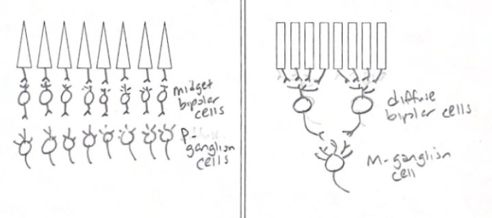
What are the different types of bipolar cells?
midget BP (cone bipolar), diffuse BP (rod bipolar)
What are the different types of ganglion cells?
ON-center (light in center of receptive field = increase firing rate); OFF-center (light in center of receptive field = decrease firing rate)
How are the diff types of bipolar & ganglion cells connected to each other? What types of photoreceptors do they get info from? How is this all related to the sensitivity-acuity tradeoff? How is it related to the size of visual receptive fields? (???)
(?) bipolar cells get info from rods & cones, split into ON/OFF-center types, synapse onto RGCs, form retina’s parallel processing stream
What is lateral inhibition, what is it good for (contrast!) and what cells accomplish it?
Input from adjacent cells is antagonistic (if ur neighbor receives input, you will not)
Achieved thru the horizontal cells (best for CONTRAST), as shine light on center of receptive field, center is activated, periphery is inhibited by horizontal cell + amacrine cells also involved but their role is unclear
Describe the connections between horizontal cells and photoreceptor cells and how the former influences the activity of the latter (?)
(?) Horizontal cells are retinal interneurons that provide inhibitory feedback to photoreceptor cells (rods and cones), playing a critical role in lateral inhibition, contrast enhancement, and adaptation to light intensity. They connect to multiple photoreceptors in the outer plexiform layer, receiving glutamatergic input and providing feedback to shape the center-surround receptive fields of bipolar cells
What is the receptive field of ganglion cells derived from? (??)
specific, circular area of the retina, encompassing inputs from numerous photoreceptors (rods and cones)
What would increase the firing of an on-center off-surround retinal ganglion cell? What would decease it’s firing? What would cause no change in its firing rate?
light hits center; light hits surround; light hits both
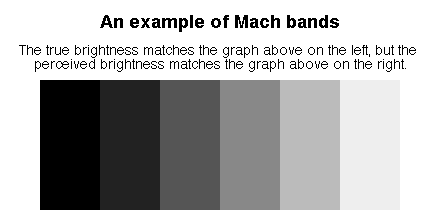
Describe the Mach band illusion and the Hermann grid in terms of ganglion cell receptive fields (/??)
the Hermann grid and Mach bands are visual illusions explained by lateral inhibition within center-surround receptive fields of retinal ganglion cells; these cells enhance contrast at edges
in grid, more inhibition occurs at intersections, producing "ghostly" grey spots, while in Mach bands, it accentuates light/dark edges
What is the pathway of visual information from the eye to primary visual cortex? What processing occurs at each stop? (/)
optic nerve → optic chiasm → optic tract → lateral geniculate nucleus → superior colliculus of midbrain
(in relation to pathway of visual info from eye → primary visual cortex) Compare the human midbrain with that of birds & fish (?)
(?) basic components of midbrain exist in all, but its relative size and functional prominence vary significantly due to different evolutionary pressures
How many cellular layers are in the LGN? What are they called? What parts of the retina do these different layers receive info from?
6 layers; magnocellular & parvocellular layers; retinal ganglion cells
What are the receptive fields of LGN neuron like? (?)
center-surround receptive fields (like retinal ganglion cells)
What and where are the “what pathway” and the “where pathway”?
what = cone pathway (parvocellular LGN)
analysis of form and colorwhere = rod pathway (magnocellular LGN)
analysis of motion and spatial relations
What visual fields go to what retina (left or right, and part of that retina: nasal or temporal)? (/)
Left visual field (periphery) projects to left (L) nasal retina & right (R) temporal retina; processed by right brain hemisphere
Right visual field projects to the right (R) nasal retina and left (L) temporal retina; processed by left brain hemisphere
To what LGN and striate cortex (left or right) does that info go? (/)
Information from the left half of the visual field goes to the Right LGN and Right Striate Cortex
Information from the right half of the visual field goes to the Left LGN and Left Striate Cortex
What would be the visual field deficit after damage to (a) the right optic nerve, (b) the right optic tract, and (c) the very center of the optic chiasm? (/?)
Results in ipsilateral monocular blindness = total loss of vision in the right eye only
Results in left homonymous hemianopia = loss of the left half of the visual field in both eyes
Results in bitemporal hemianopia = loss of the outer (temporal) half of the visual field in both eyes
How many layers does striate cortex have?
6
What is cortical magnification?
more neurons in cortex to process info from fovea compared with info from peripheral retina; visual acuity decreases with eccentricity
What is visual crowding?
when more objects in periphery it's more difficult to identify objects
Where in striate cortex is the fovea represented? ; Where is the fovea represented in striate cortex?
parvocellular layers
How is striate cortex organized?
topographically (aka retinotopically)
What types of stimuli do cells in striate cortex respond to?
cells in striate cortex: linear, respond to lines (bars, edges, gratings)
for each cell in striate cortex, the line has to be a particular angle (orientation tuning)
each line must be a particular width
some cells respond best when that line is moving in a particular direction
Compare and contrast simple cells and complex cells. (?)
Simple cells: simple receptive fields; responds best to object of given shape and orientation; best stimuli are bars, lines, rectangles with definite edges gratings, tuned to spatial frequency which corresponds to line width
Complex cells: orientation specific, larger receptive fields than simple cells, stimulus can occur in wider range of visual field; Best response to movement if stimulus is properly oriented and movement is in particular direction
What are columns in cortex and how do the response properties of neurons in a given column compare to each other? (/)
vertical, functional units of neurons, spanning the 6 layers of the cerebral cortex that act as the fundamental repeating unit of brain processing
types: orientation, ocular dominance, hypercolumns
all cells in a column respond to the same type of stimulus VS. neighboring columns respond to similar (not identical) stimuli
What are ocular dominance columns? (/)
alternating stripes of neurons that get preferential input from either right or left eye; all share the same eye preference
What is a hypercolumn? (/)
2 sets of columns, each covering every possible orientation (0-180) w/ 1 set preferring input from left eye and the other set preferring input from the right eye
Describe the tilt aftereffect (/?)
demonstrates orientation tuning of cortical neurons → visual illusion where prolonged viewing of an oriented stimulus (e.g., tilted lines) causes a subsequently viewed vertical or horizontal stimulus to appear tilted in the opposite direction
adapt the cells that respond to -20 & 20 →
new stimulus (vertical) → cells that now respond best are those to 10 or 20 (top) & -10 or -20 (bottom) →
image tilted in opposite direction
How does early experience play a role in the development of columns and of visual experience later in life? (/)
critical period for dev't: 1st 3-8 years in humans
cortical neurons are still wiring with rest of visual system
if one eye doesn't receive appropriate stimulation, the neurons destined to respond to that eye don't properly connect
cataracts: reduce/blur image into one eye
strabismus: (1 eye turned out) leads to 2 diff images on 2 foveas
ampblyopia: reduced visual acuity in 1 eye due to abnormal visual experience
“There is no red in a 700 nm light, just as there is no pain in the hooves of a kicking horse.” What does that mean? (/)
it's all in the mind
What does hue refer to?
chromatic (colorful) aspect of light; each point on spectrum defines a different hue;
perception is color
What is additive color mixing (/)
perception of colors results when lights of various wavelengths are combined in the visual system (ex: in a TV/phone, yellow is actually red + green); with each wavelength you're adding more cones to the response
What is the trichromatic theory of color vision? (/)
the color of any light is defined in our visual systems by the relationships of 3 numbers, the outputs of 3 receptor types (cones)
S, M, L cones, determined by particular opsin within a photoreceptor;
color perceived through the relative rates of respons of each cones (CROSS FIBER PATTERNING)
Our 3 cones are called L-, M- and S-cones. Why?
named for peak sensitivity (Red = long wavelength, Green = medium wavelength, Blue = short wavelength)
Why do color afterimages occur? (/)
retinal cone cells adapt to sustained stimulation, becoming fatigued and insensitive to a specific color
What are scotopic and photopic vision?
scotopic: rod vision. light intensity is bright enough to stimulate the rods but not the cones
photopic: cone vision. light is intense enough to stimulate the cones and saturate the rods
Describe “the problem of univariance”? (/)
2 variables to light: intensity (brightness) and wavelength (color)
but photoreceptors can only provide 1 piece of information to cortex: firing rate (sensitive to intensity (rate law) and wavelength)
so there is an infinite set of different wavelength-intensity combinations that can elicit exactly the same response from a single photoreceptor
for color: the output of 1 photoreceptor is completely ambiguous (explains lack of color in dimly lit scenes)
What is the opponent processes theory of color vision? (/)
color vision is controlled by three opposing, inhibitory systems: red vs. green, blue vs. yellow, and black vs. white; theory posits that cells in the visual system are excited by one color and inhibited by its opponent, meaning we cannot perceive combinations like reddish-green
Describe the different contributions of the trichromatic theory and the opponent processes theory to our understanding of color perception (/) (??)
Trichromatic theory explains that the retina uses three types of cones sensitive to short (blue), medium (green), and long (red) wavelengths to detect color; Opponent-process theory explains that neural cells in the thalamus/visual cortex process colors in opposite pairs (red-green, blue-yellow, black-white)
What is then nature of the color-sensitive receptive fields in the LGN? In other words, what types (colors) of opponent cells are seen in the LGN? What are opponent processes useful for? (???)
?
What is color constancy? Why does it occur? (/)
feature of the human visual system that ensures objects are perceived as having a consistent color, even when the illumination, wavelength, or intensity of light reflecting from them changes significantly
Describe the study we discussed on color constancy (???)
?
Describe evidence that all people DO, DON’T or MIGHT see color the same (??)
?
Describe the disorders of color vision
?
Why is red-green colorblindness more common in men than in women?
x-linked gene
What part of LGN do photoreceptors project to?
Cones → midget bipolar → p ganglion → parvocellular layer of LGN
Rods → diffuse bipolar → m ganglion → magnocellular
(P ganglion have smaller dendritic trees than m ganglion cells, but many more p ganglion cells)
What do hypercomplex cells respond to? (??)
used to be called HC cells, now considered subtype of simple and complex cells
Like simple: orientation specific
Like complex: orientation specific, movement specific
Also LENGTH specific. End stopping.
Good for detecting edges, corners, and borders
What does saturation refer to?
the amount of hue present in a light
pure white = zero saturation
blood-red = fully saturated red
full saturation = equivalent of a pure tone (only 1 wavelength present)
What does brightness refer to?
perceptual consequence of the physical intensity of a light (more photons)
What is subtractive color mixing (/)
like with paints, or pigments. the paint splotch absorbs (subtracts) all but the blue wavelengths of light, so subtracting cones from the response.