UARK Anatomy Exam 2 - Becker
1/147
There's no tags or description
Looks like no tags are added yet.
Name | Mastery | Learn | Test | Matching | Spaced | Call with Kai |
|---|
No analytics yet
Send a link to your students to track their progress
148 Terms
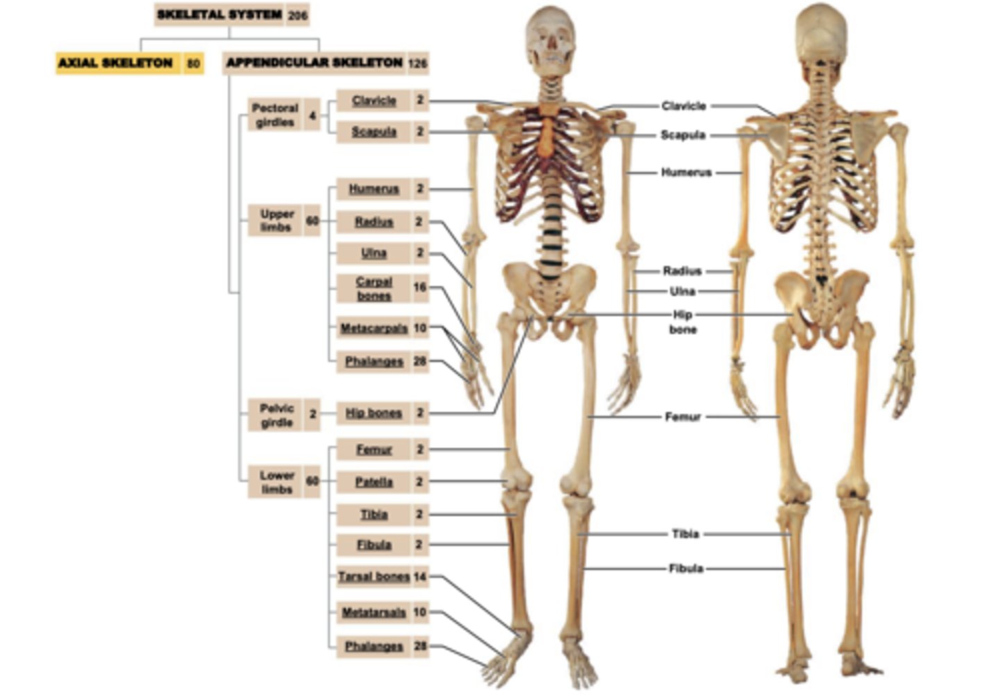
Skeletal system
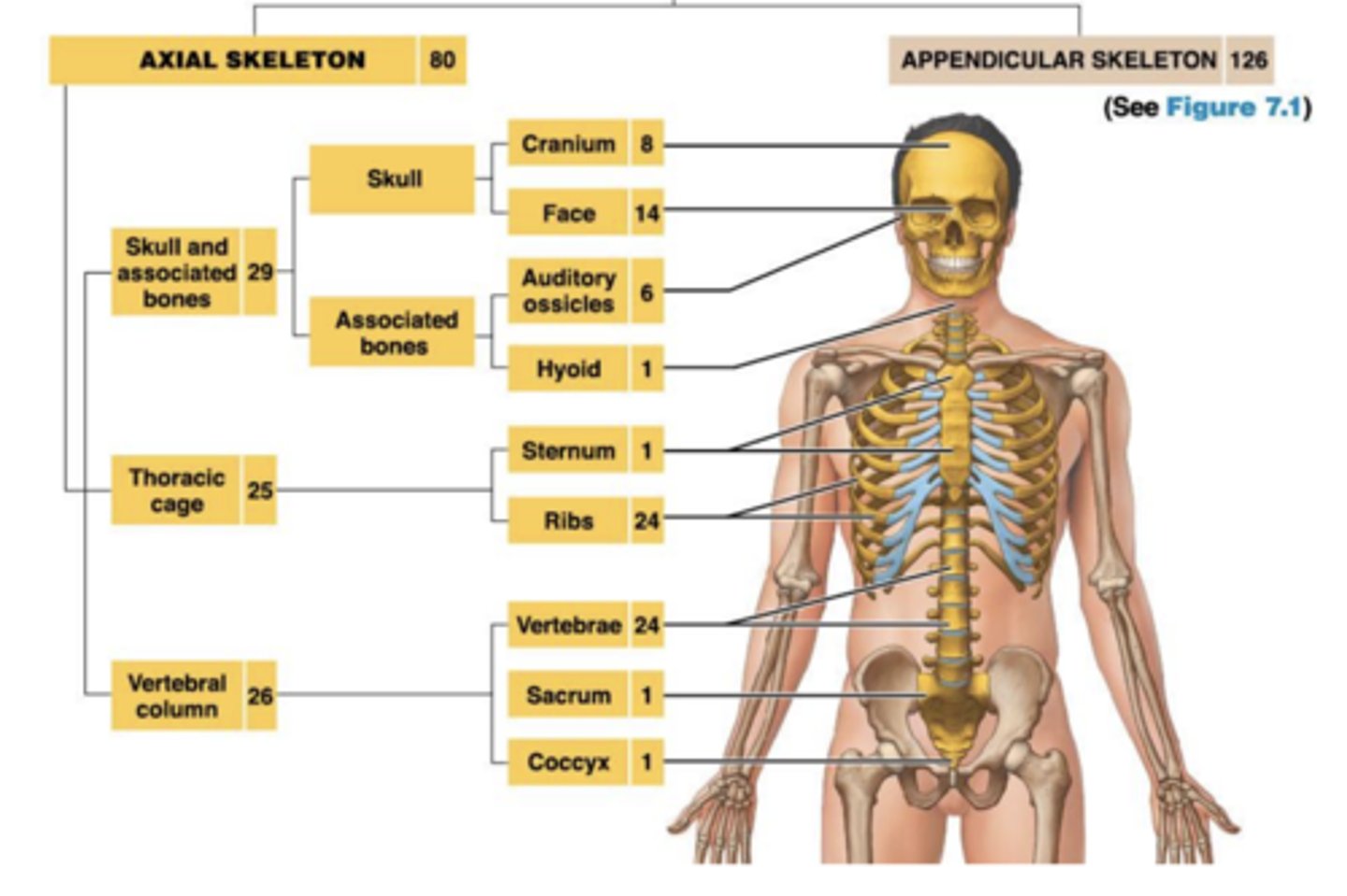
Axial
Appendicular
Cartilage
Ligaments
axial
skull, spine, thoracic cavity
Appendicular
limbs, pectoral girdle, pelvic girdle
Support
framework for attachment of other organs
Movement / locomotion
muscles use bones as levers
Storage of minerals
Calcium ions
phosphate ions
Blood cell production (hematopoiesis)
bone marrow produces erythrocytes, leukocytes, & platelets
protection
Ribs protect heart & lungs
Skull protects brain
Vertebrae protect spinal cord
Pelvic bones protect reproductive organs
Matrix of bone consists of
Hydroxyapatite crystals: mainly calcium phosphate (and calcium hydroxide) will resist compression, but inflexible
Calcium phosphate makes up ~2/3 of bone mass
Collagen fibers
Make up ~1/3 of bone mass
Contribute to tensile strength of bones
Imparts limited flexibility to matrix
Bone cells
Contribute only ~2 percent of bone mass
Osteocytes
maintains matrix
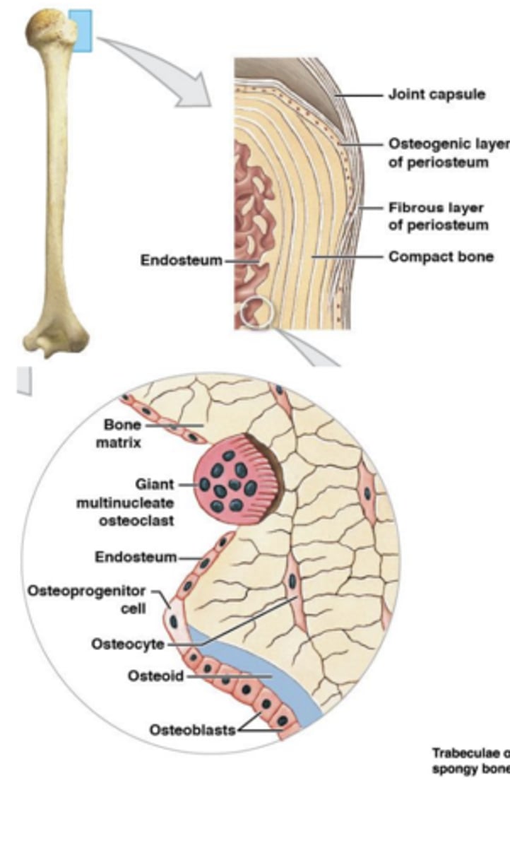
Osteoblasts
produces matrix
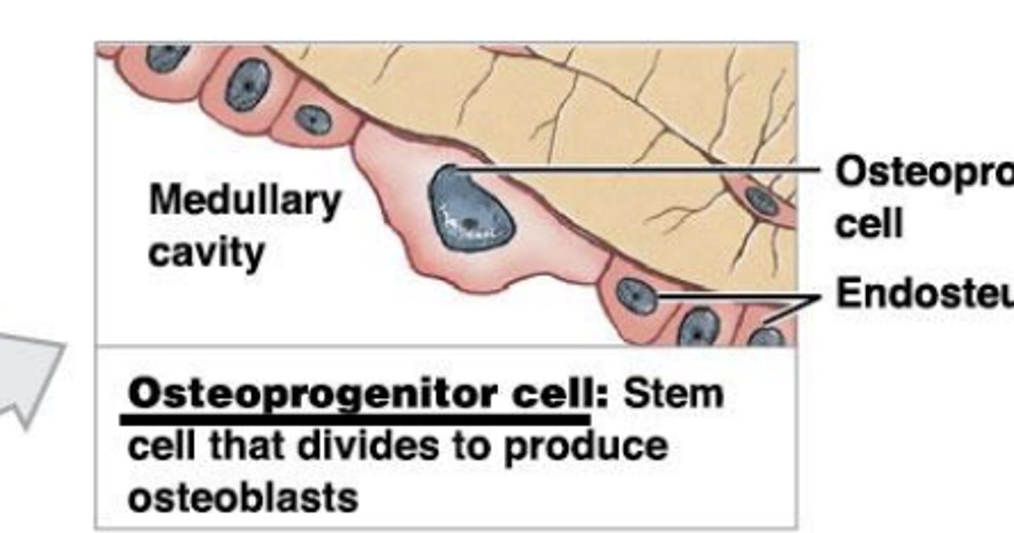
Osteoprogenitor cells
produce osteoblasts
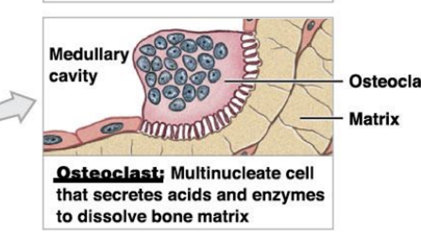
Osteoclasts
break down matrix
Osteoprogenitor cells: bone stem cells
Innermost layer of periosteum & inner lining of endosteum
Differentiate to form new osteoblasts
Involved in repair of bones after a fracture
Osteoclasts: multinucleated cells
osteolysis: secrete HCL, dissolving bones causing release Ca2+ & PO4 3- into blood
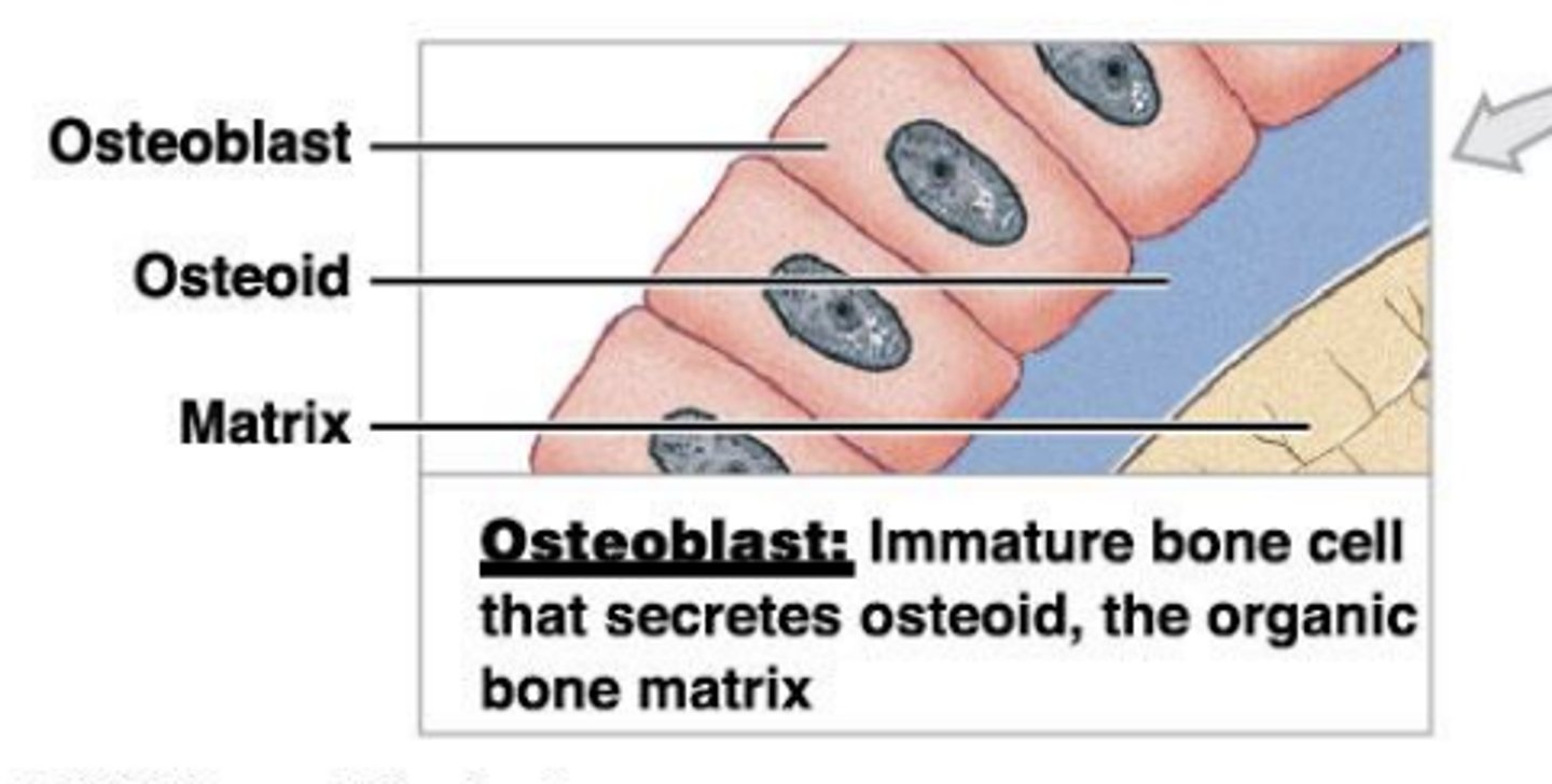
Osteoblasts: Immature bone cells
Found on inner & outer surfaces of bones
Produce osteoid: organic mix dumped into matrix
Osteogenesis: new bone formation via calcification of osteoid leading to ossification
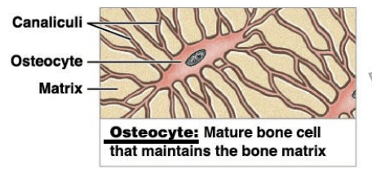
Osteocytes: Mature bone cells
Maintain protein & mineral content of matrix
Controls release & deposition of Ca2+ in/out of bone
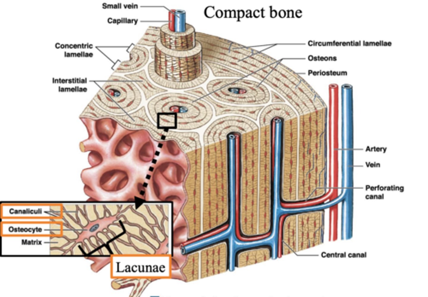
Lacunae
depressions where osteocytes reside
lamella
osteocytes matrix
Canaliculi:
small channels from osteocytes to bone capillaries
Osseous tissue types
1. Compact bone (dense bone)
2. Spongy bone (trabecular bone)
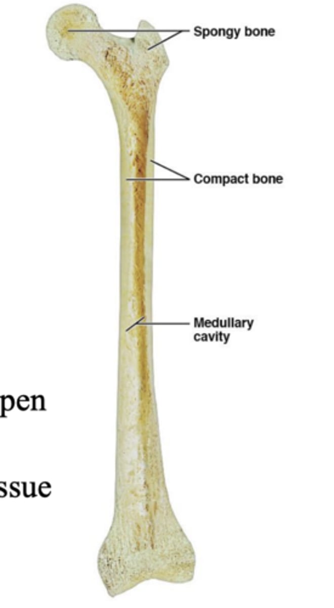
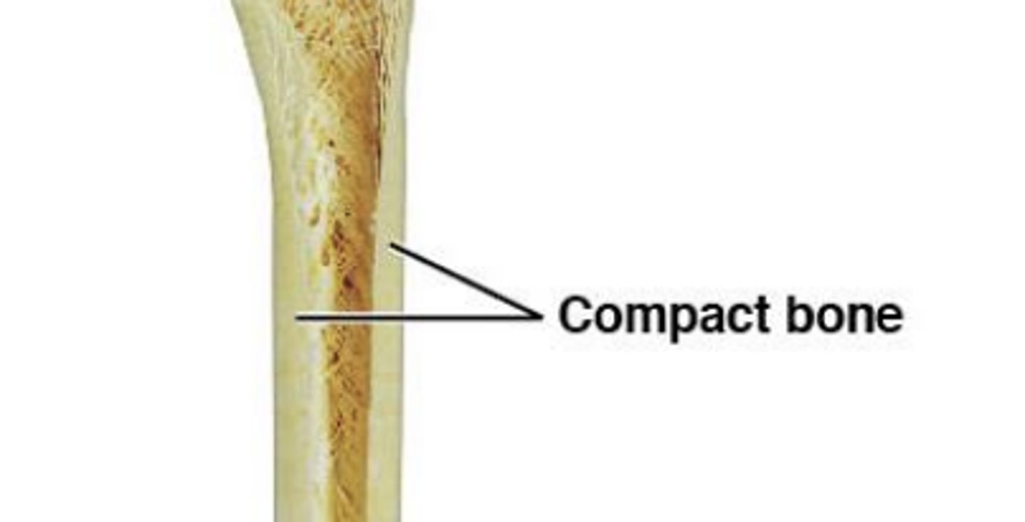
Compact bone (dense bone)
- dense & solid
- Forms walls of bone
- Parallel compression
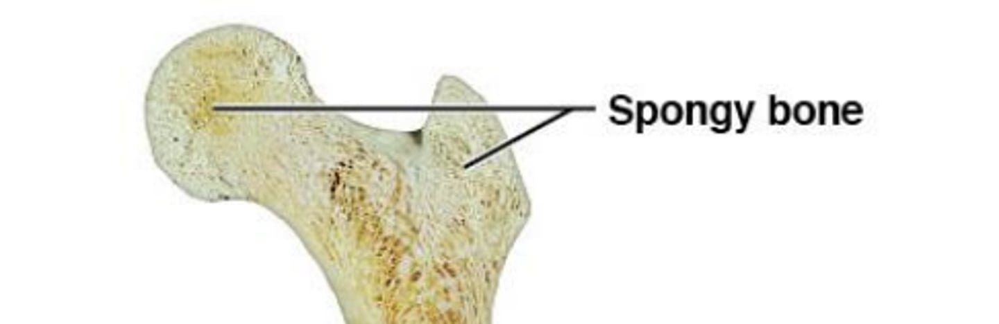
Spongy bone (trabecular bone)
- Open network of plates
Multidirectional or light strain
- Surrounds medullary cavity (open space)
bone marrow: connective tissue in medullary cavity
Epiphysis
ends of long bones
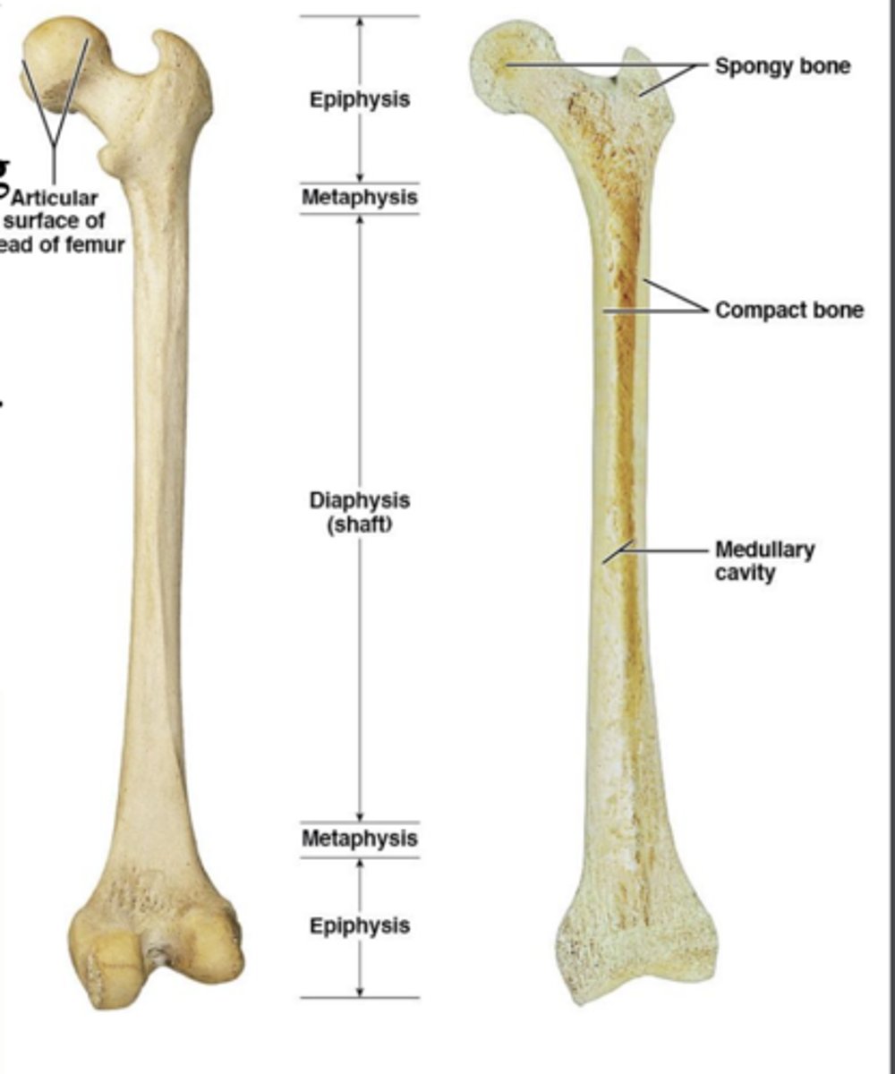
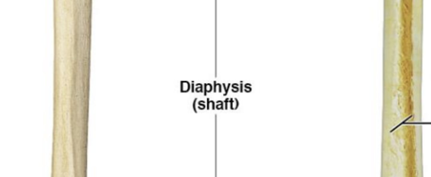
Diaphysis
shaft of long bones
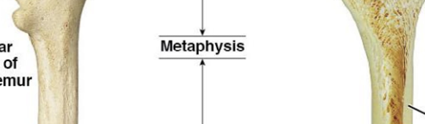
Metaphysis
transition
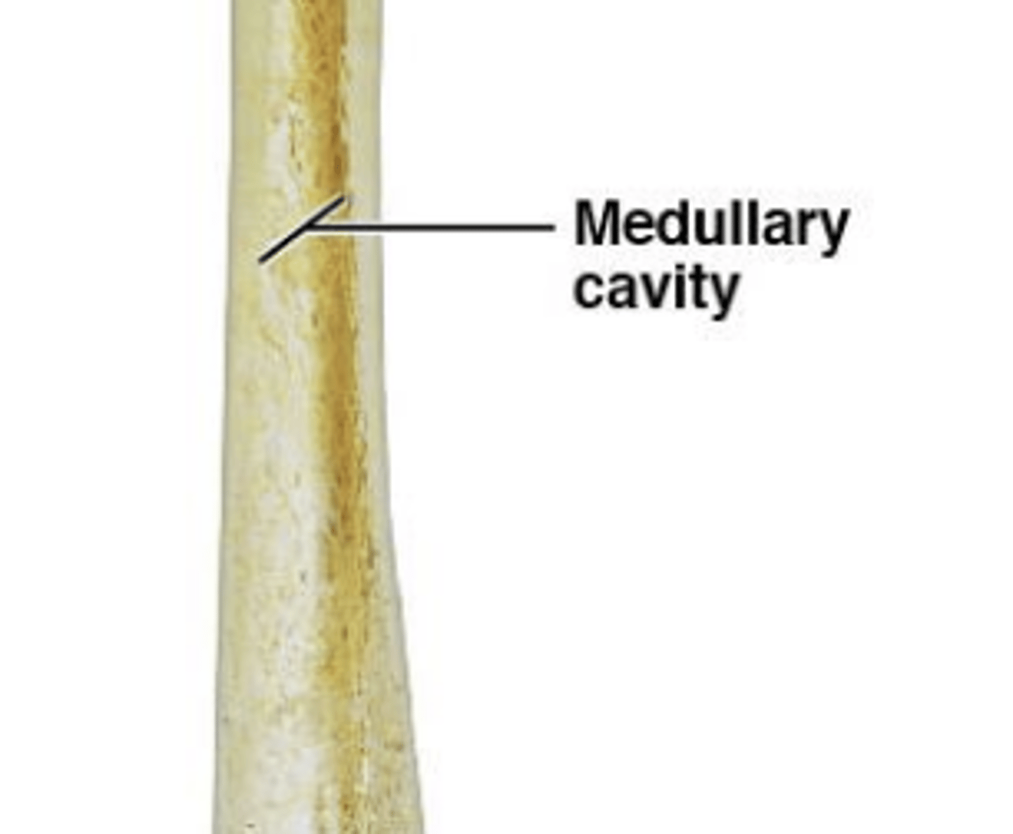
Medullary cavity:
inner cavity of diaphysis
Epiphyseal line
"growth line"

gross anatomy of the humerus
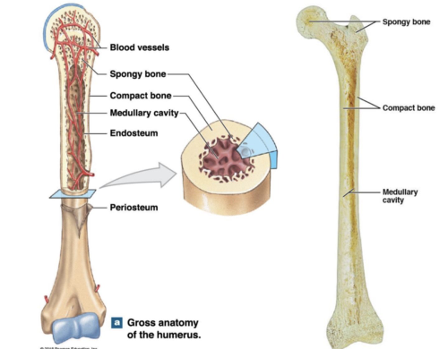
the organization of compact and spongy bone
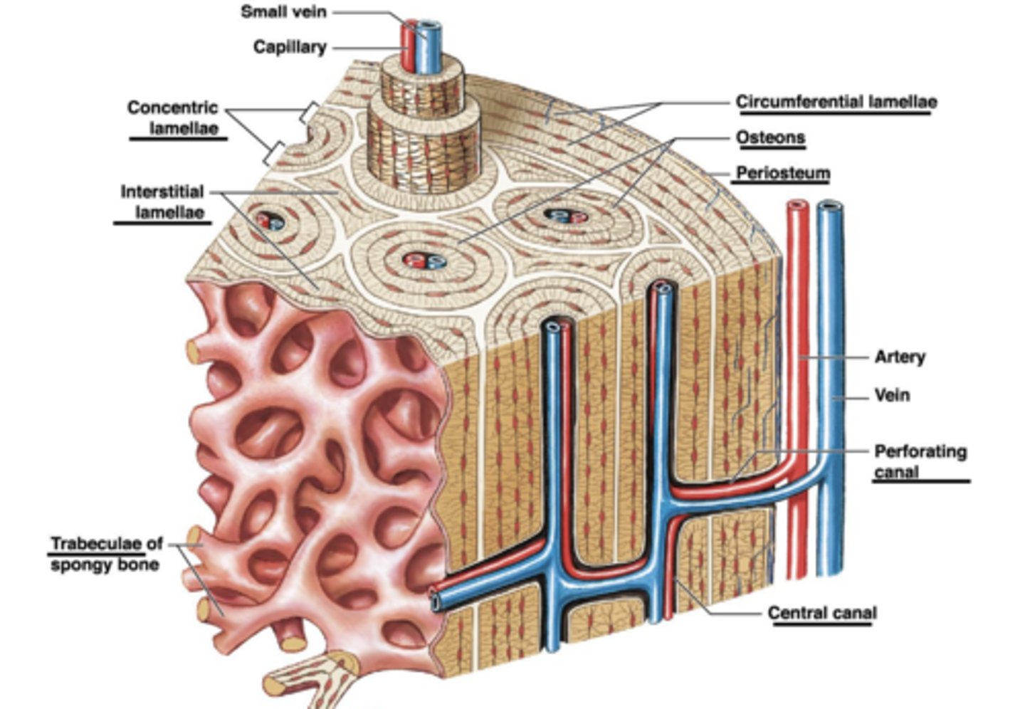
compact bone
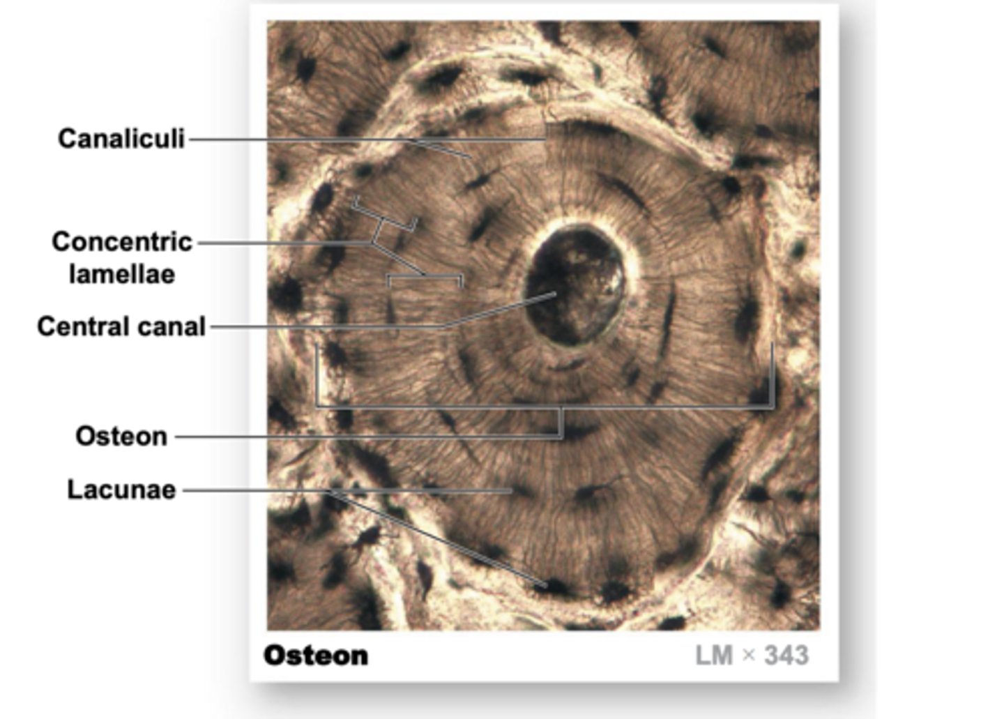
A single osteon at higher magnification. The central canal appears black on this section
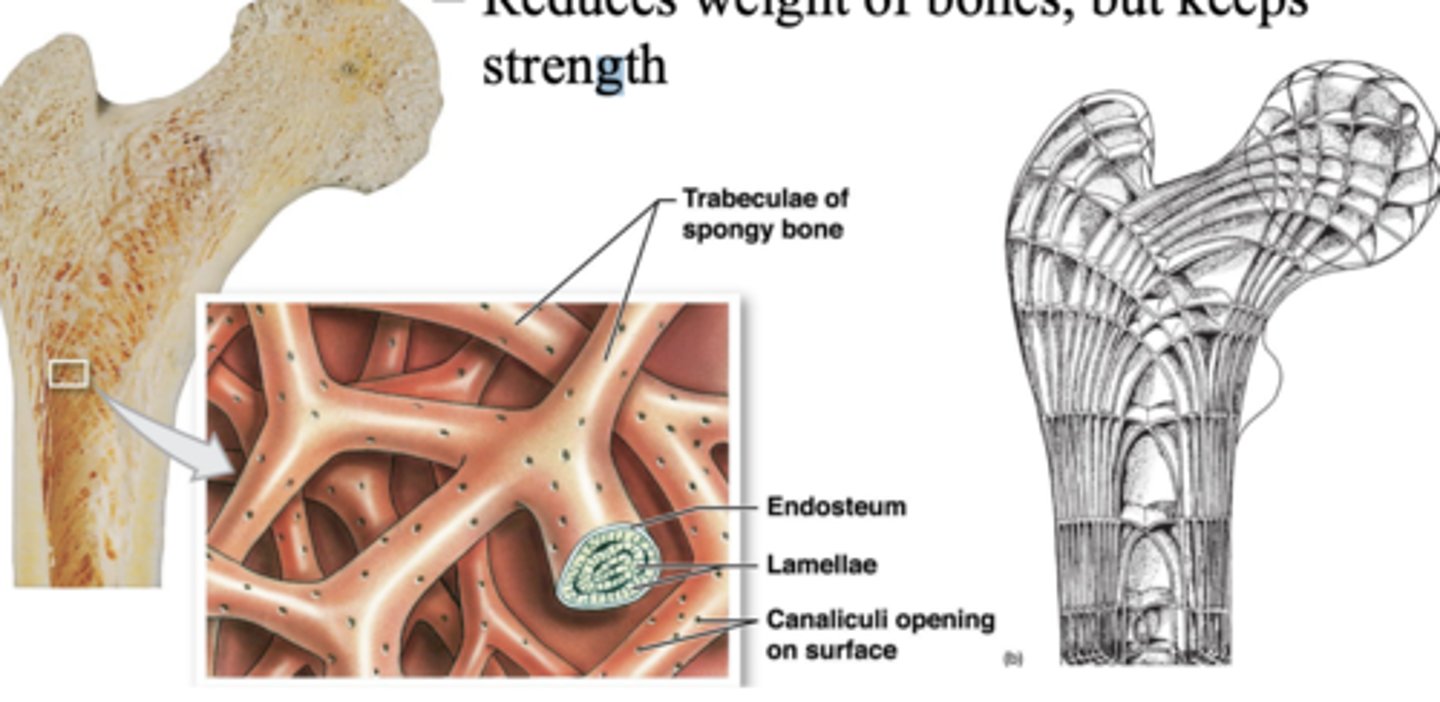
Spongy bone
- Arranged in parallel struts
- Trabeculae: branching plates
- Trabeculae form an open network
- Large trabeculae can have osteons
- Reduces weight of bones, but keeps strength
Functional Differences • Compact bone
Transmission of stress parallel to bone axis
- Osteon arrangement is parallel to bone axis
- Weak bone strength perpendicular to bone axis
Functional Differences Spongy bone
- Multidirectional or light strain
- Trabeculae are oriented along stress lines
- Has extensive cross-bracing
- Supports marrow
Yellow marrow
mainly adipose, energy store
- Medullary cavity
Red marrow
Production & storage of leukocytes, erythrocytes, & thrombocytes
- In epiphysis or spaces between trabeculae -not restricted to just long bones
Periosteum: Membrane on outer surface of bone
Fibrous layer & osteogenic layer (complete, multi cell layer)
• Isolates & protects bone from surrounding tissue
• Attachment for circulatory & nervous supply
• Actively participates in bone growth & repair
• Perforating fibers: anchors periosteum to bone & other connective tissues
• Attachment site for tendons & ligaments
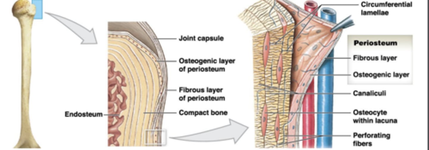
Endosteum
membrane on inner surface of bone; single, incomplete cell layer
Lines medullary cavity, perforating canals & central canals
Both endosteum & periosteum have
- Osteoblasts: produces matrix
- Osteoprogenitor cells: produce osteoblasts
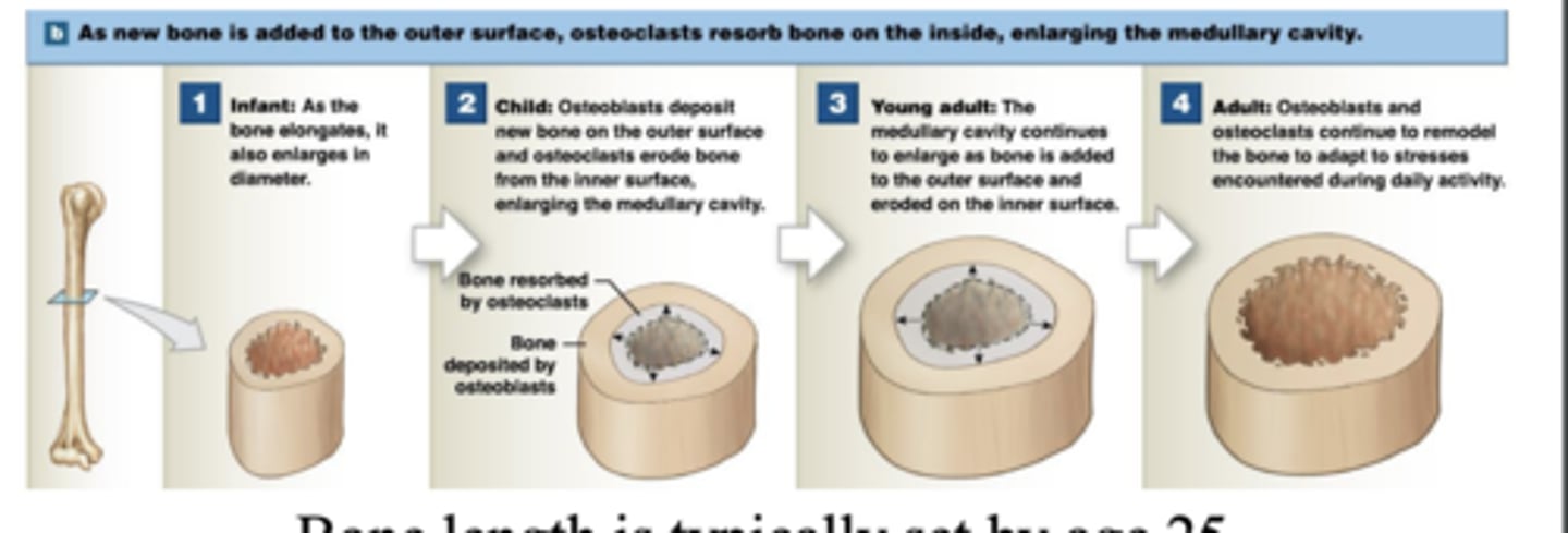
Appositional growth
Osteoblasts in periosteum add bone matrix to surface
• Forming circumferential lamellae on outer surface
• Osteons are formed
• Osteoclasts break down layer below endosteum to enlarge medullary cavity
- bone length typically set by age 25
Osteoblast & osteoclast activity is
equal in bones that aren't changing shape
Bones change shape in response to strain
Increased muscular development during exercise
- Inactivity of bones can cause degeneration
- After a few weeks, unstressed bones can lose ~1/3 mass; shape & density
Factors Regulating Bone Growth
Minerals: calcium, phosphate, magnesium, citrate, carbonate, sodium
Vitamins:
- A:
stimulates osteoblasts
-C:
collagen formation & osteoblast differentiation
- D3 :
used for calcitriol by kidneys
• Calcitriol: increase Ca2+ & PO4 3- absorption in small intestines
Hormones:
work to regulate plasma Ca2+ levels
Parathyroid hormone (PTH)
- Stimulates osteoclasts
• Increases circulating Ca2+
• Influences production of calcitriol in kidney
- Increase Ca2+ absorption from small intestines
Calcitonin (produced by thyroid gland)
Inhibits osteoclasts
- Decreases circulating Ca2+
• Removing Ca2+ from blood & deposit as bone
Thyroxine (thyroid gland) & Growth hormone (anterior pituitary gland)
- Influence basal metabolic rate of bone cells
-Maintain activity in epiphyseal region for growth
Estrogen & Testosterone
Stimulate osteoblast activity causing growth spurts during puberty
-Maintain bone density in adults
skeletal system
Houses central nervous system (CNS)
• Integration centers for reflex arcs
• Controls skeletal muscle
Houses senses
Sight • Hearing • Taste • Smell • Balance
CNS
Allows for sound production & communication
• Feeding
• Breathing
• Attachment points for appendicular system
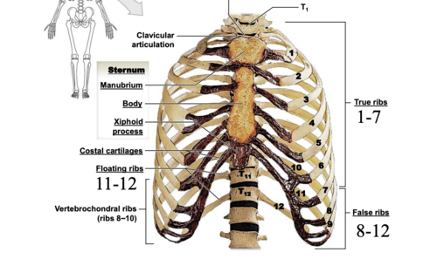
Thoracic cage (thorax)
Ribs
• Sternum
• Thoracic vertebrae
• Costal cartilage
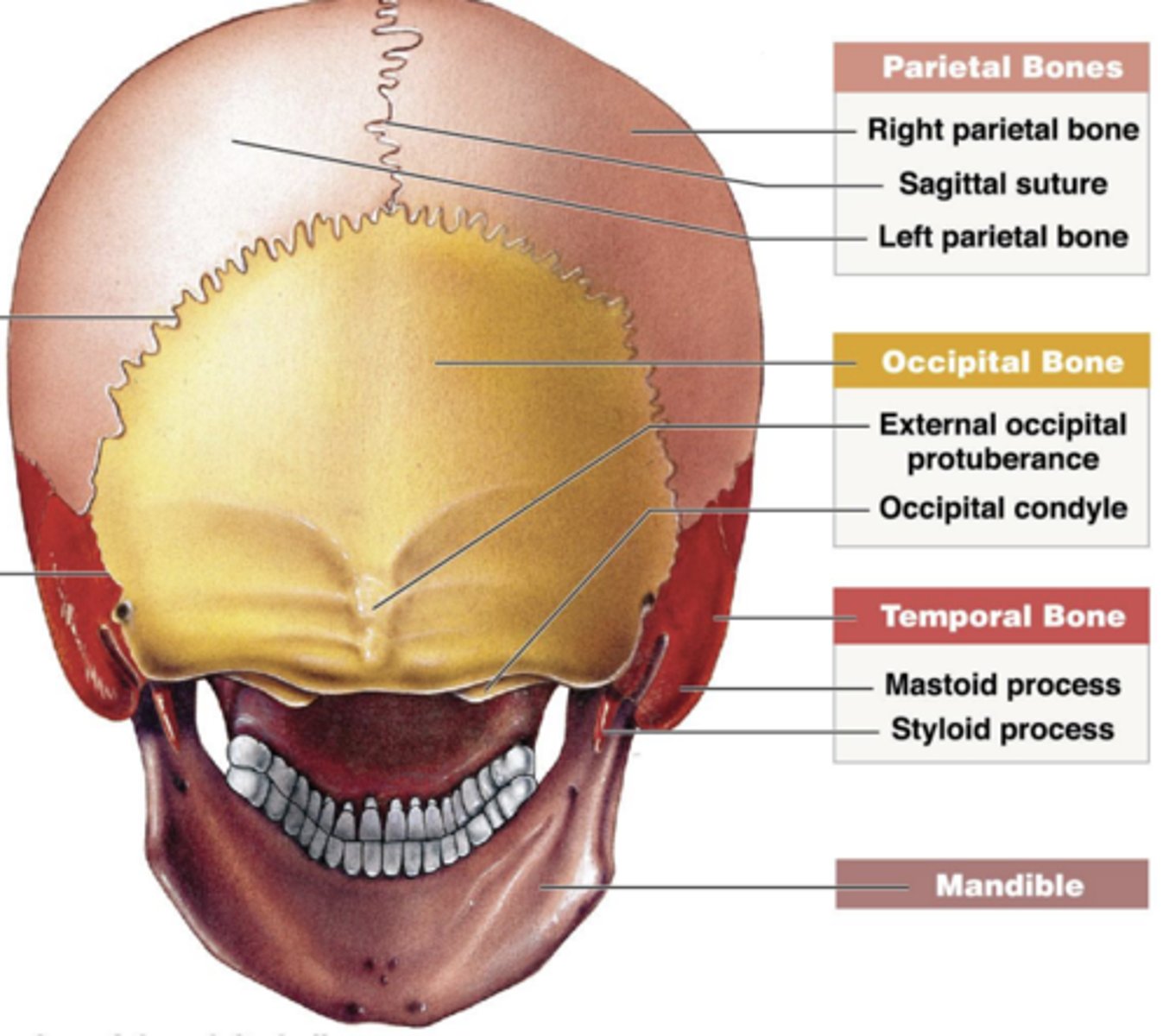
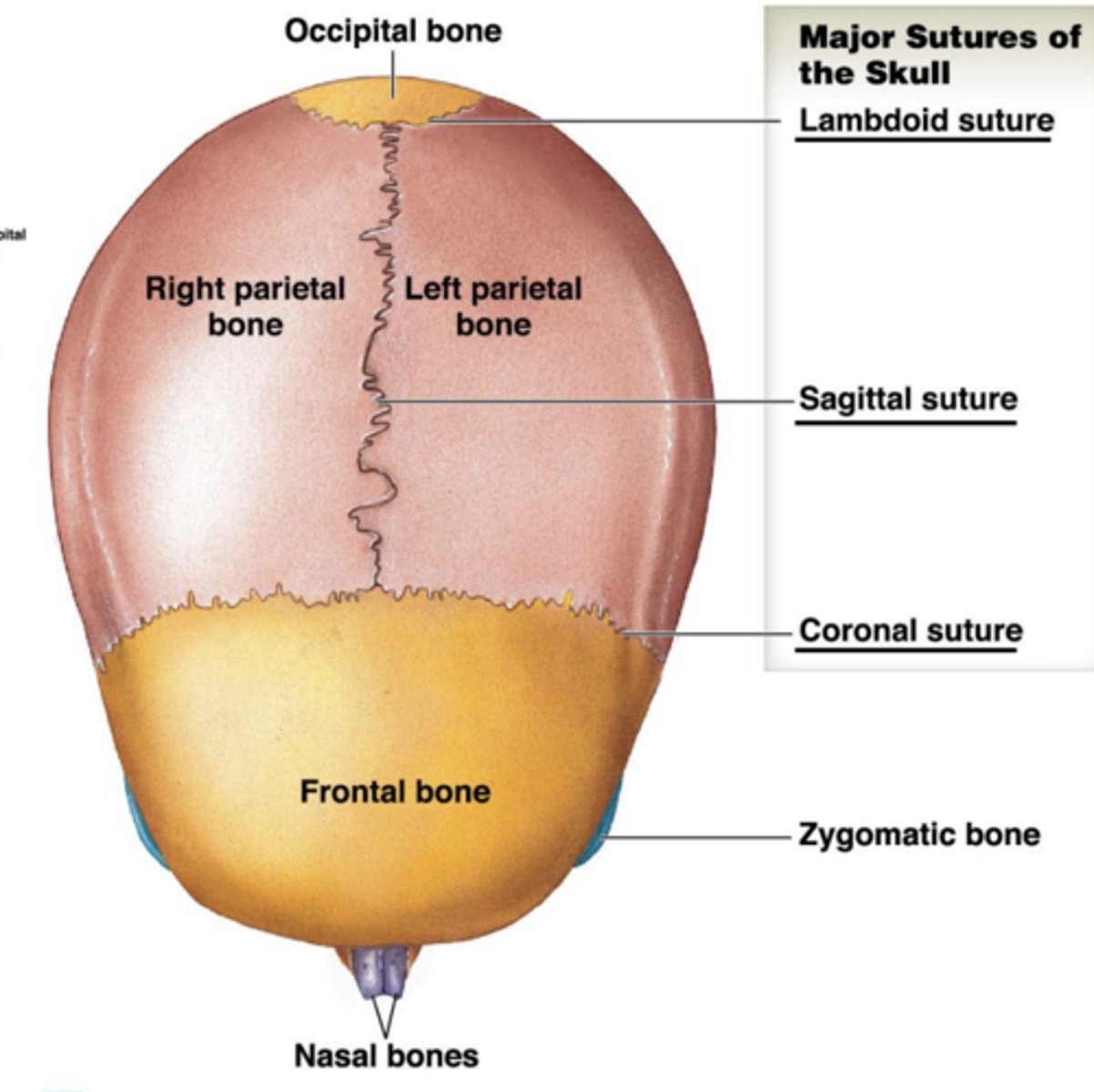
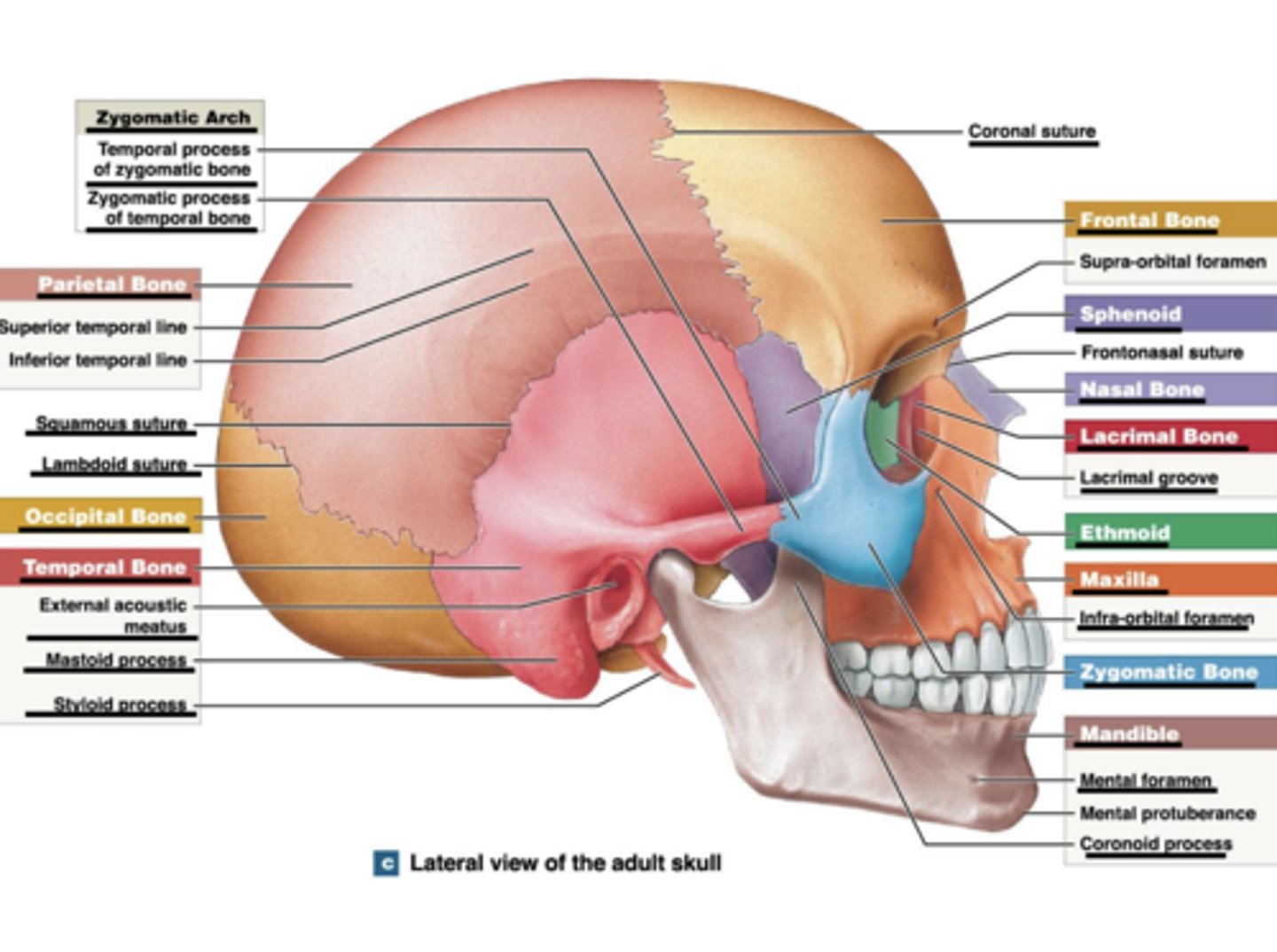
skull
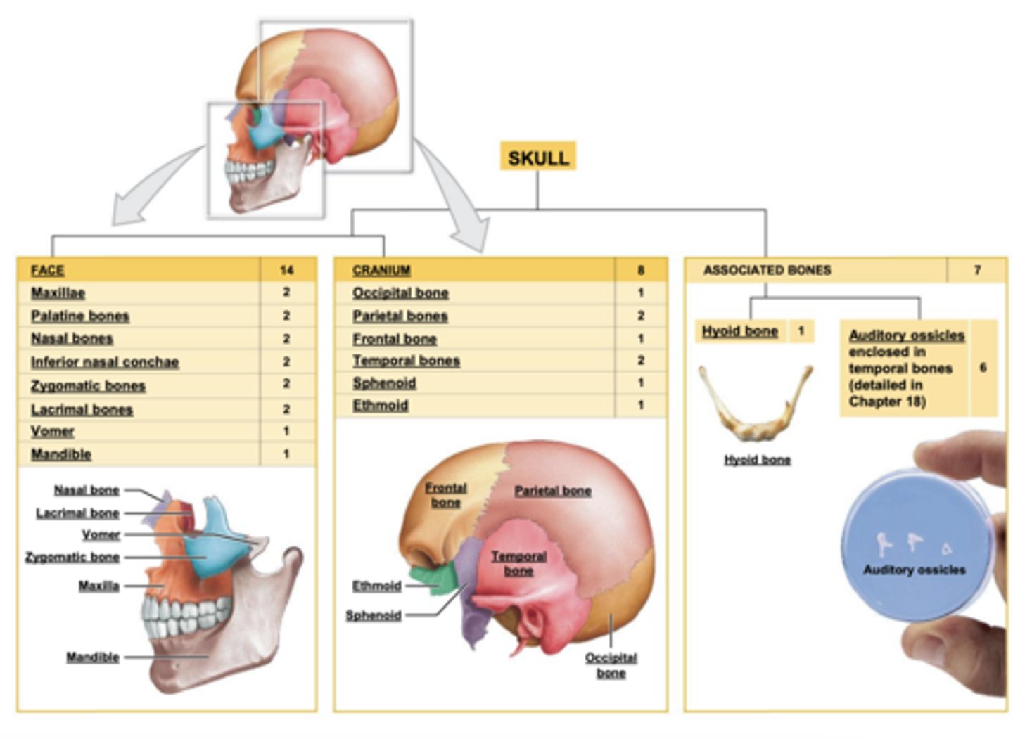
Suture
dense fibrous connective tissue
Fontanelles
gaps between cranial sutures; gives flexibility to skull during parturition
lateral view of adult skull
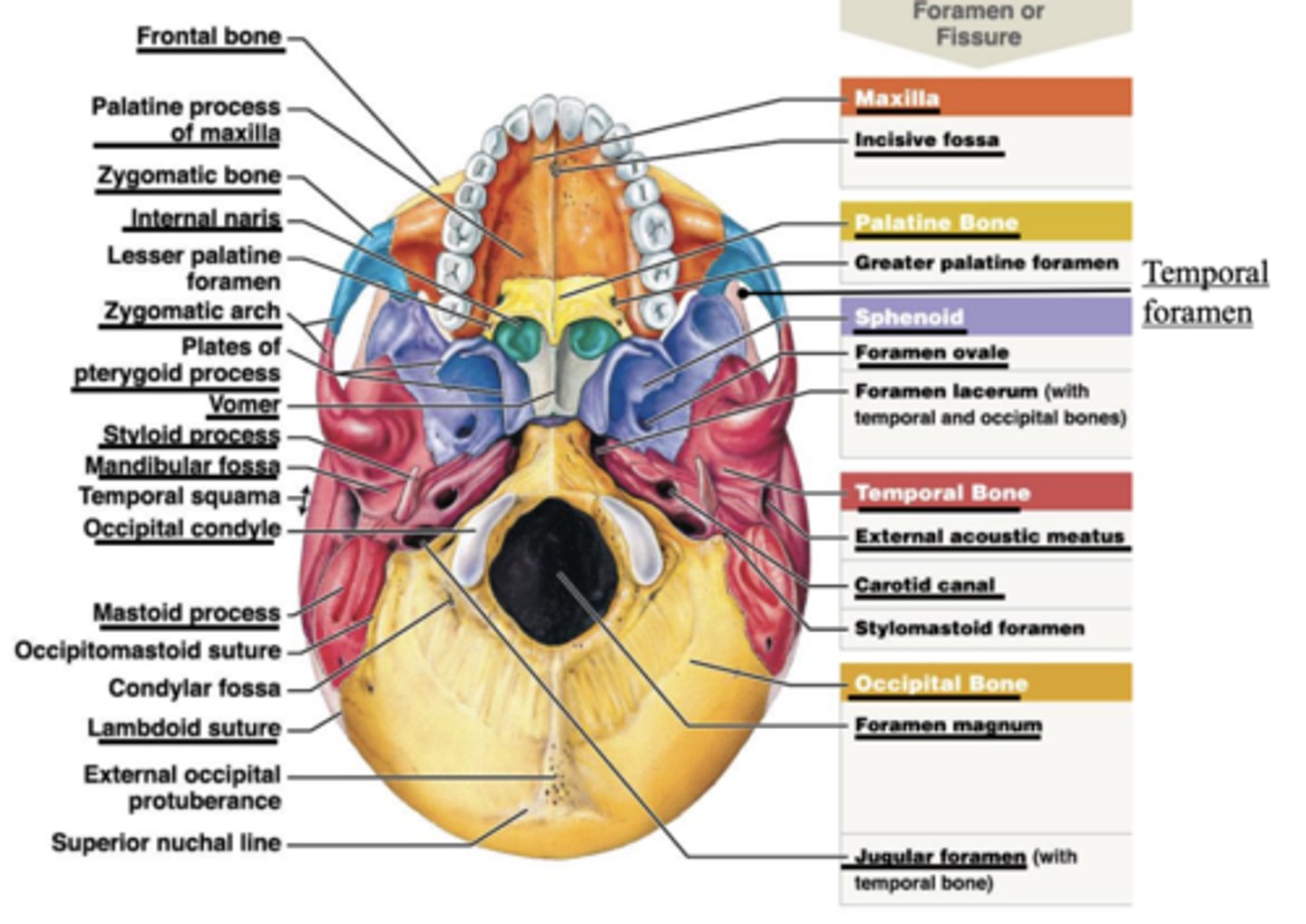
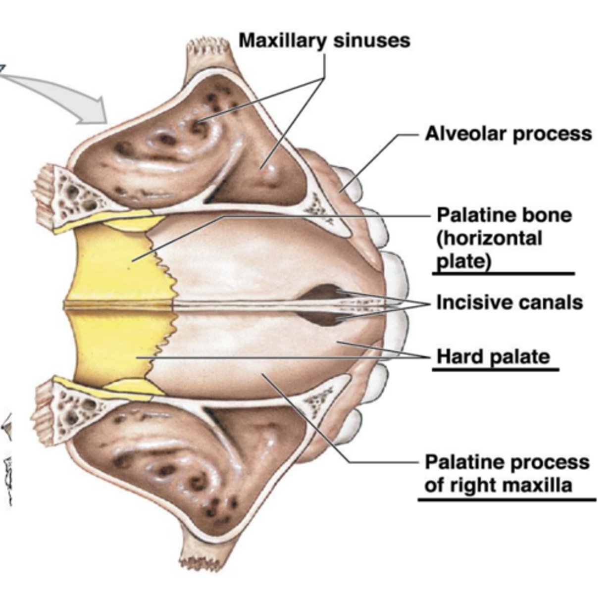
inferior view mandible removed
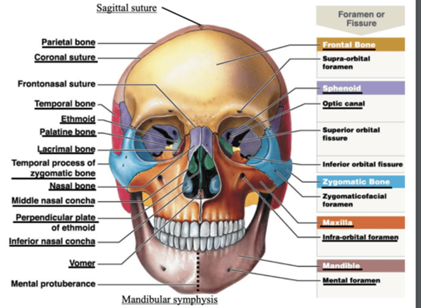
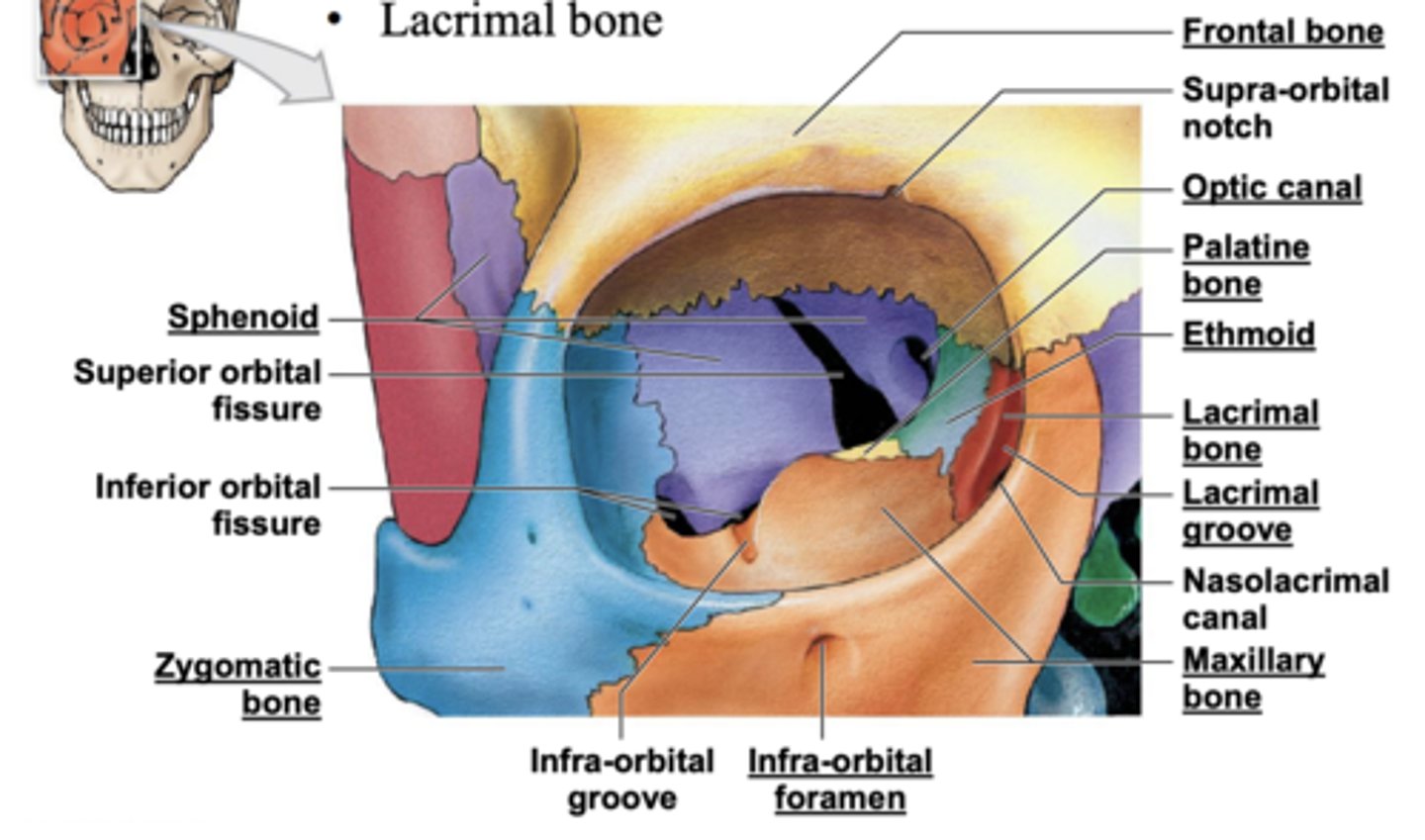
Orbit made of
Frontal bone
• Maxilla bone
• Lacrimal bone
Ethmoid bone
• Sphenoid bone
• Zygomatic bone
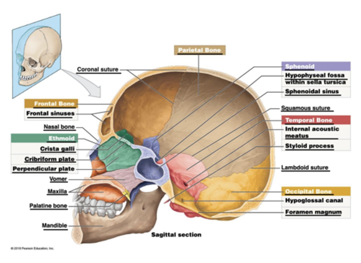
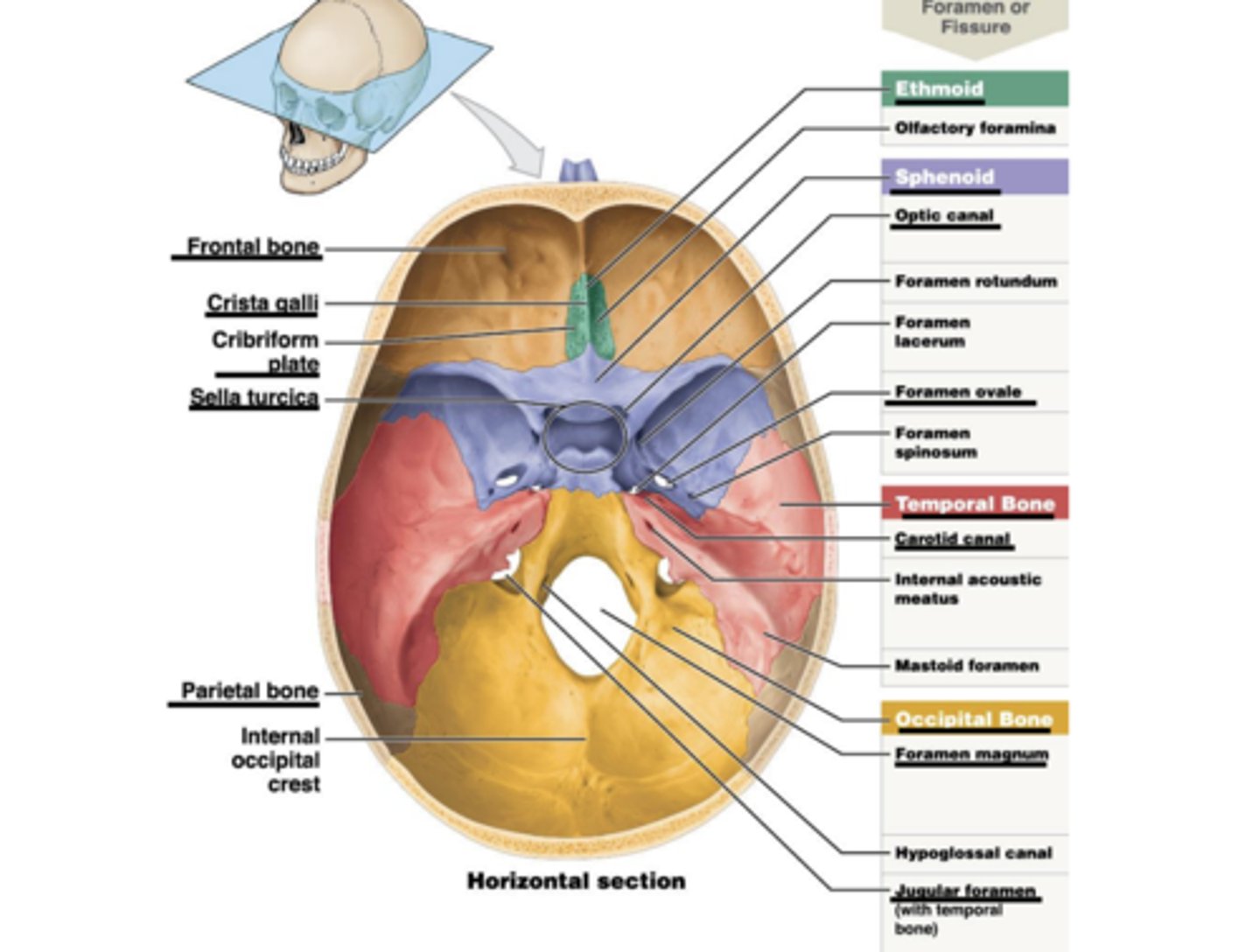
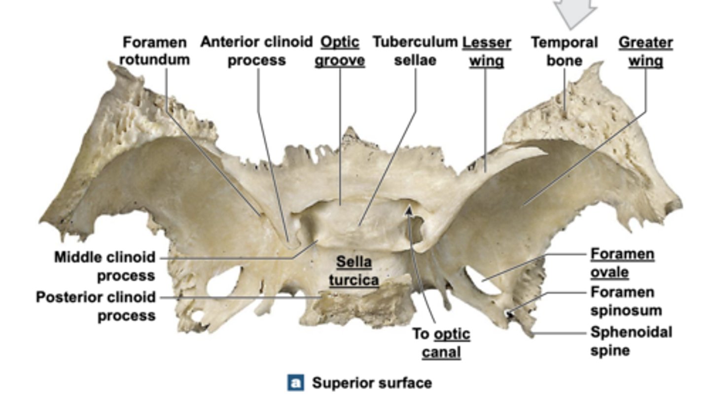
sphenoid
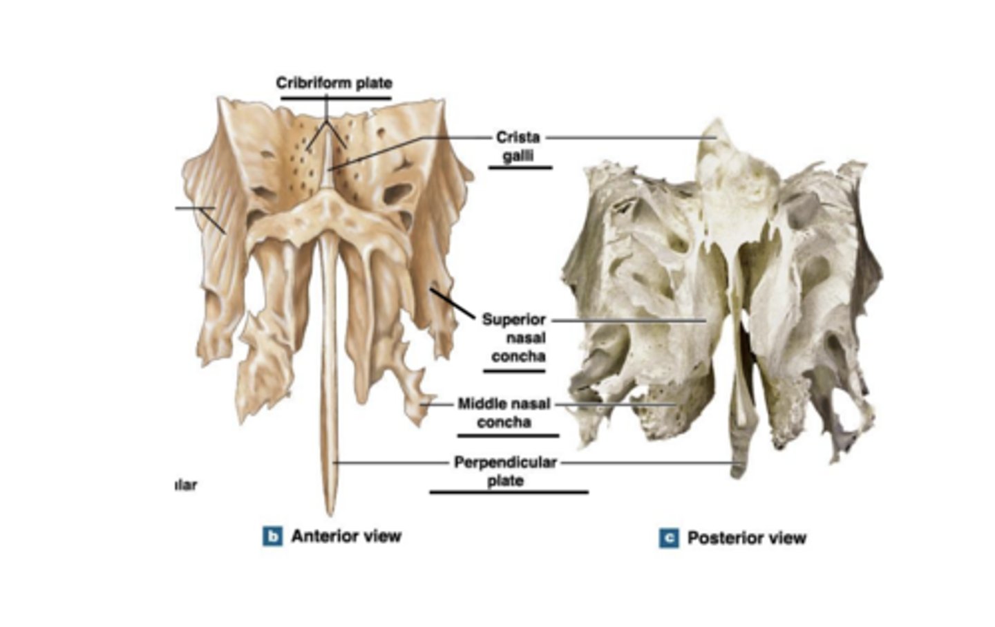
ethmoid
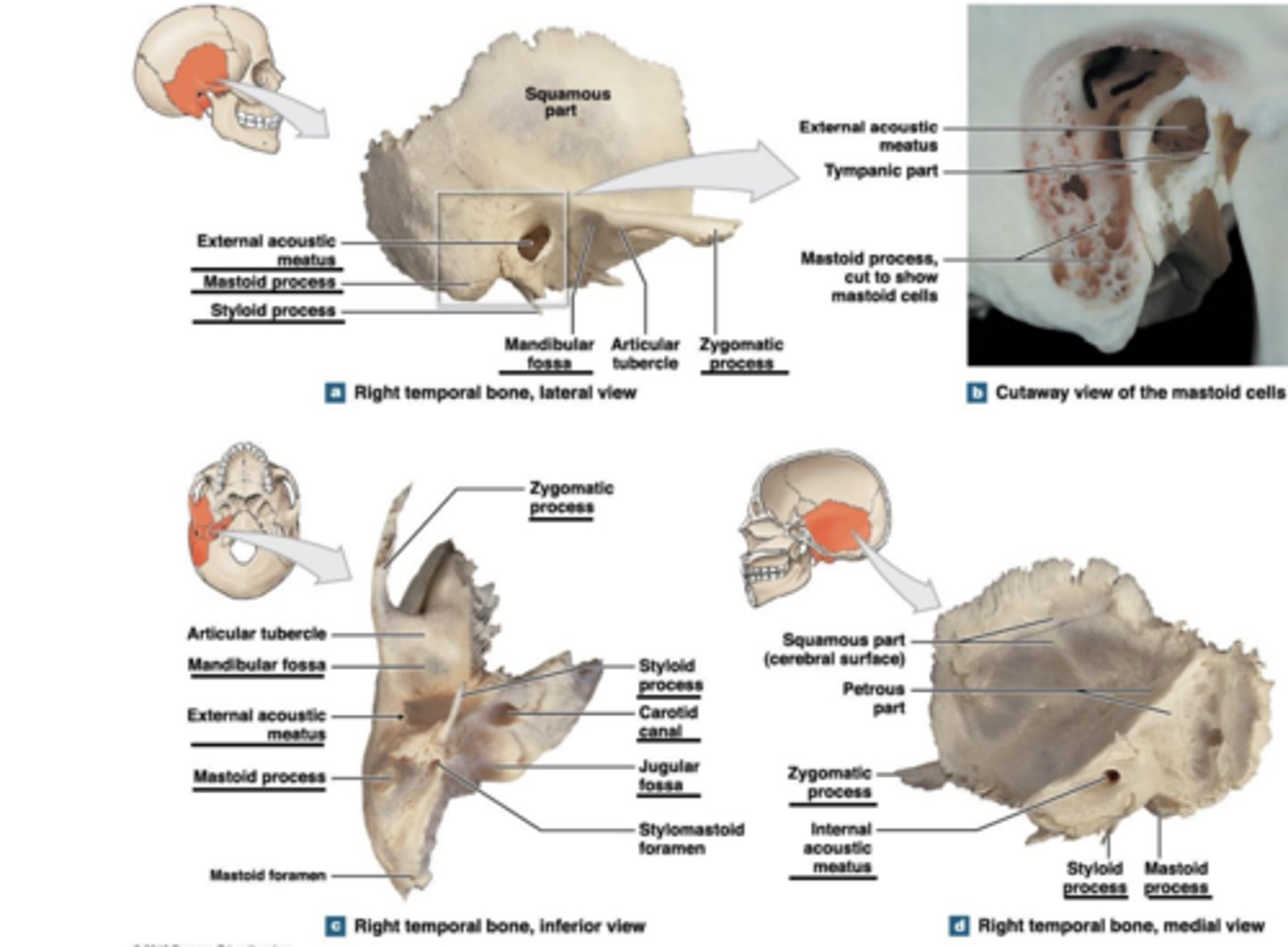
right temporal bone
maxilla
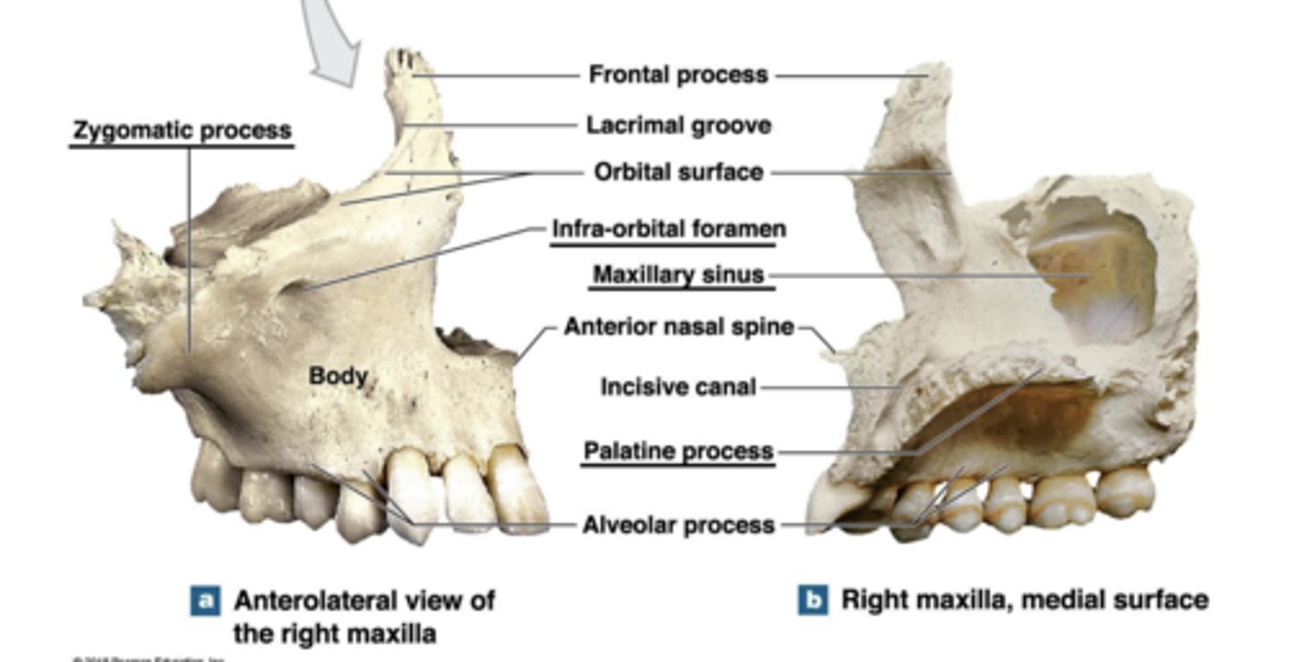
palatine
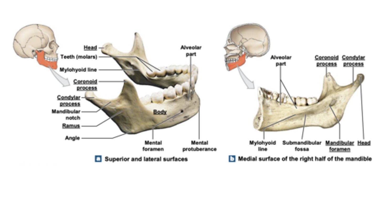
mandible
sinuses
Produce mucus
• Resonate sound
• Lighten skull
• Humidifies air
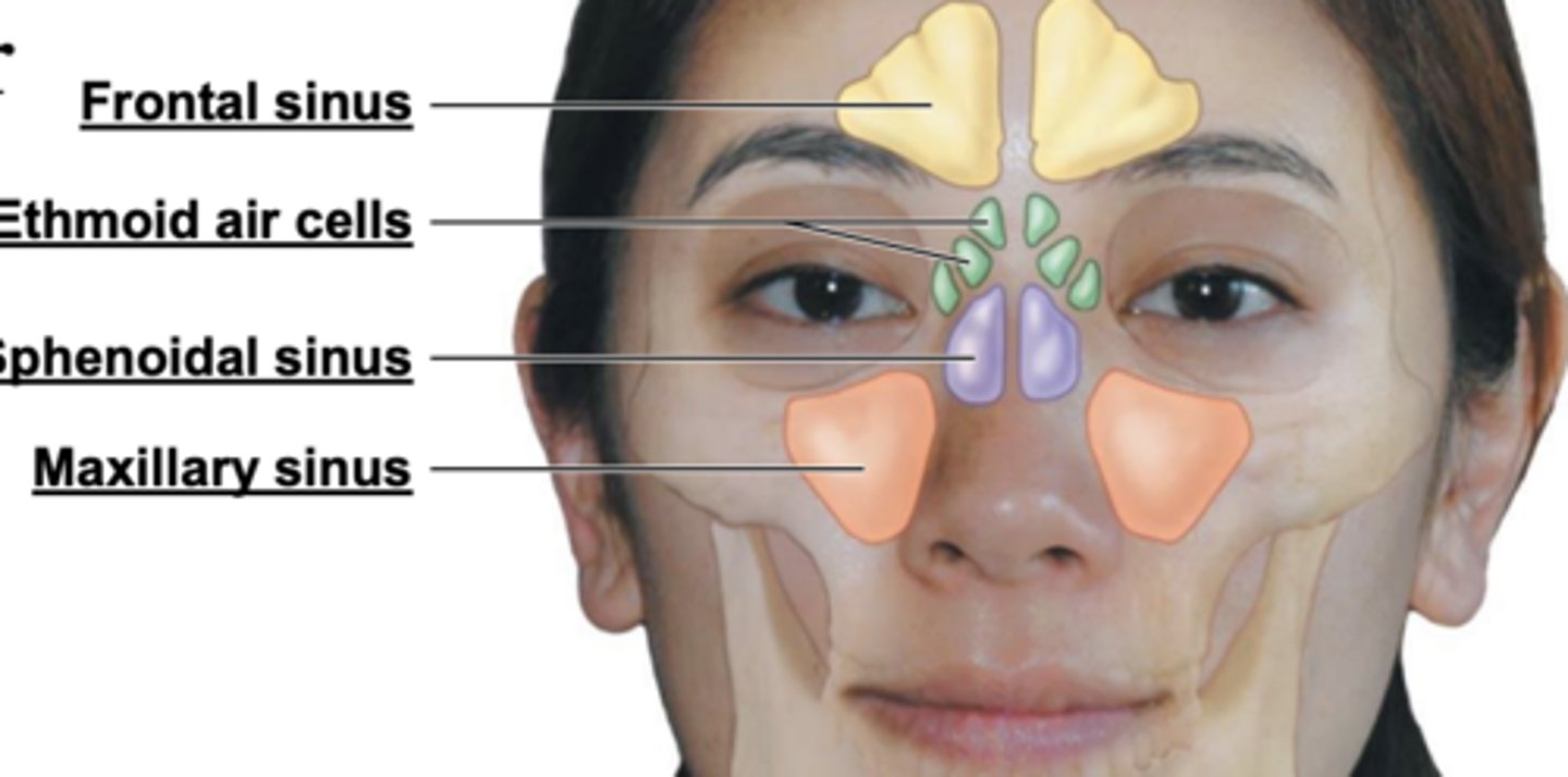
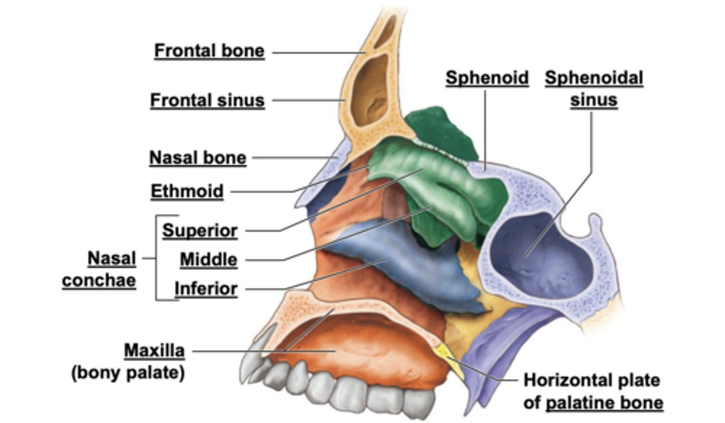
section showing the positions of the paranasal sinuses

sagittal section with the nasal septum removed to show major features of the wall of the right nasal cavity
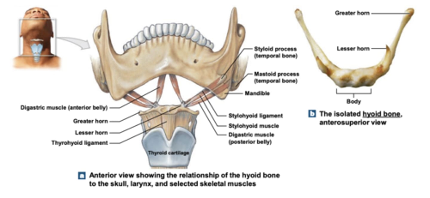
hyoid
Anchor point for muscles of:
• Tongue • Larynx
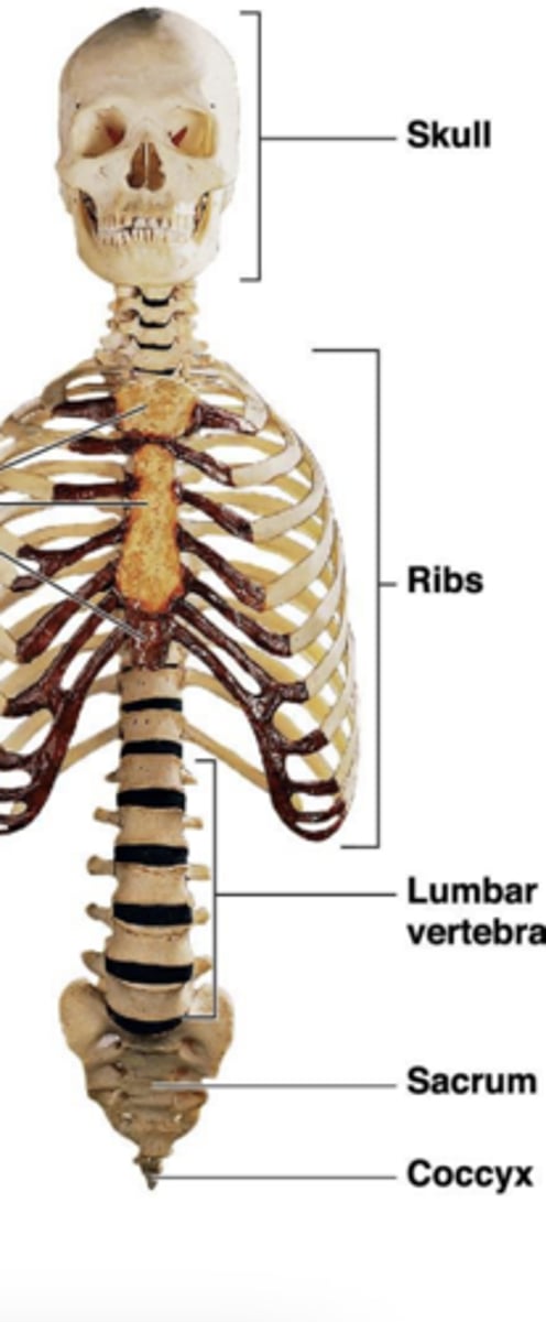
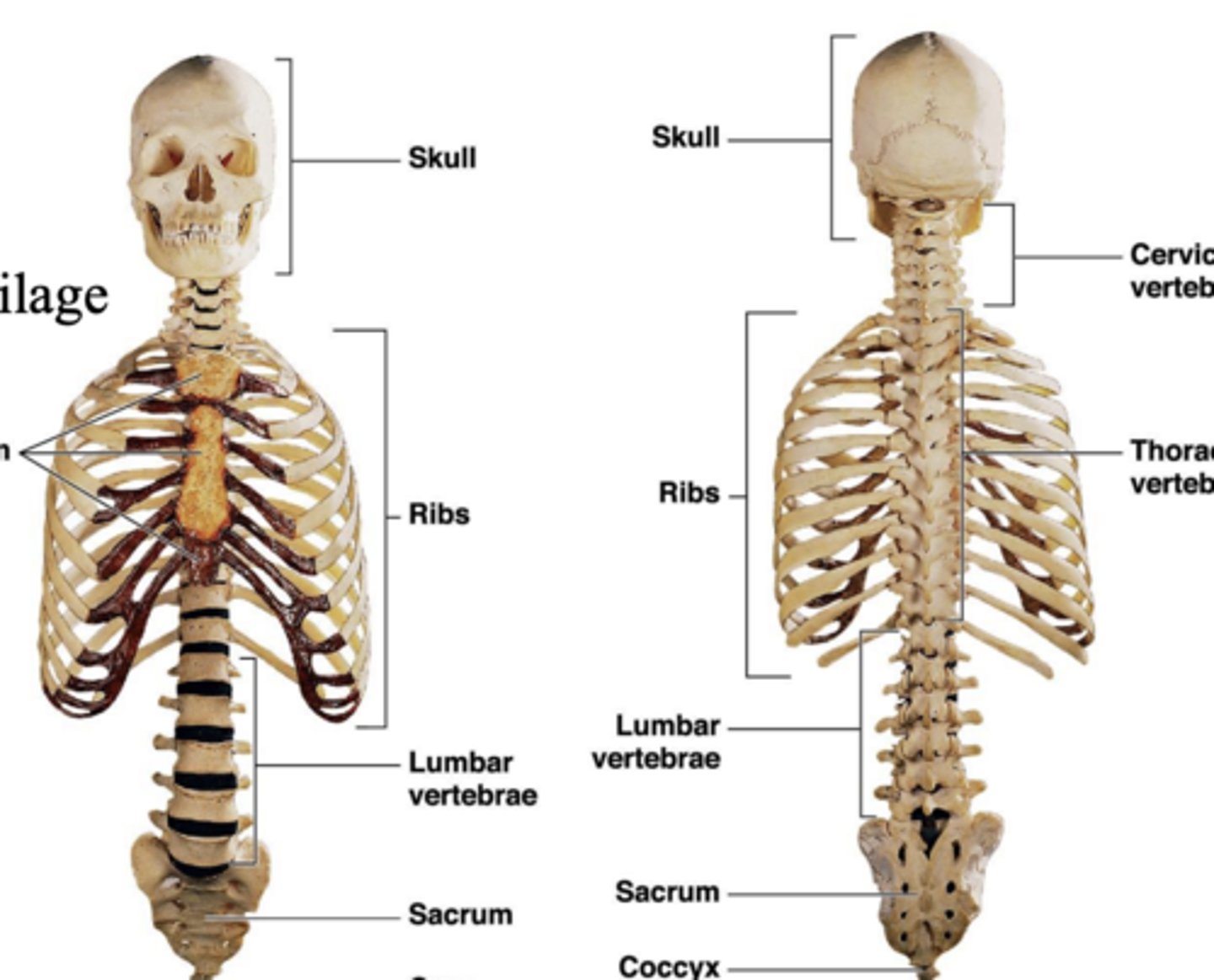
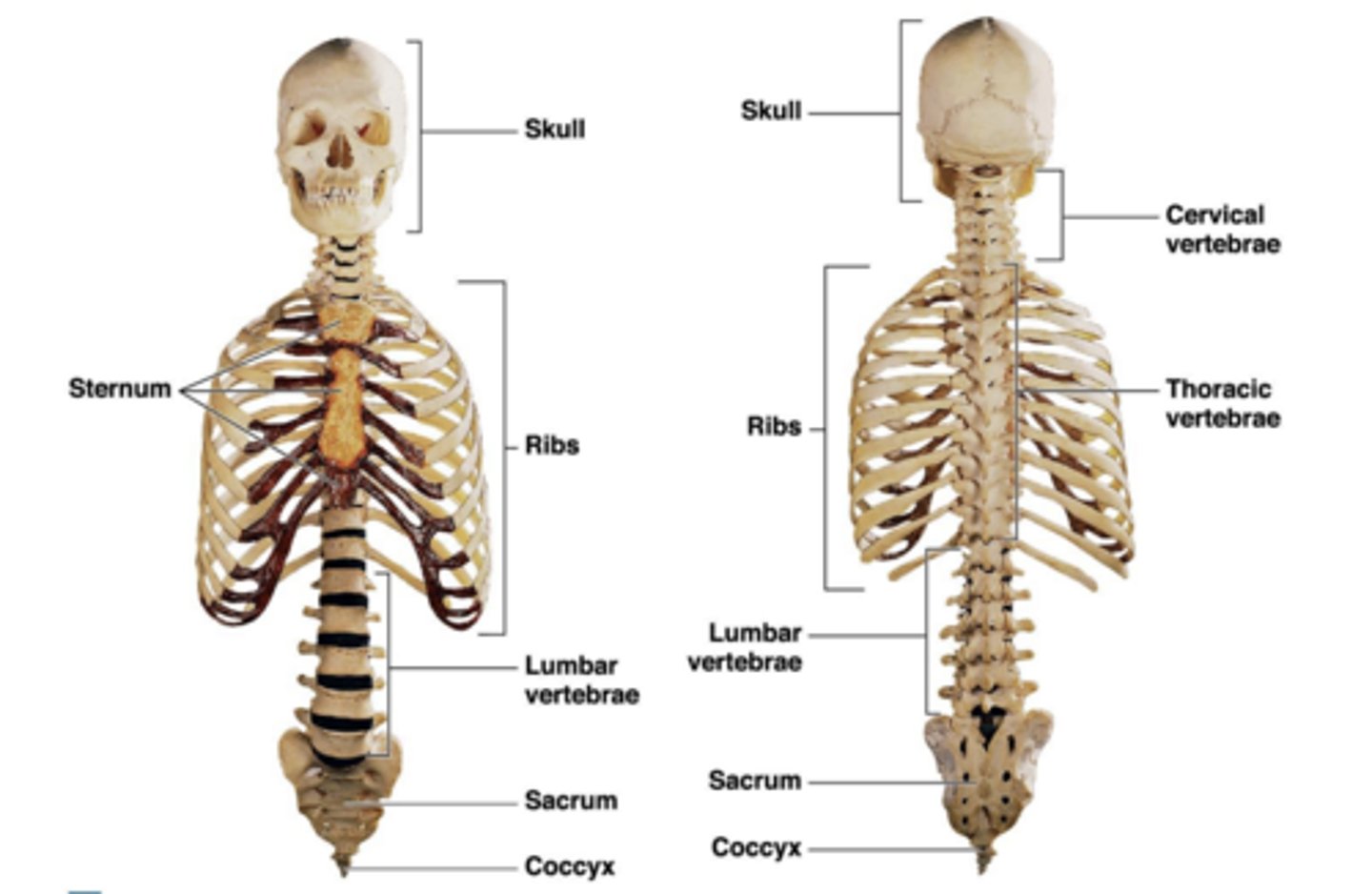
anterior and posterior views of the bones of the axial skeleton
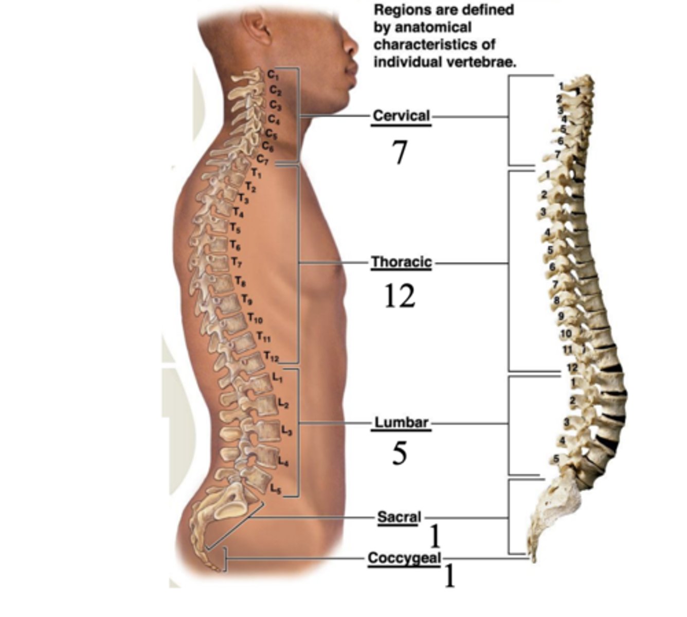
vertebral regions
regions are defined by anatomical characteristics of individual vertebrae
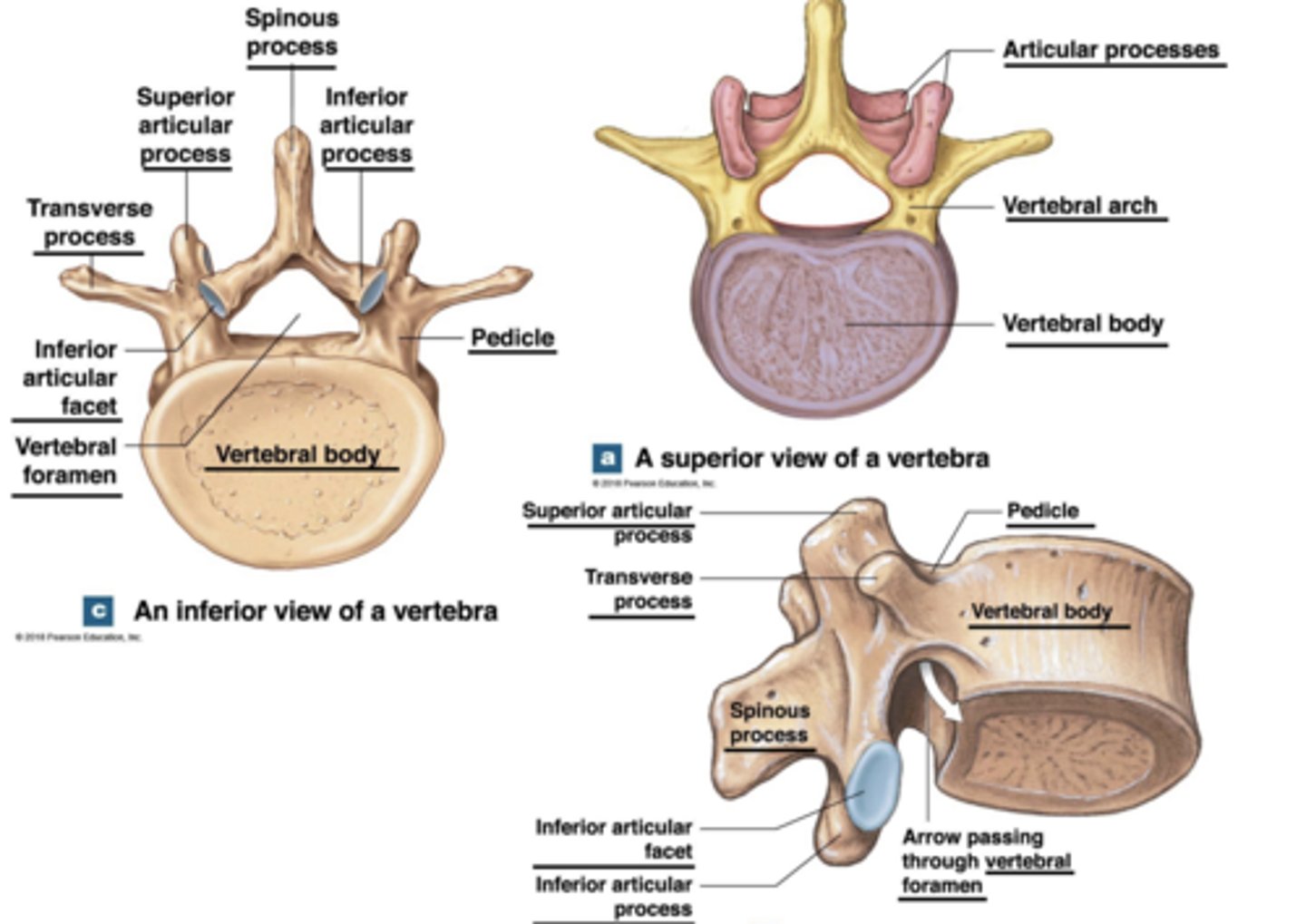
views of vertebra
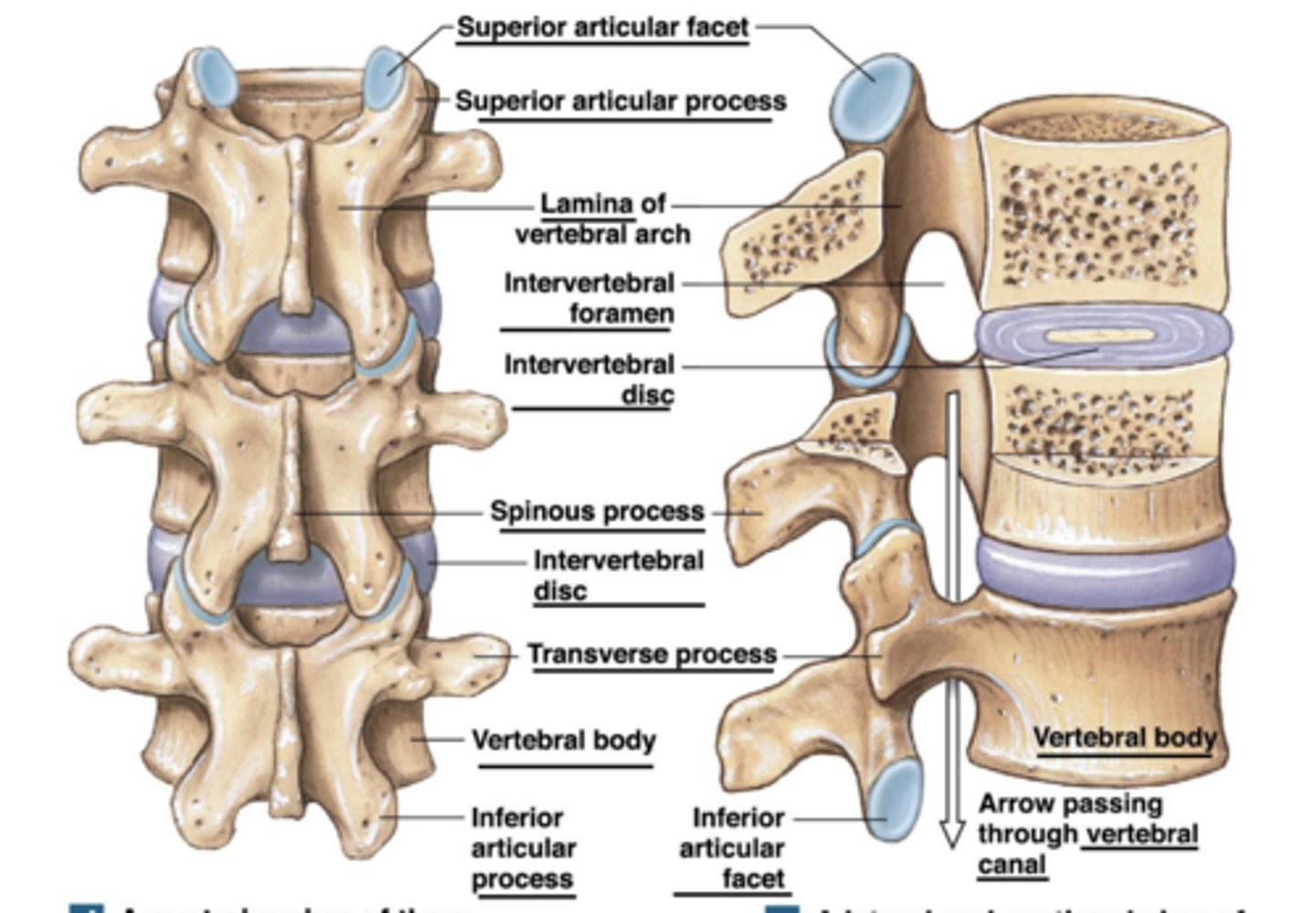
articulated vertebrae views
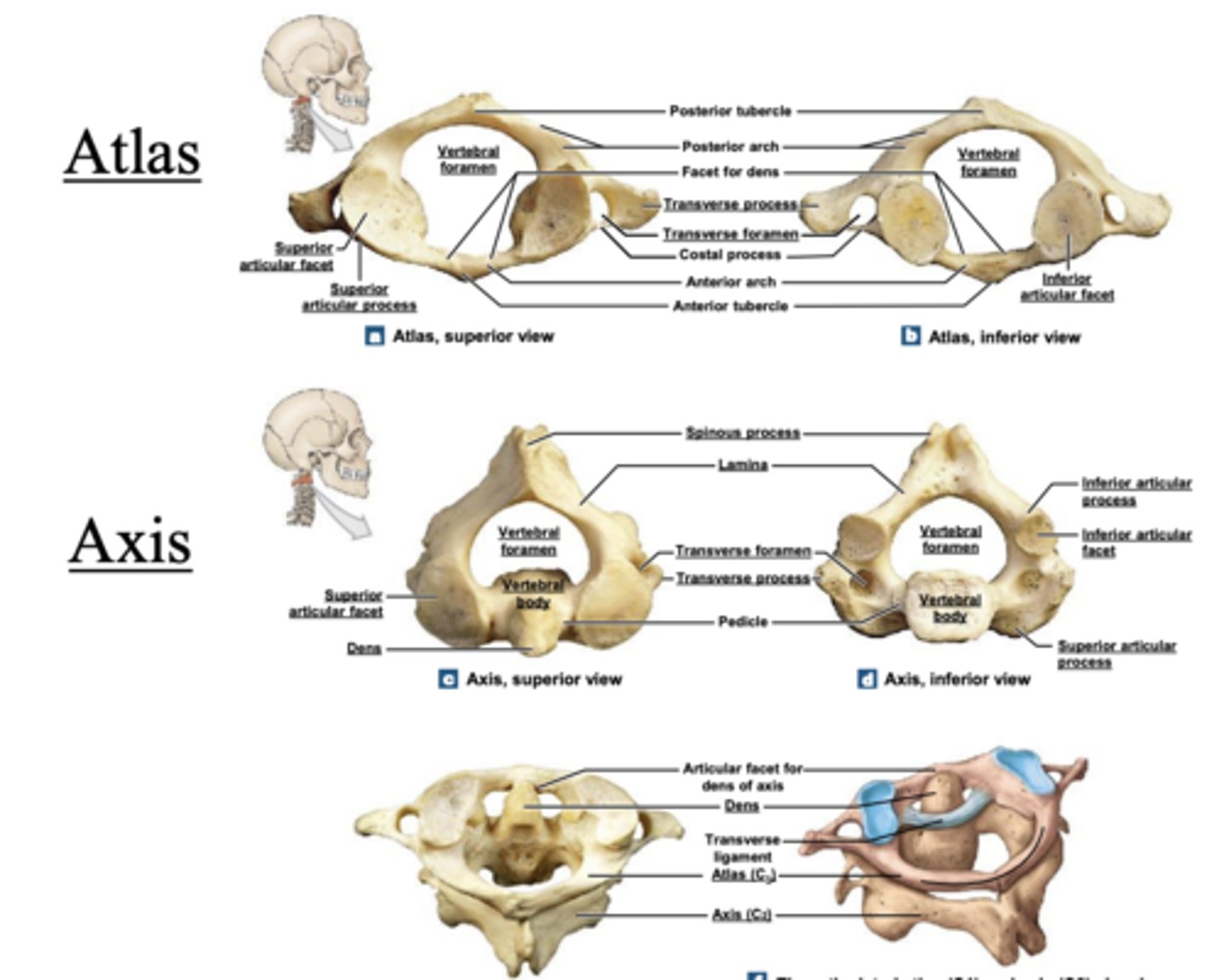
atlas & axis
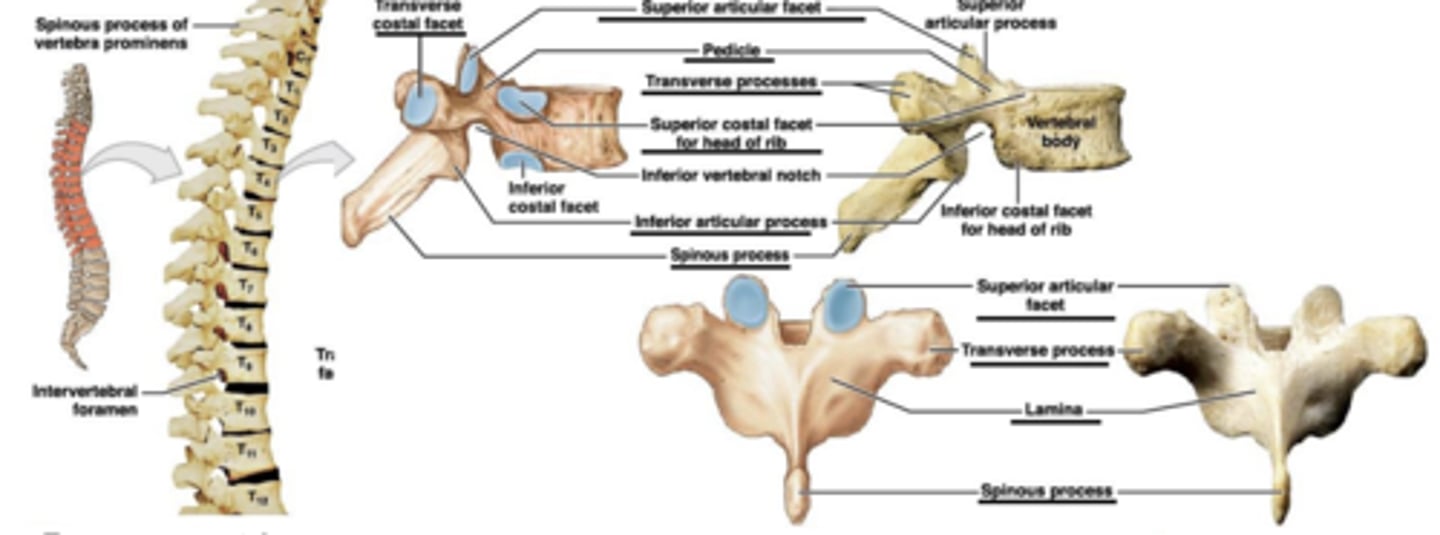
spinous process of vertebra prominens
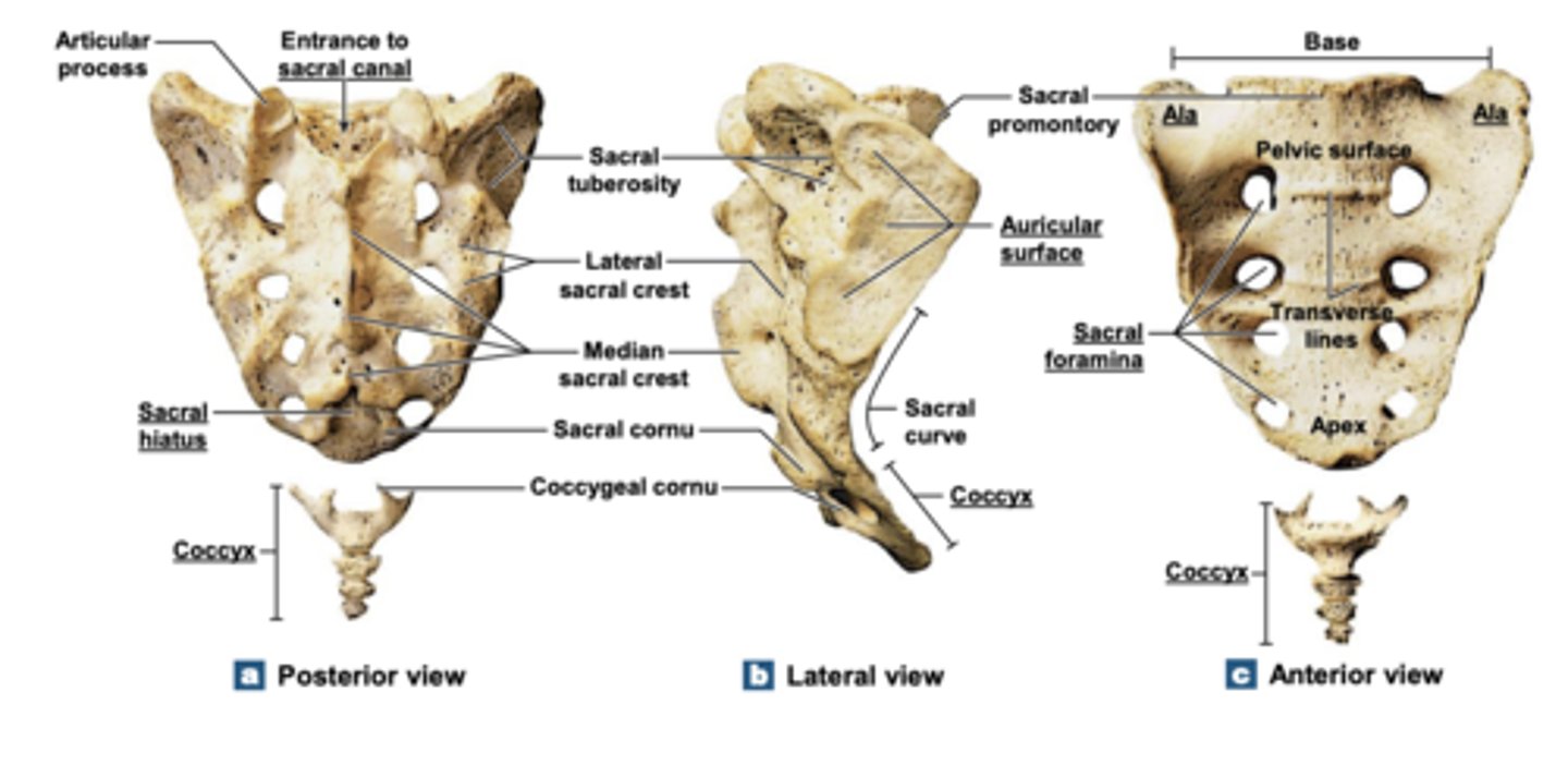
Sacrum
Anterior view of the rib cage and sternum
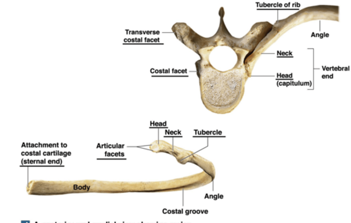
posterior and medial view showing major anatomical landmarks on an isolated right rib
axial skeleton
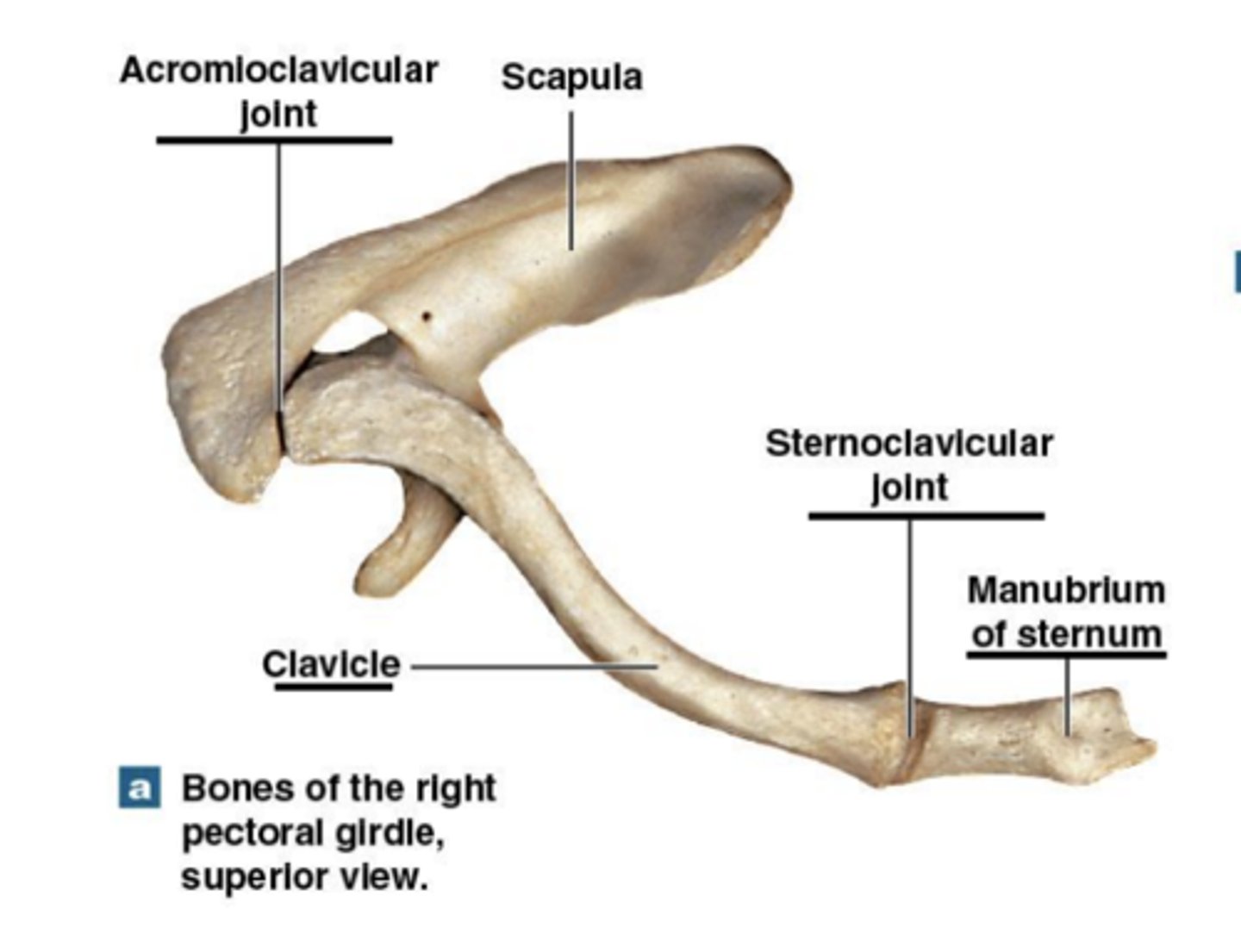
All of these examples are from right side of body
Sternoclavicular joint:
only direct connection between pectoral girdle & axial skeleton
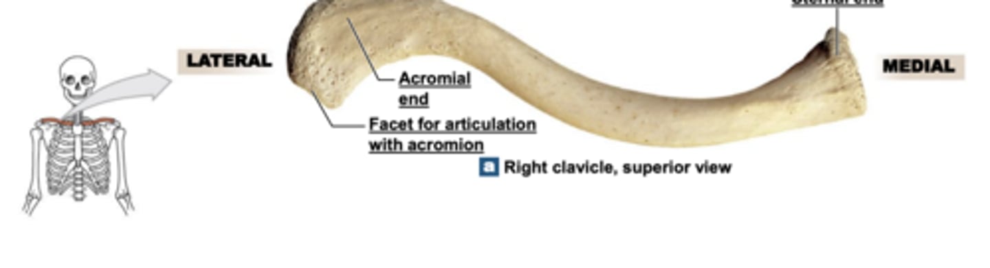
Right clavicle, superior view
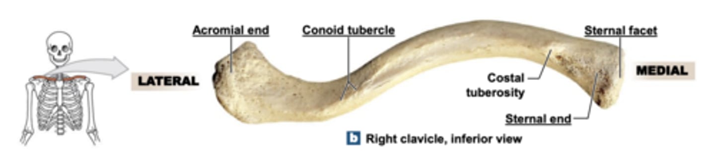
Right clavicle, inferior view
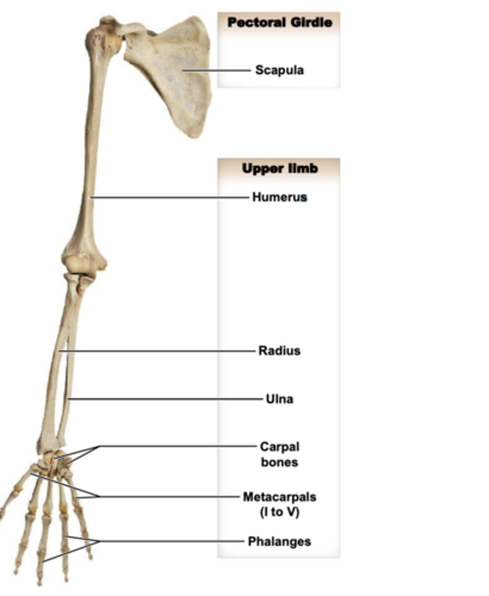
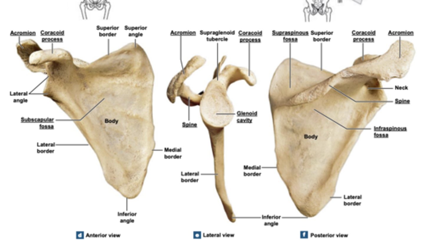
Scapula
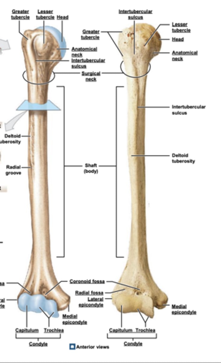
Humerus
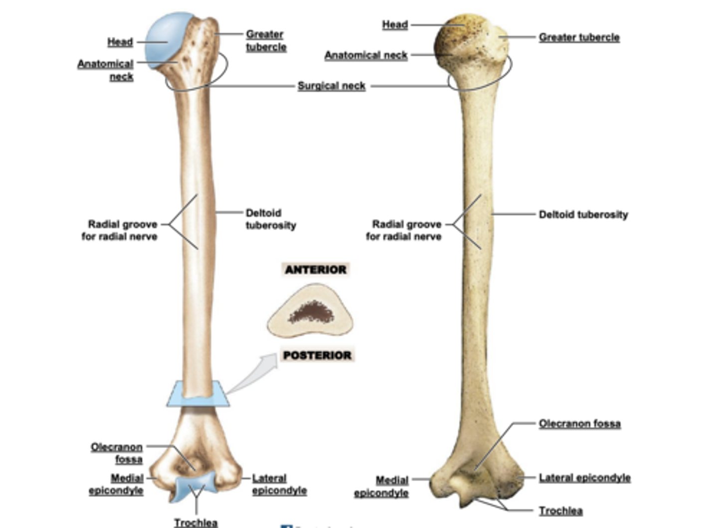
humerus posterior view
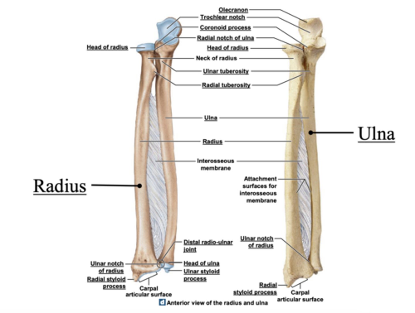
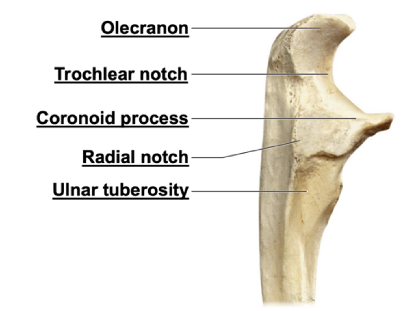
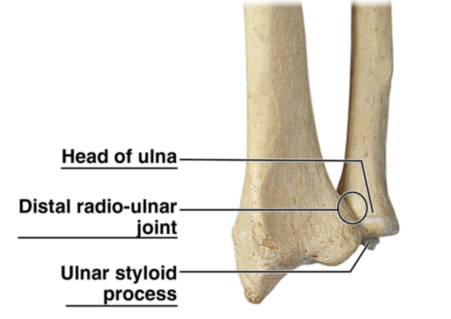
radius & ulna
Lateral view of the proximal end of the ulna
anterior view of the distal ends of the radius and ulna and the distal radio-ulnar joint
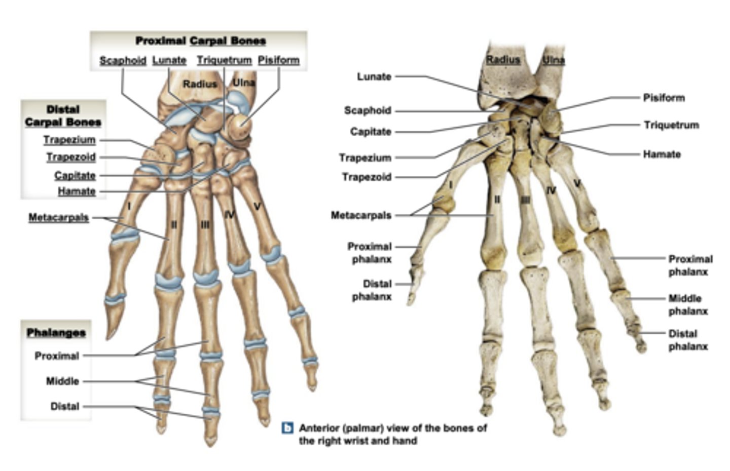
Anterior (palmar) view of the bones of the right wrist and hand