ultrafast ultrasound
1/8
There's no tags or description
Looks like no tags are added yet.
Name | Mastery | Learn | Test | Matching | Spaced | Call with Kai |
|---|
No analytics yet
Send a link to your students to track their progress
9 Terms
B-mode vs ultrafast
B mode:
image is the sum of A lines
groups of elements are fired to produce a focused beam and the rest of the elements capture the returning echoes
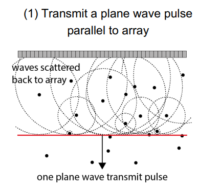
Ultrafast:
all elements are fired → produces an unfocused plane wave parallel to the probe
backscattered wave is measured by all elements at once
algorithm is sued to reconstruct all the A line at once
how does ultrafast US work?
measure the backscattered scan line using the whole array
find the amplitude of each constituent back scattered plane wave
relate the amplitudes to plane wave we transmitted
relationship tells use the scattering distribution
how is ultrafast ultrasound modelled in maths
incident wave is modelled as a plane wave with x (lateral) and z(depth) dimensions:
P(x,z,t)=∫0Tp^(Kx,Kz,w)ei(Kxx+Kzz)dx (sum of incident wave amplitudes and angular frequencies)
amplitude of the received signal is p^∗s^ (multiplying the fourier domains → same as subtracting scattered K from incident K)
since p^ is known you can divide the fourier by it to isolate the tissue’s fourier spectrum
spatial frequency fourier spectrum (s^) of the backscattered wave is inverse fourier transformed to find the scatterer distribution:
S(x,z,)=∫S^(Kx,Kz,)ei(Kxx+Kzz)dx
advantages of ultrafast
image formed from 1 transmit pulse → faster frame rate
disadvantages of UFUS → how is it mitigated
no transmit focusing → backscattered amplitudes are low -~> low signal to noise ratio → grainy image
poor lateral resolution
mitigated using coherent-angle or incoherent-angle compounding:
US plane wave is sent at different transmit angles
frames are averaged
speckle varies and is therefore removed
reduces frame rate
what is ultrafast doppler?
instead of scanning multiple a line, a single horizontal scan line allows you to take more doppler samples in the same amount of time
greatly reduces motion artefacts in power doppler as images can now be taken at 80Hz instead of 20Hz
what is functional US
Power Doppler signal-to-noise ~50x higher than conventional systems, so finer details can be obtained
Due to:
Ultrafast systems taking massive temporal averaging (coherent compounding), several thousands of frames are taken per second
Power doppler integrates the signal energy
Multiple plane wave angle compounding reduces noise and clutter
Short pulse repetition intervals -> strong sensitivity to slow flow
localisation microscopy
contrast agent is injected → usually microbubbles
if you can image a single microbubble and know it is just one → positional accuracy can be determined more accurately
allows us to replace the blurry image with a single sharp point
used in vascular imaging to image very small vessels
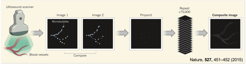
what are hte 2 ways you can perform localisation microscopy
low microbubble conc and low frame rate→ individual microbubble can be seen on the image
high microbubble conc and frame rate (500fps) → compare images to see the absence of microbubbles that have disintegrated → easier to use as no need to dilute bubbles