Week 2 abdominal ultrasound
1/35
There's no tags or description
Looks like no tags are added yet.
Name | Mastery | Learn | Test | Matching | Spaced | Call with Kai |
|---|
No analytics yet
Send a link to your students to track their progress
36 Terms
Portal veins have more hyperechoic walls than hepatic veins
When viewing the liver on an ultrasound, what vessels will have echogenic walls and what vessels will have similar echogenicity to that of the liver?
Spleen
Which organ on an ultrasound is more hyperechoic: the spleen or the liver?
Gall bladder sludge
What hyper echoic material (when compared to the rest of the gallbladder lumen) can often be found in the gallbladder?
cats
Which species is a bilobed gall bladder common?
Hepatomegaly
What can be said about the liver if it fully encompasses the right kidney?
Lipidosis, lymphoma, or cirrhosis
What are some reasons as to why the liver could have an increased echogenicity when compared to the spleen?
Acute hepatitis and passive congestion. Will see inc echogenicity of hepatic vessels
What are some causes to hypo-genicity of the liver and what are some ultrasound findings?
Homogenous and of a fine echo texture
How can you one describe the echogenicity of the parenchyma of the liver?
Multiple hypo echoic nodules
What could cause the spleen to have a Swiss cheese appearance on an ultrasound?
The medulla is more hypo echoic compared to the cortex
What is the echogenicity of the renal medulla compared to the cortex?
Acute renal failure often secondary to ethylene glycol toxicity
A thick hyper echoic cortex of a kidney on an ultrasound could indicate what?
Leptospirosis/ acute interstitial nephritis
An increase in echogenicity and size of the kidney could be caused by which disease?
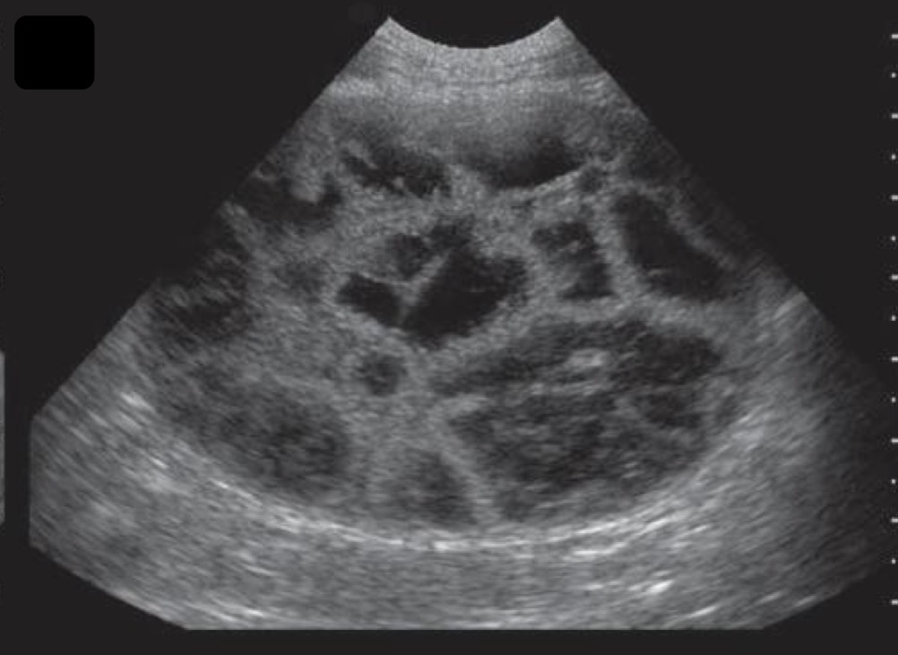
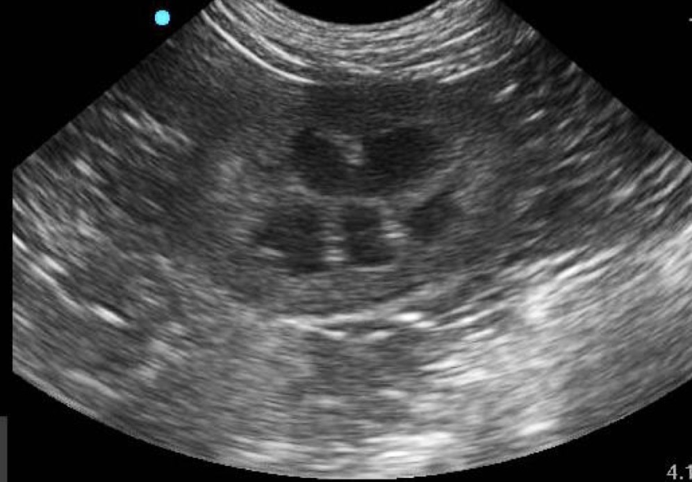
Polycystic kidney disease
Multiple anechoic structures occupying the renal cortex can indicate what?
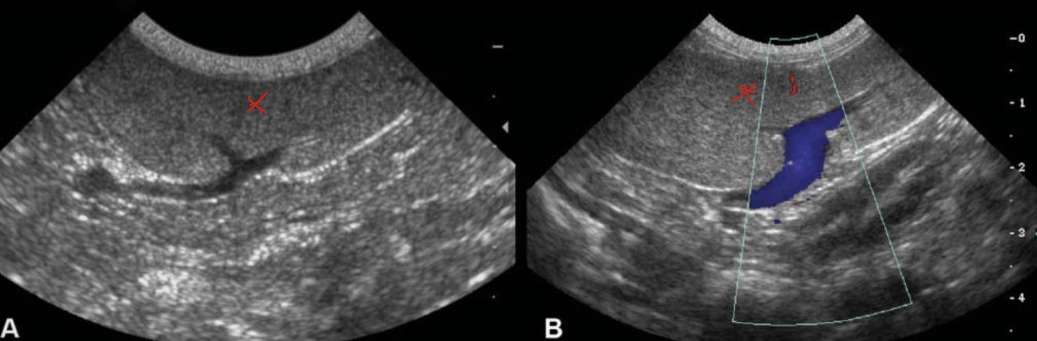
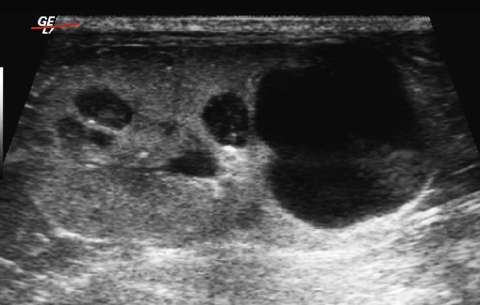
Hydronephrosis
A dilated renal pelvis with an anechoic fluid that can cause renomegaly is termed what?
Lumen/mucosa interface -hyper
Mucosa- hypo
Submucosa- hyper
Muscularis- Hypo
Serosa- hyper
When looking at a ultrasound of small intestinal loops, what are the differing layers from center to outer and their echogenicities?
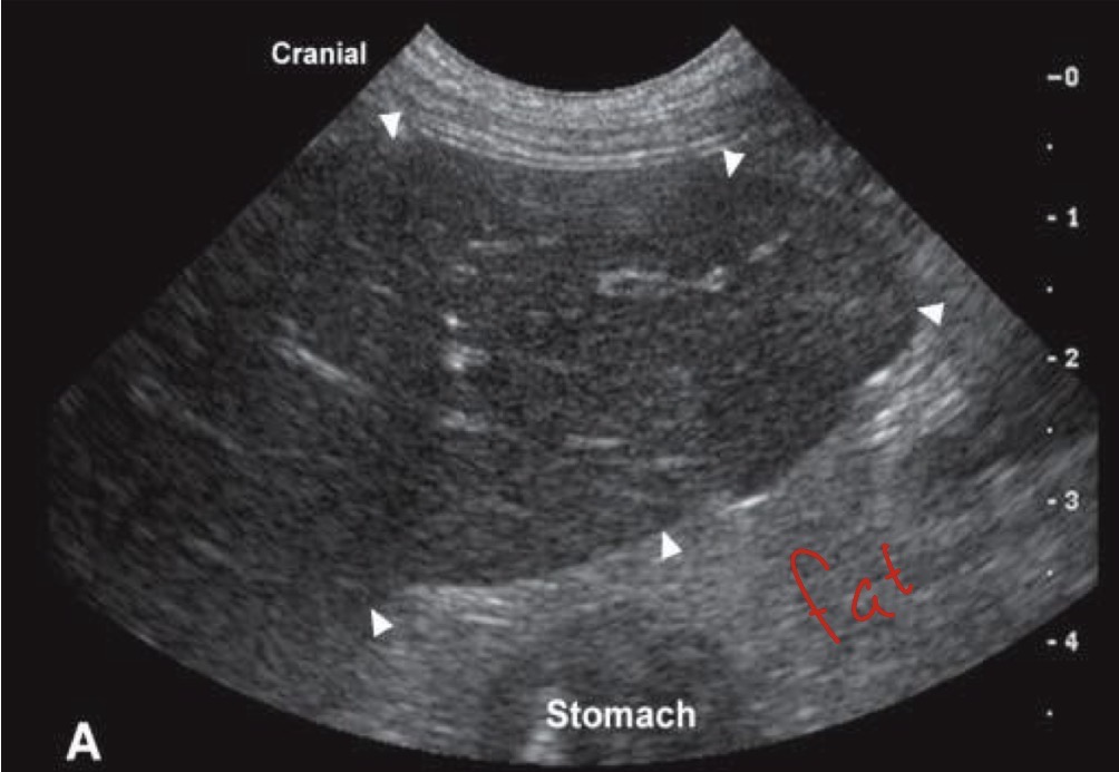
Rugal folds
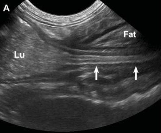
When looking at an ultrasound, what are internal structures that can confirm the stomach is in view?
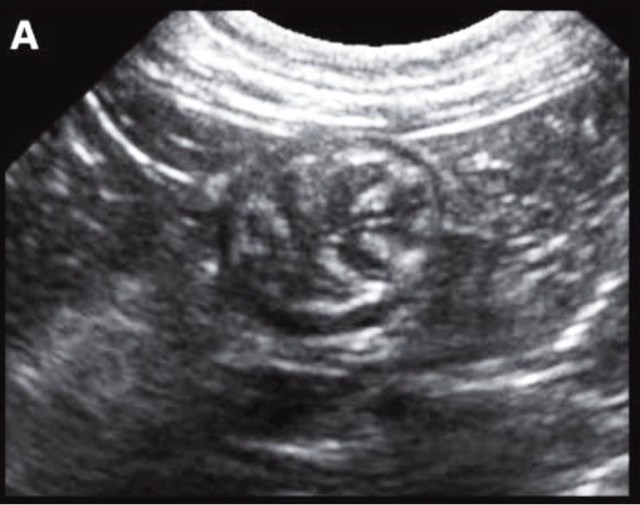
bullseye
What shape does intussusception of the bowel often take the shape of on an ultra sound?
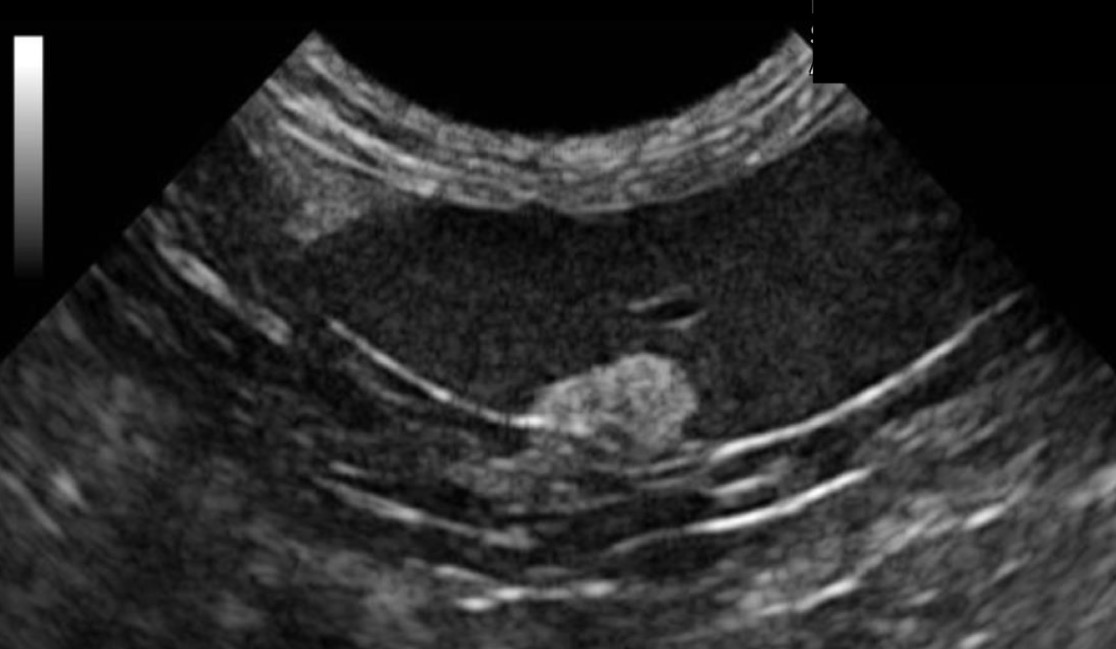
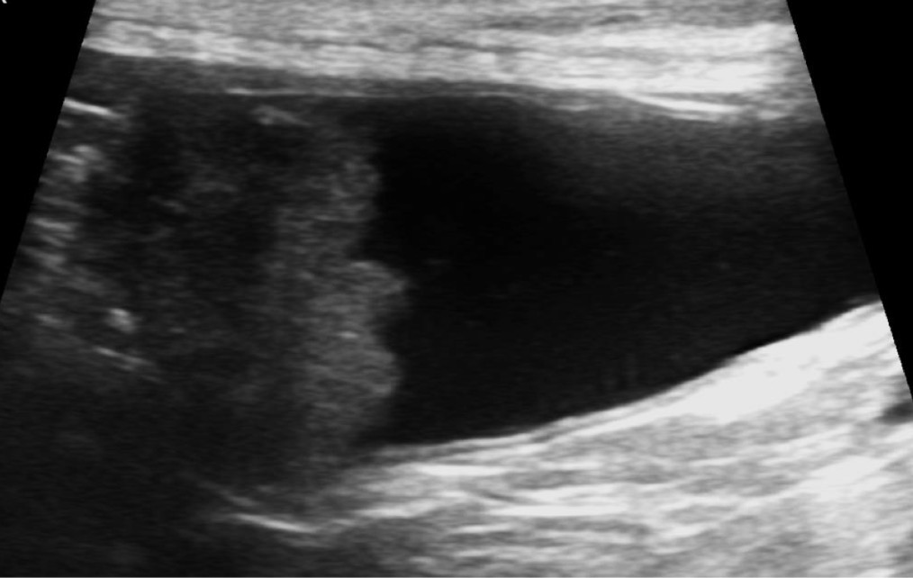
Liver
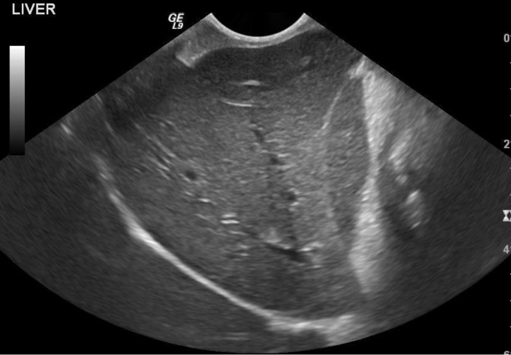
What organ is most prominent in this ultrasound?
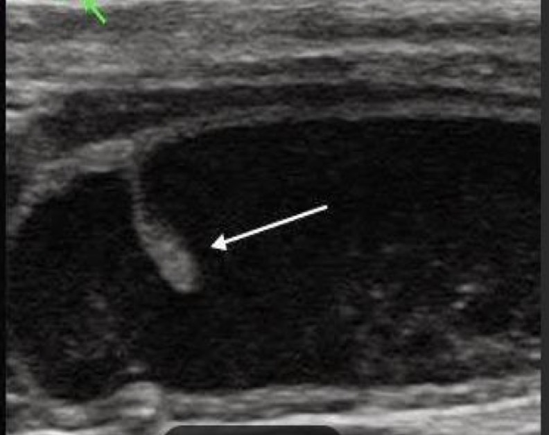
Bilobed gall bladder
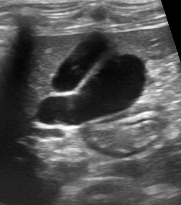
What organ is this?
Left gall bladder
Right liver
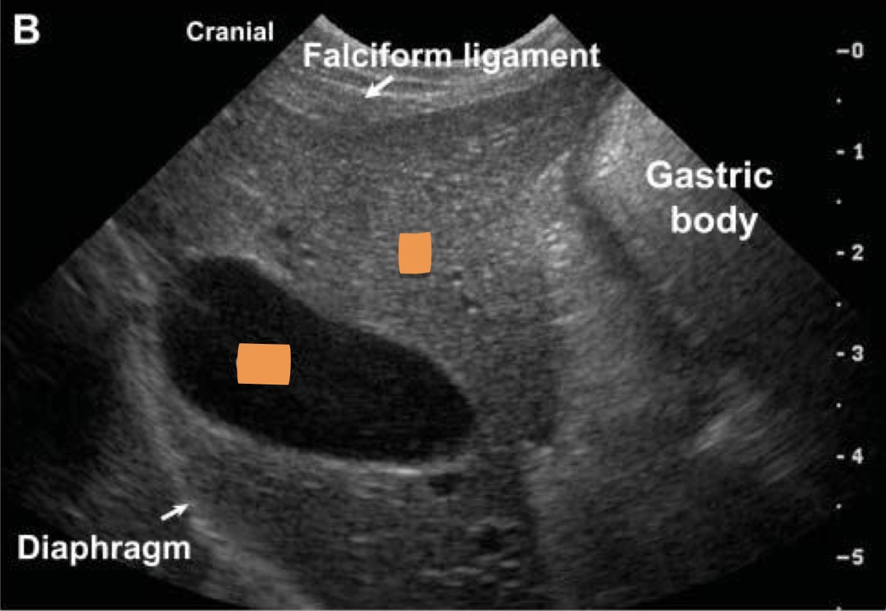
What organs are marked?
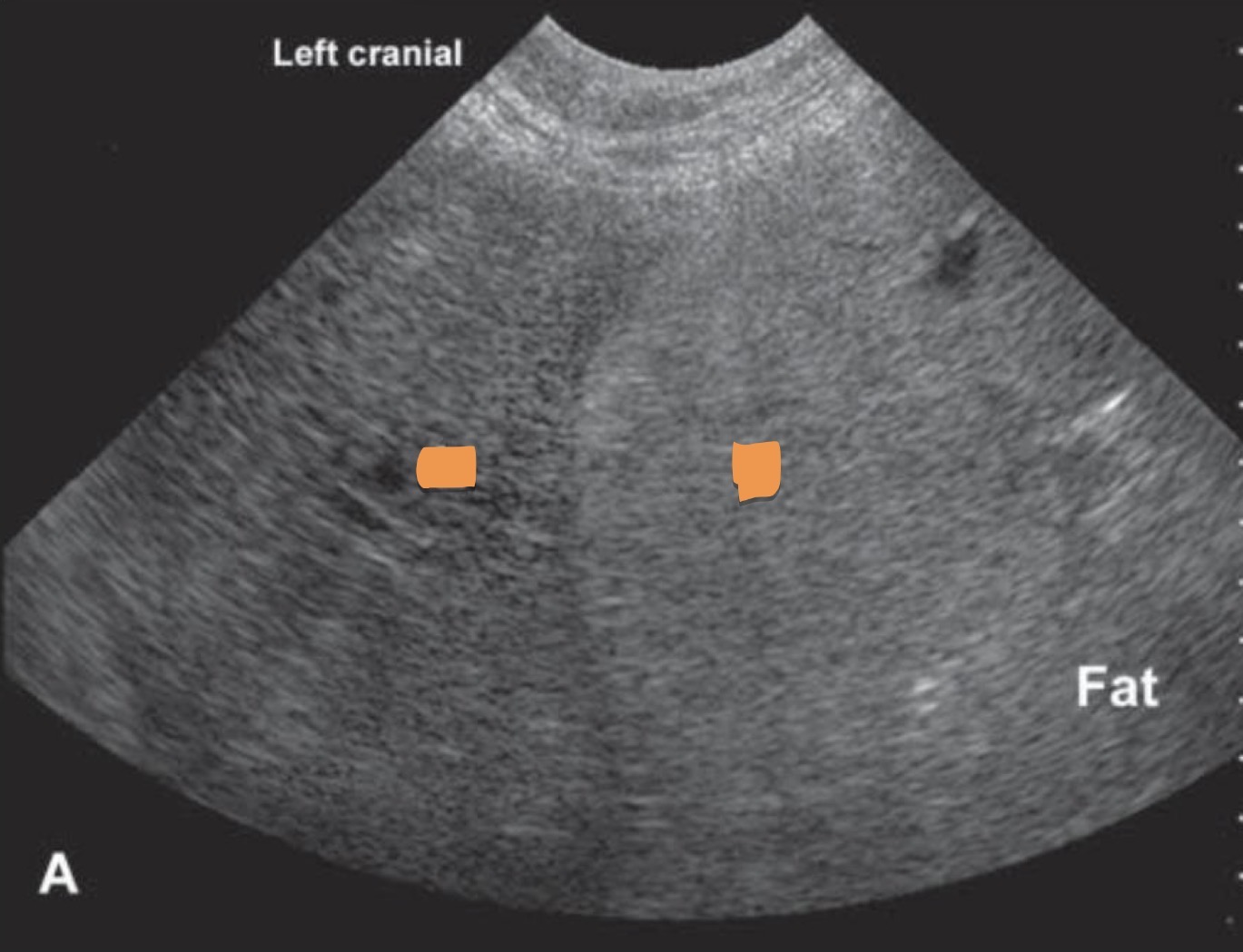
Portal veins
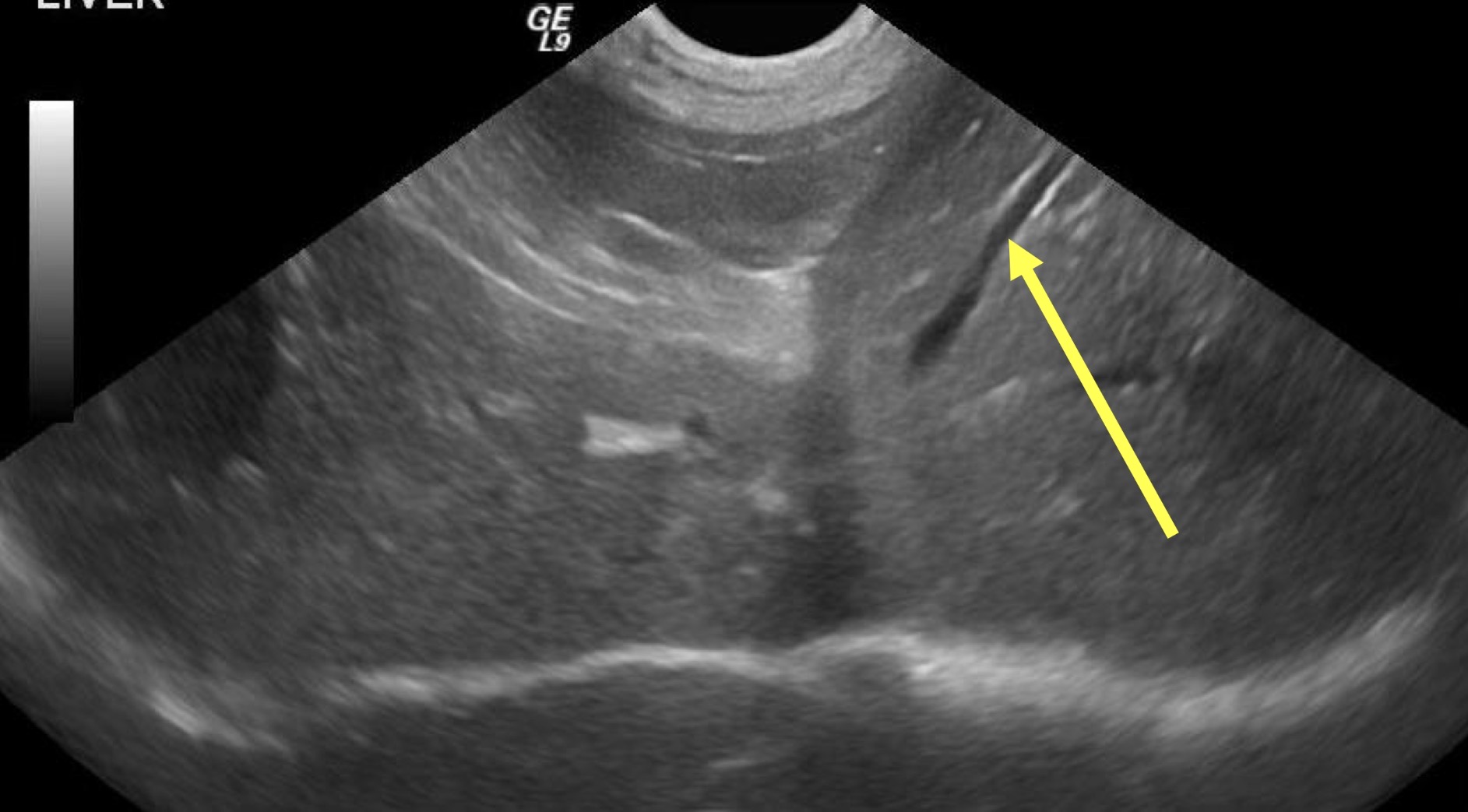
What structure is the yellow arrow pointing to ?
Left liver more hypoechoic
Right spleen more hyper echoic
What organs are labeled here and what is their echogenic pattern?
Hepatomegaly
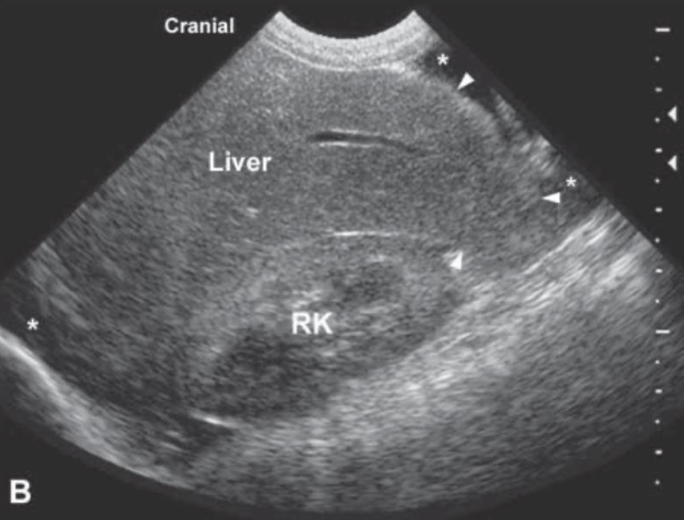
What is occurring in this patient?
Hypoechoic liver due to acute hepatitis or passive congestion
What organ is this, describe the echo texture and what can be causing it?
Spleen
What organ is this a ultrasound of?
Hyperechoic splenic mass
What anomaly is being observed in this ultrasound?
Splenic hemangiosarcome
What organ is being affected and by what?
Kidney
What organ is prominent in this ultrasound?
Hyper-echoic cortex and medullary rim sign

What anomalies are occurring in this kidney
Polycystic kidney disease
What is occurring in this patient?
Hydronephrosis
What is this arrow pointing to?
Stomach
what organ is prominent in this ultrasound?
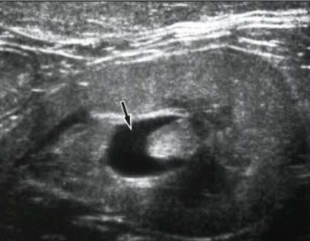
Small intestine
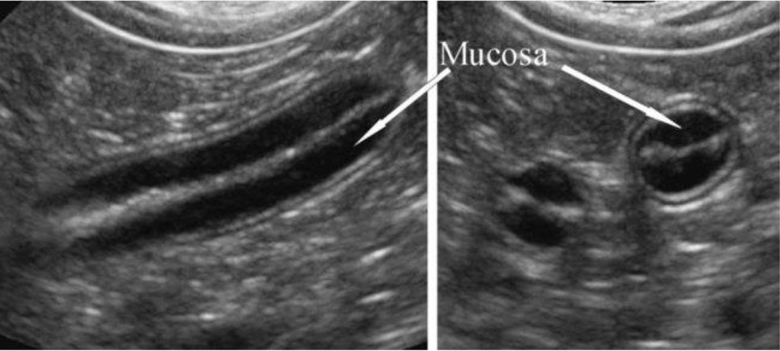
Organ?
Intussusception of the small intestine
What anomaly are these arrows pointing to?
TCC
What is being depicted here?
Polyp in the bladder
What are that?