Respiratory System
1/59
There's no tags or description
Looks like no tags are added yet.
Name | Mastery | Learn | Test | Matching | Spaced | Call with Kai |
|---|
No analytics yet
Send a link to your students to track their progress
60 Terms
Upper respiratory tract
includes
nasal cavity
oral cavity
pharynx
function: filters, warms and humidifies air
Lower Respiratory Tract
includes:
larynx
trachea
bronchi
lungs
function: transport and exchange of gases
Conducting Portion
Includes:
Nasal cavity
• Oral cavity
• Pharynx
• Larynx
• Trachea
• Bronchi
• Bronchioles
• Terminal bronchioles
Function: filters, warms and humidifies air
Respiratory Portion
Includes:
• Respiratory bronchioles
• Alveolar ducts
• Alveolar sacs
• Alveoli
Function: gas exchange
respiratory system cell type
pseudostratified ciliated columnar epithelium

External Nares (nostrils)
blue
allows air to enter and exit system

Internal Nares (choanae)
yellow
allow air to pass from nasal cavity to throat

hard palate
pink
interior, boney, roof of mouth
rigid structure
allows for speech, and food manipulation,
separates oral and nasal cavities
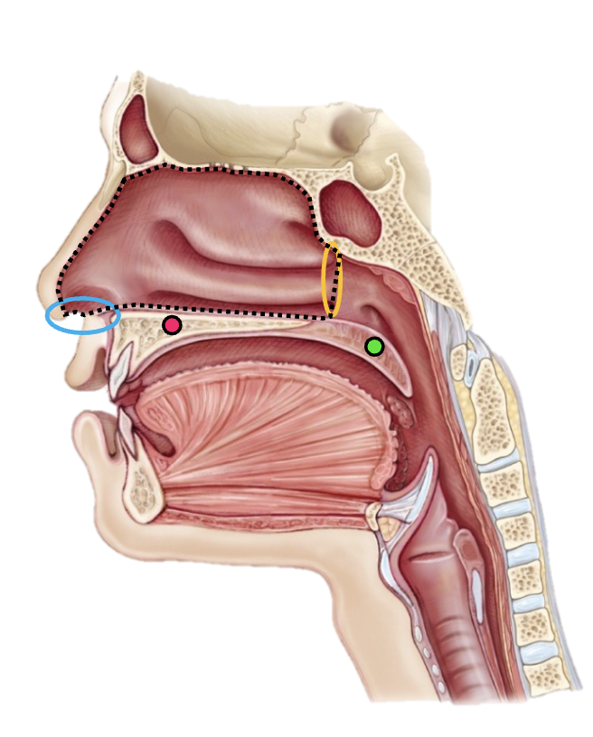
soft palate
green
posterior, muscular, flexible
closes nasal cavity during swallowing and speech

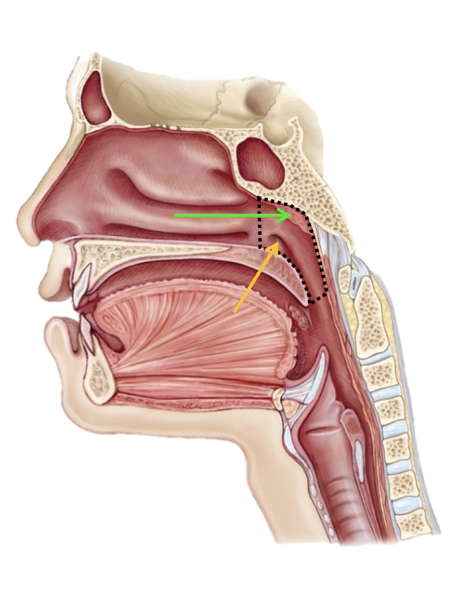
nasal cavity
pink
Function: conditions inhaled air by warming, moisterizing, and filtering it

Nasal Chochae
green
function: produce turbulence
• Fluctuates, moves around
• Increases the length of time air stays in the nasal cavity
• Helps warm and humidify air
Nasal septum
black
provides structural support to nose
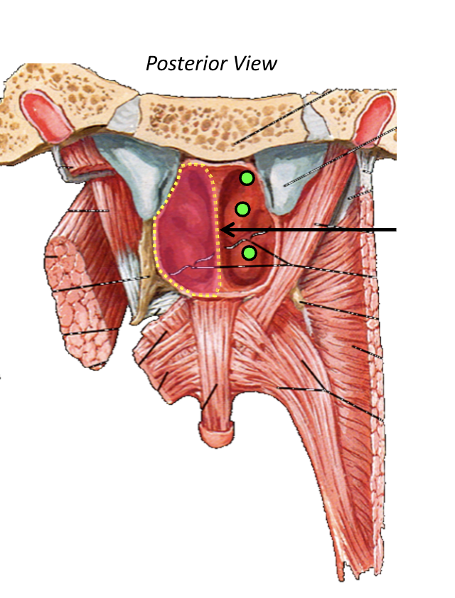
Pharynx
tube for food and air
contains skeletal muscle
lined with mucosa

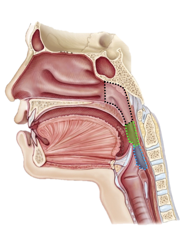
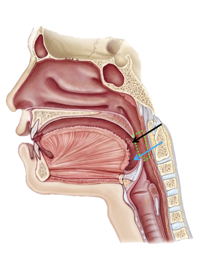
Nasopharynx
black
serves as airway and regulates pressure

Oropharynx
Green
pathway for food and air

Laryngopharynx
blue
protects airway and redirects food to esophagus
Opening of auditory tube
yellow
connects ear to nasopharynx

Lingual Tonsil
blue
filter pathogens passing through mouth
Palatine Tonsil
black
first line of defense against inhaled pathogens
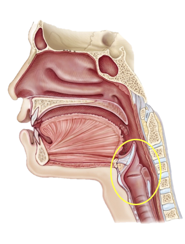
larynx
functions: breathing, phonation (producing sound), protection of respiratory system

hyoid bone
red
functions: enable swallowing, enable speech, secure airway and support tounge movement
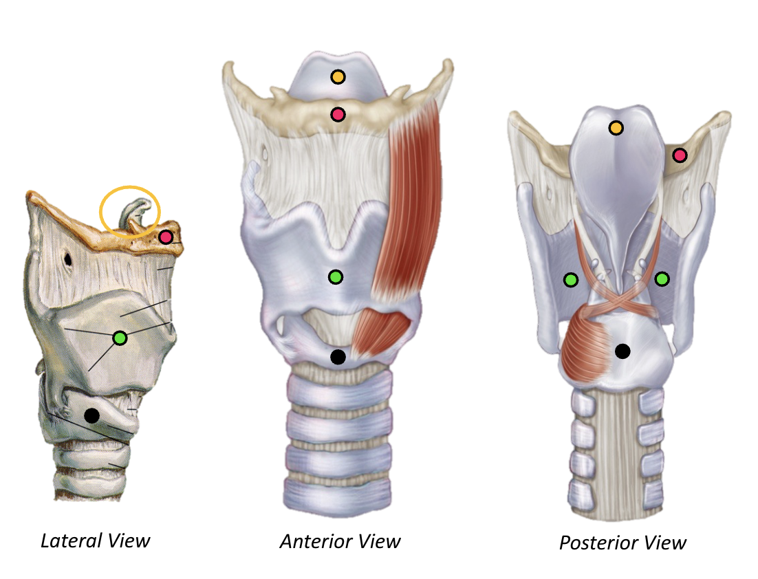
epiglottis
yellow
function: sorts food and air by closing airway when swallowing and esuring food and liquid go to esophagus and not lungs

Thyroid Cartilage
Green
function: protects vocal cords

cricoid cartilage
black
function: keeps airway open and serves as an attachment point for other muscles and ligaments

Arytenoid Cartilage
pink
controls movement of vocal cords

ventricular fold
yellow
allow for coughing and gagging, along with protecting airway while swallowing

vocal fold
green
phonation and airway protection
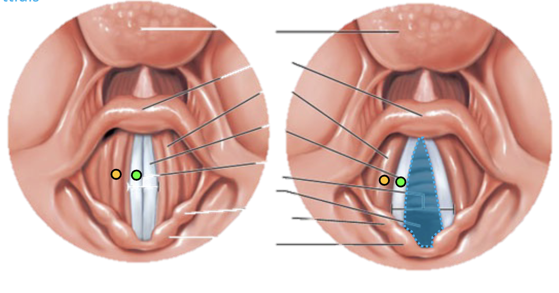
Rema Glottides
blue
narrows and widens to regulate airflow and produce sound
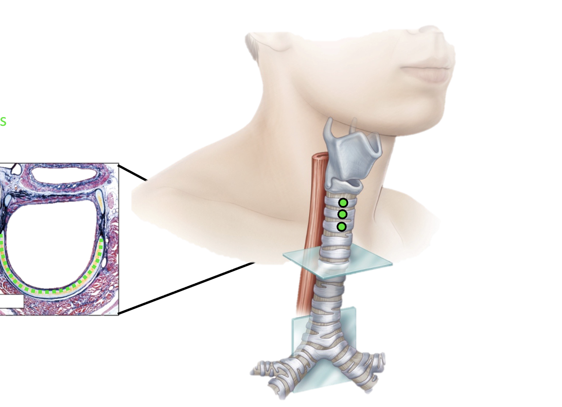
trachea
“windpipe”
allows air to travel in and out of lungs
tracheal cartilage
provide trachea support and flexibility

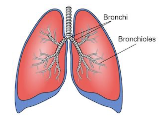
bronchial tree
airway system conducting air between the trachea and the lungs

primary bronchi
green
largest tubes, main entry point for air from trachea

secondary bronchi
blue
deliver and separate air into different lobes
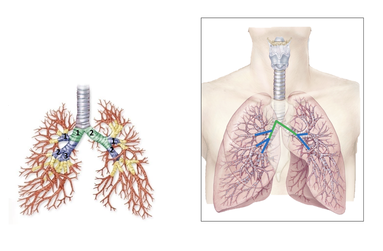
segmental bronchi
yellow
branch from secondary bronchi - distribute air to different, independant sections called bronchopulmonary segments

smaller bronchi
red
narrower branches of segmental bronchi - regulate air distribution through contraction and relaxation
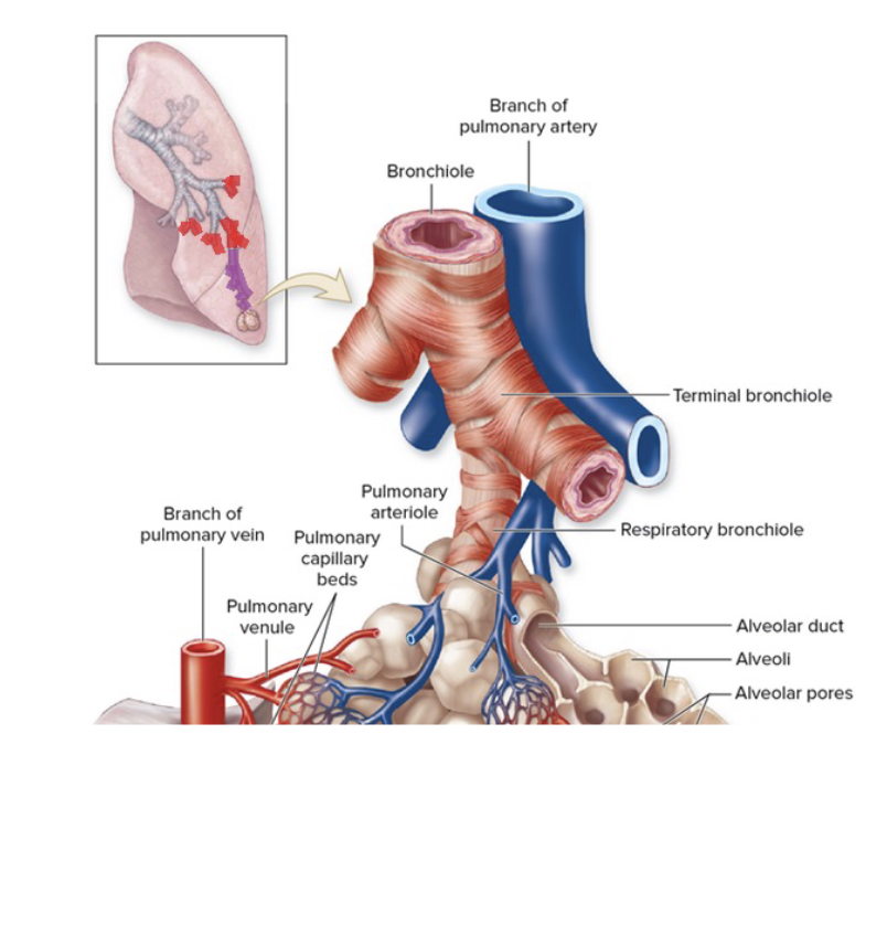
bronchioles
conduct air between bronchi and alveoli
regulate airflow through contraction and dilation
Terminal bronchioles
deliver air to respiratory bronchioles (where gas exchange begins)
Bronchi vs bronchioles
bronchi-
incomplete rings of cartilage
psuedostratified ciliated columnar epithelium
Bronchioles:
no cartilage
simple columnar/cuboidal epithelium
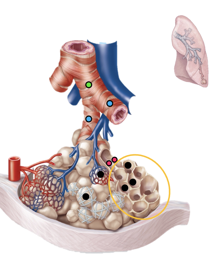
resiratory bronchioles
blue
conduct air and initiate gas exchange

alveolar ducts
pink
connect respiratory bronchioles to alveolar sacd

alveolar sacs
yellow
primary location for gas exchange
alveoli
functional units of lung tissue
• Gas exchange location between air and blood
type 1 alveoli
simple squamous epithelium. Rapid gas diffusion
type 2 alveoli
secrete fluid to reduce surface tension and prevent alveoli collapse
Alveolar Macrophanges
clear airspaces. Fixed or free
visceral pleura
green
directly covers lungs

parietal pleura
blue
lines inner chest wall

lung
gas exchange
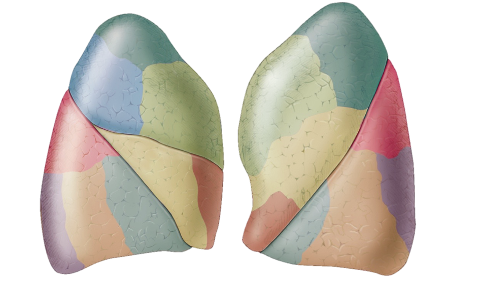
superior lobe
provide air to upper portion of lungs
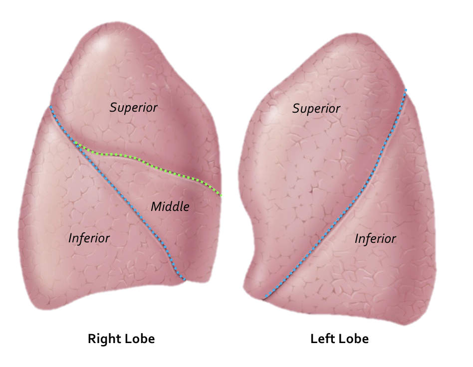
middle lobe
provide air to medial portion of lungs

inferior lobe
provide most air - lower portion of lungs


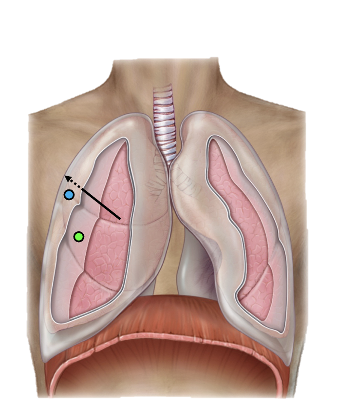
oblique fissure
blue
separates lung lobes

Transverse fissure
green
separates lung lobes
bronchopulmonary segments
Supplied by a single segmental bronchus, branch of a pulmonary
artery, and branch of a pulmonary vein
pulmonary circulation
• Transportation of deoxygenated blood from the heart (right side) to the lungs
• Returns oxygenated blood to the heart (left side)
Bronchial Circulation
Part of the systemic circulation
• Smaller than the pulmonary circulatory system
• Supplies the alveolar cells
• Bronchial arteries and veins supply the bronchi and bronchioles
• Arteries: Thoracic aorta à capillary beds à bronchial tree
• Veins: drain into the azygos/hemiazygous systems
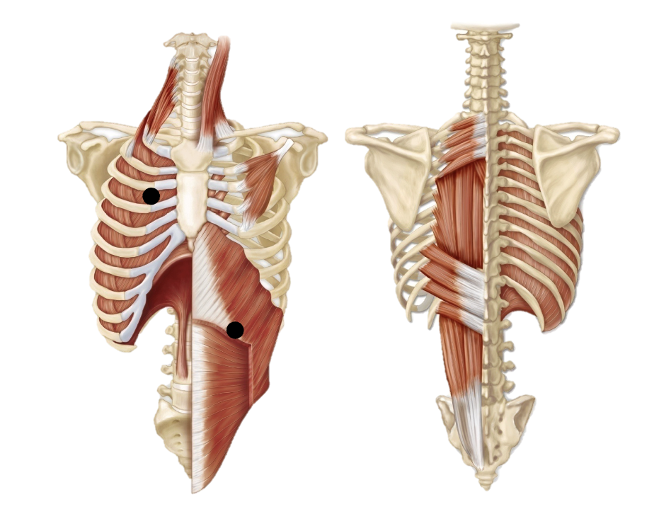
Pulmonary ventilation
“quiet” breathing
quiet breathing
• Diaphragm
• External intercostals
Moves air in and out of lungs
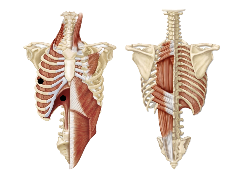
forced inhalation
Accessory muscles:
• Sternocleidomastoid
• Pectoralis minor
• Erector spinae
Increases thoracic cavity volume
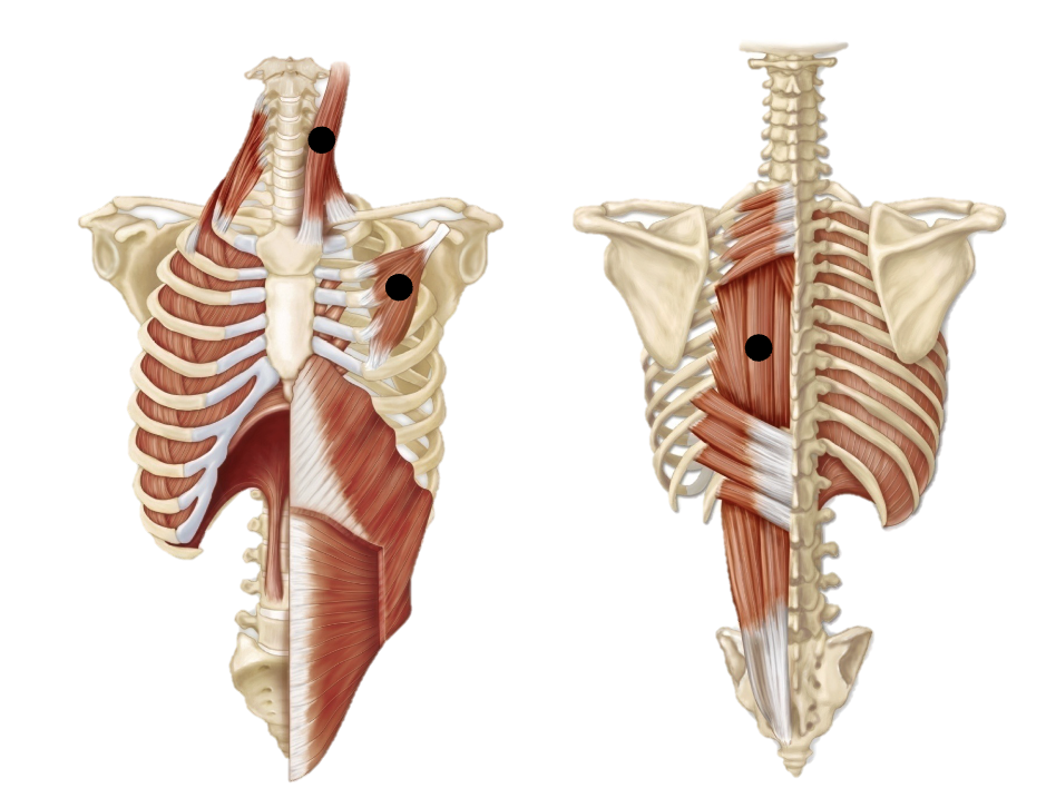
forced exhalation
• Internal intercostals
• Abdominal muscles
Decreases thoracic cavity volume