Digestive system
1/58
Earn XP
Name | Mastery | Learn | Test | Matching | Spaced | Call with Kai |
|---|
No analytics yet
Send a link to your students to track their progress
59 Terms
The organs pf the digestive system are separated into what categories? (2)
accessory digestive organs
organs of the gastrointestinal (digestive) tract

Describe the digestive tract
continuous muscular tube composed of skeletal muscle and smooth muscle
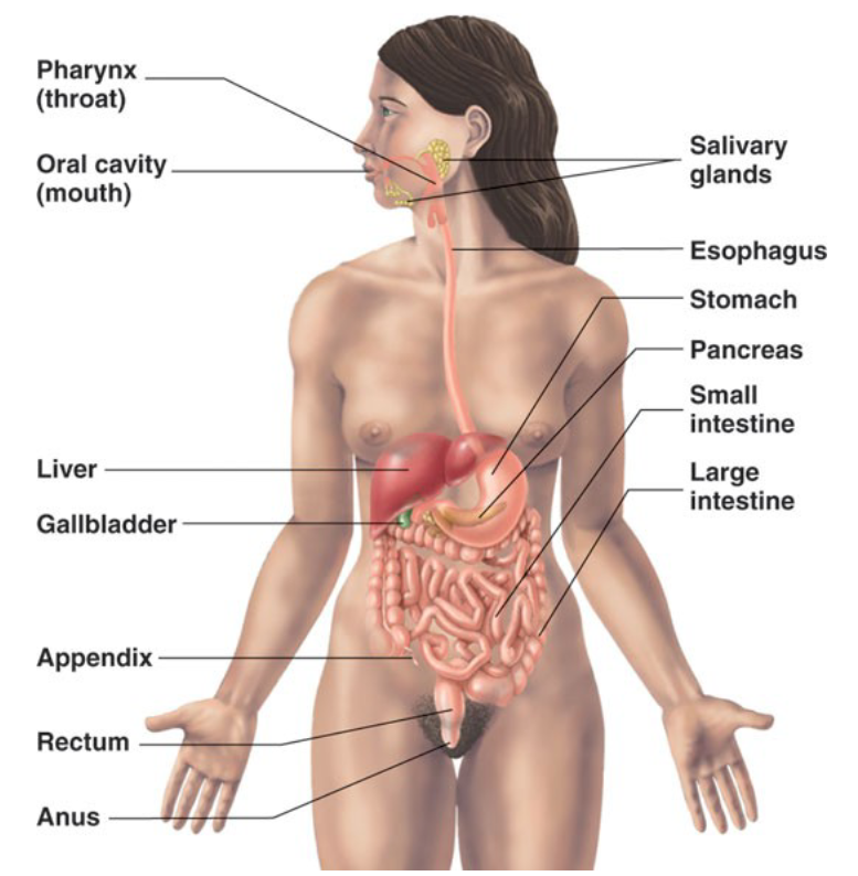
List the organs of the digestive tract (6)
mouth
pharynx
esophagus
stomach
small intestine
large intestine
List the accessory digestive organs (8)
teeth
tongue
gallbladder
digestive glands
liver
pancreas
salivary glands
parotid glands
submandibular glands
sublingual glands

True or False: food does not pass through the accessory digestive organs
True
List the functions of the digestive system (8)
ingestion
mastication
propulsion
mixing
secretion
digestion
absorption
excretion
Describe the peritoneum
membrane that lines the walls and organs of the abdominal cavity
two portions
visceral peritoneum
parietal peritoneum
Describe the visceral peritoneum
covers the external surface of the organs within the abdominal cavity
Describe the parietal peritoneum
lines the internal surface of the walls of the abdominal cavity
The space between the two layers of the peritoneum is called the…
peritoneal cavity
What is the peritoneal cavity filled with?
peritoneal fluid
Many organs of the abdominal cavity are held in place by what structures?
mesenteries
Describe mesenteries
fused double layer of peritoneum that extends from the body wall to a digestive organ
Describe retroperitoneal organs
organs that lie against the posterior abdominal wall (not held in place by a mesentery)
List the associated structures of the oral cavity (6)
lips
cheeks
palate
tongue
teeth
salivary glands
Describe the lips and cheeks
composed mainly of muscles
orbicularis oris (lips)
buccinator (cheeks)
involved with processes of mastication and speech
Describe the palate
consists of two portions
hard palate
forms the roof of the mouth
tongue forces food against the hard palate during chewing
also plays a role in speech
soft palate
posterior to the hard palate
composed of skeletal muscle
closes off the nasopharynx when we swallow
Describe the tongue
aids in
chewing
swallowing
speech
Describe the salivary glands
produce and secrete saliva
three pairs
parotid (one anterior to each ear)
submandibular (inferior to mandible)
sublingual (inferior to tongue)
List the functions of saliva (3)
cleanses the mouth
dissolves food chemicals so that they can be tasted
contains the enzyme salivary amylase (begins breakdown of starchy foods)
Describe the pharynx
throat
food from the oral cavity passes through the fauces
food then passes into the laryngopharynx
Describe the esophagus
food passes from the laryngopharynx into the esophagus (carries food into the stomach)
lined with stratified squamous epithelium
no digestion “starts” in the esophagus
Describe the stomach
food is broken down mechanically and chemically
three layers of smooth muscle mechanically digest the food
gastric glands (located in the lining of the stomach) release substances that chemically digest the food
List the major structures of the stomach (9)
cardiac sphincter
cardiac region
fundus
body
greater curvature
lesser curvature
pyloric region
pyloric sphincter
rugae
Describe the histology of the stomach
the internal surface of the stomach is lined with simple columnar epithelium
the epithelium lining of the stomach contains gastric pits
gastric pits are the openings for the gastric glands
produce gastric juice
the highly acidic gastric juice (pH 1.5 to 3.5) contains substances that aid in chemical digestion
the substances that compose gastric juice are produced by the cells that form gastric glands
mucous cells
parietal cells
chief cells
endocrine cells
What is the function of simple columnar epithelium?
allows for absorption and secretion
List the cells that form the gastric glands (4)
mucous cells
parietal cells
chief cells
endocrine cells
Describe the small intestine
digestion is completed and nearly all absorption occurs
extends from the stomach to the large intestine
~20 ft long in a cadaver (shorter during life due to muscle tone)
divided into three parts
duodenum
jejunum
ileum
contains modifications that increase surface area, which allows for increased absorption
Describe the duodenum
first portion of the small intestine
wraps around the head of the pancreas
lined with simple columnar epithelium
contains glands which produce an alkaline substance that neutralizes acidic chyme
Describe the jejunum
middle portion of the small intestine
extends from duodenum to ileum
Describe the ileum
last portion of the small intestine
joins the cecum of the large intestine
Describe the modifications of the small intestine that increase surface area
the overall length of the small intestine
the lining of the small intestine is folded (circular folds) and has tiny fingerlike projections called villi
villi are lined with simple columnar epithelial cells
the cells contain microvilli which increase surface area
Describe the liver
second largest organ in the body
largest gland in the body
located in the upper right quadrant of the abdomen
List the major structures associated with the liver (5)
right lobe
left lobe
caudate lobe
quadrate lobe
hepatic porta
List the functions of the liver (3)
stores glucose as glycogen
takes waste products out of the blood
hepatocytes of the liver produce bile
bile is stored in the gallbladder
List the ducts of the liver (5)
right hepatic duct
left hepatic duct
common hepatic duct
cystic duct
common bile duct
Describe the ducts of the liver
the right and left hepatic ducts unite to form the common hepatic duct
common hepatic duct carries bile away from the liver
common hepatic duct and cystic duct (from gallbladder) unite to form the common bile duct
the common bile duct unites with the pancreatic duct to form the hepatopancreatic ampulla (ampulla of vater)
the hepatopancreatic ampulla drains via the major duodenal papilla into the duodenum of the small intestine
Describe the pancreas
located in the abdomen, partially behind the stomach
is an exocrine and endocrine gland
substances produced by the endocrine portion of the pancreas include
insulin and glucagon
substances produced by the exocrine portion of the pancreas include
enzymes that aid in digestion of carbohydrates, fats, and proteins in the small intestine
HCO3- that neutralize acidic chyme in small intestine
exocrine pancreatic secretions exit the pancreas via the pancreatic duct
Describe the large intestine
extends from the small intestine to the anus
List the functions of the large intestine (2)
absorption of water
elimination of waste products (feces)
List the divisions of the large intestine (4)
cecum
colon
rectum
anal canal
Describe the cecum of the large intestine
first part of the large intestine
joins with ileum of small intestine
forms the ileocecal junction
the vermiform appendix is attached to the cecum
Describe the vermiform appendix
plays a role in immunity
can collect bacteria
Describe the colon of the large intestine
consists of the
ascending colon
right colic (hepatic) flexure
transverse colon
left colic (splenic) flexure
descending colon
sigmoid colon
Describe the rectum of the large intestine
portion of large intestine located between the sigmoid colon and anal canal
stores feces
Describe the anal canal
last 2-3 cm of the digestive tract
extends from the rectum to the anus (external opening of anal canal)
surrounded by internal and external anal sphincters
internal composed of smooth muscle
external composed of skeletal muscle
Describe the digestive pathway
mouth
fauces
oropharynx
laryngopharynx
esophagus
cardiac sphincter of stomach
cardia region of stomach
body of stomach
pyloric region of stomach
pyloric sphincter of stomach
duodenum of small intestine
jejunum of small intestine
ileum of small intestine
ileocecal valve
cecum of large intestine
ascending colon of large intestine
hepatic flexure of large intestine
transverse colon of large intestine
splenic flexure of large intestine
descending colon of large intestine
sigmoid colon of large intestine
rectum of large intestine
anal canal
toilet
Describe the biliary pathway
liver - produces and excrete bile
right and left hepatic ducts
common hepatic duct
cystic duct
common bile duct
ampulla of vater & sphincter of oddi
major duodenal papilla

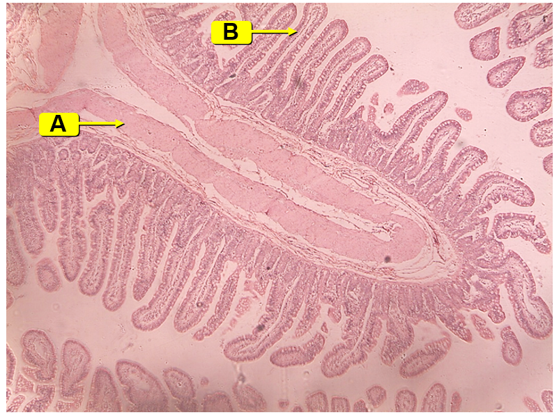
Identify the image along with the labeled structures and their functions
gastro-esophageal junction
A: esophagus
lined with stratified squamous epithelium
protection
B: stomach
lined with simple columnar epithelium
allows for absorption and secretion
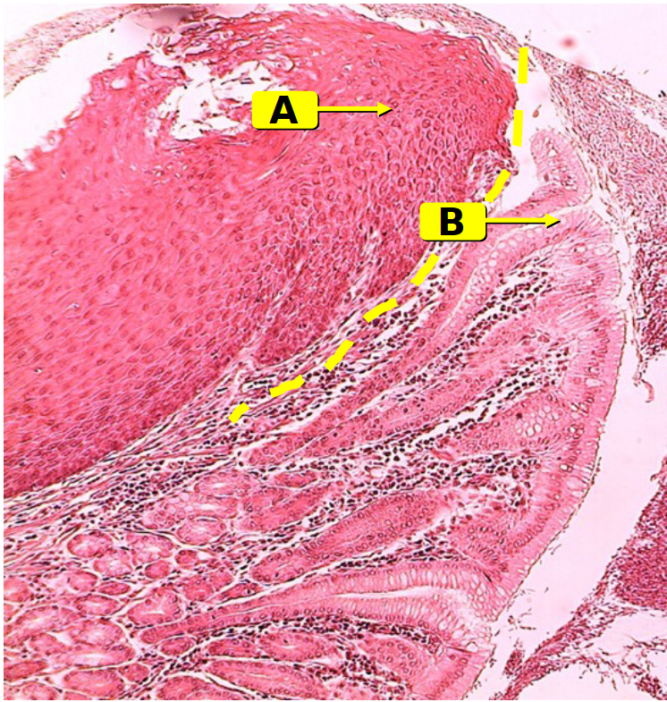
What protects the simple columnar epithelium (lining of the
stomach) from the harsh acidic environment of the lumen of
the stomach?
Alkaline mucus made by the mucous cells

Identify the image along with the labeled structures
stomach wall
A: gastric pits
B: gastric glands
C: mucous cells
line the stomach and gastric pits
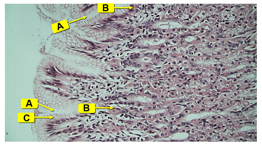
What is the function of mucous cells?
Produce alkaline mucus to protect the stomach lining from
the acid

Identify the image along with the labeled structures and their functions
stomach wall
A: parietal cells
make HCl and intrinsic factor
B: chief cells
make pepsinogen (precursor to pepsin)
C: enteroendocrine cells
release hormones that help with digestion
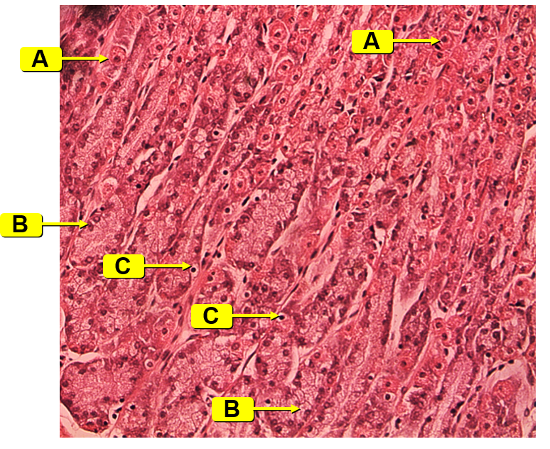

Identify the image along with the labeled structures and their functions
stomach wall
A: parietal cells
make HCl and intrinsic factor
B: chief cells
make pepsinogen (precursor to pepsin)
C: enteroendocrine cells
release hormones that help with digestion


Identify the image along with the labeled structures and their functions
intestinal lining
A: circular folds
increase surface area
line the small intestine
B: villi
lining the circular folds of the small intestine
increase surface area
lined with simple squamous epithelium which contain microvilli
*The circular fold is the whole structure extending off of the intestinal wall and it is lined with villi
What is the function of the circular folds, villi, and microvilli?
Increase surface area for absorption

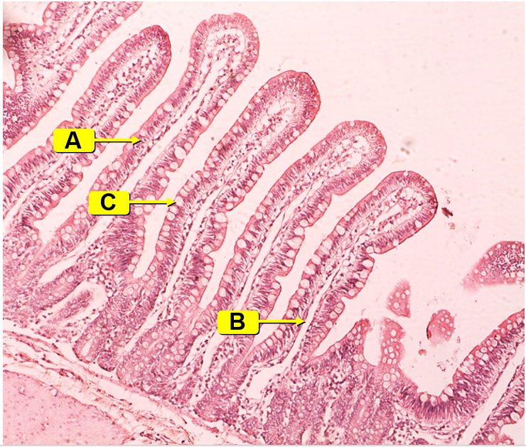
Identify the image along with the labeled structures and their functions
intestinal lining
A: villi
lining the circular folds of the small intestine
increase surface area
lined with simple squamous epithelium which contain microvilli
B: lacteals
absorb fats in the small intestine
C: goblet cells
produce and secrete mucus

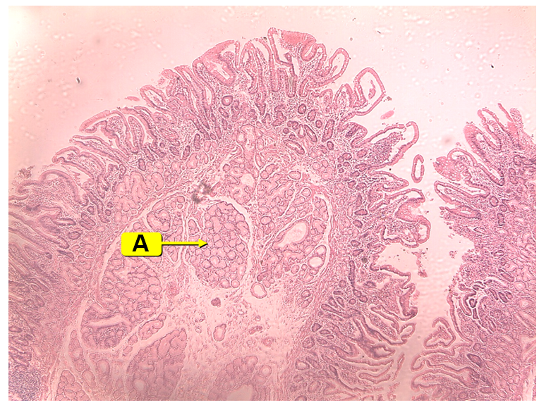
Identify the image along with the labeled structure and its function
duodenum
A: Brunner’s gland
secretes an alkaline substance into the lumen to neutralize the acid of the chyme from the stomach

Identify the image along with the labeled structure and its function
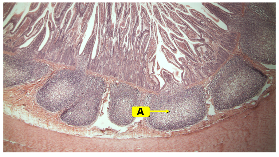
ileum
A: Peyer’s patch
masses of lymphatic tissue that play a role in the immune system
contain lymphocytes and macrophages