Dogs & cats necropsy
 Call Kai
 Learn
Learn Practice Test
 Spaced Repetition
 Match
Match Flashcards
 Knowt Play
1/167

There's no tags or description
Looks like no tags are added yet.
Name | Mastery | Learn | Test | Matching | Spaced | Call with Kai |
|---|
No analytics yet
Send a link to your students to track their progress
168 Terms
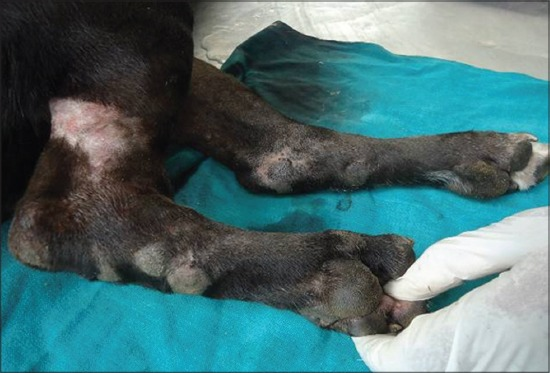
Thickening and callus formation on limb and food pad
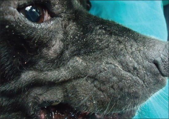
Loss of elasticity, wrinkling and thickening of skin on face region
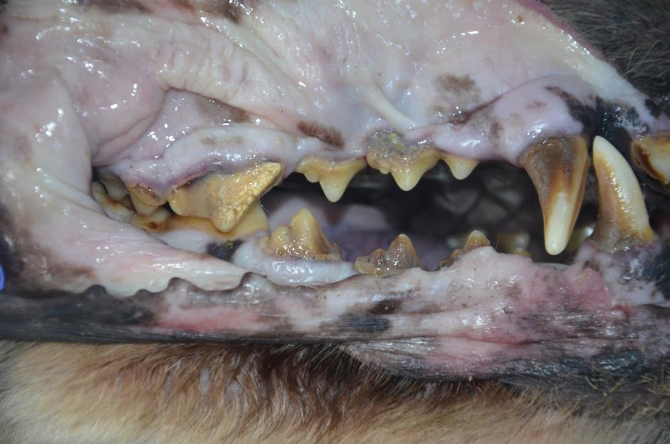
Dog, dental calculus
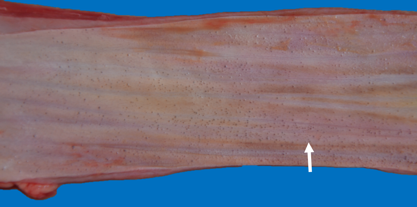
Dog, esophagus
Cystic dilation of the esophageal glands
incidental findings
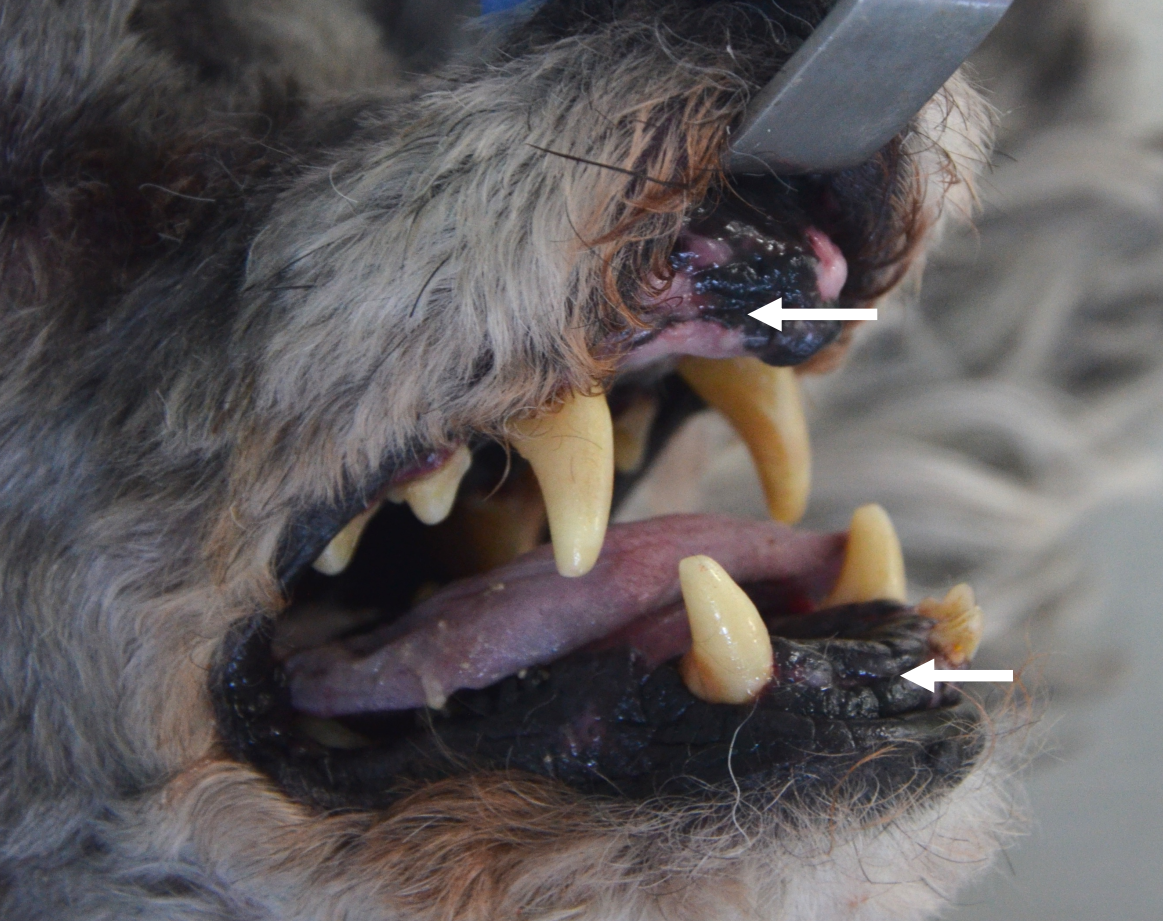
Dog, oral cavity
Acquired oligodontia - loss of teeth
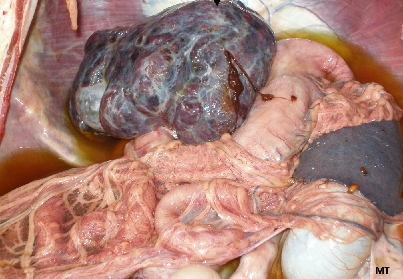
Dog, Liver, Atrophy and fibrosis
Liver: irregularly shaped, atrophied with extensive fibrosis
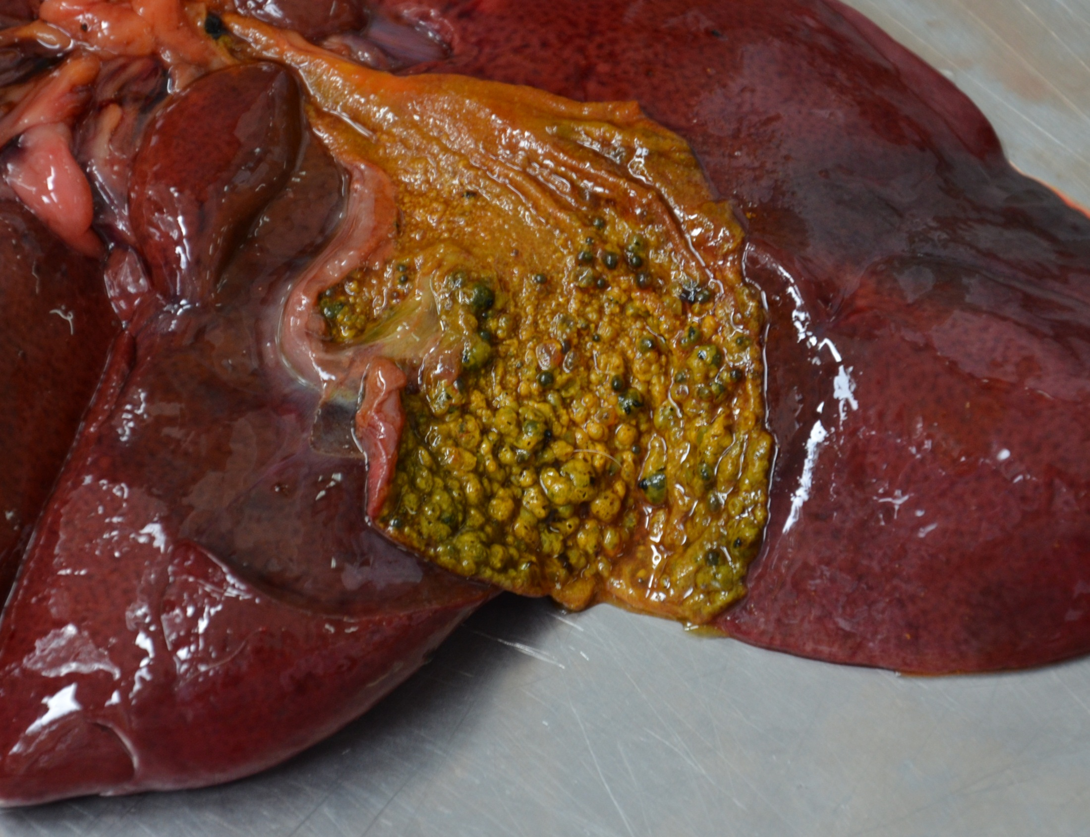
Dog
Liver, cystic mucinous hyperplasia of the mucus-producing glands in the mucosa of the gallbladder
Gallbladder: irregularly shaped because of small grey cystic nodules which represent cystic dilation of the gallbladder glands
Liver: is fine, incidental finding and animal died due to pneumonia
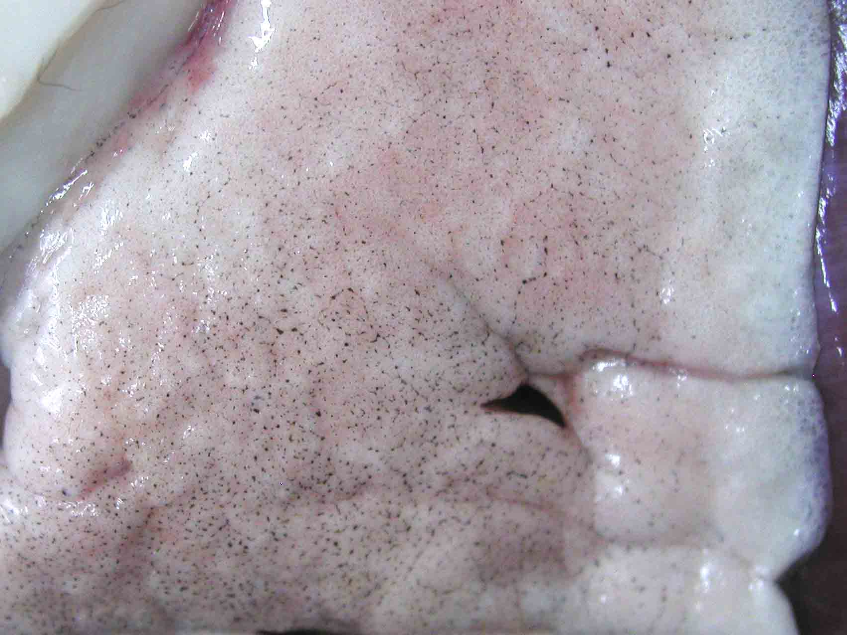
Pulmonary anthracosis
Incidental finding
Lungs: multifocal fine, black, pinpoint spots (accumulation of large amount of carbon particles mostly in the cytoplasm of macrophages)
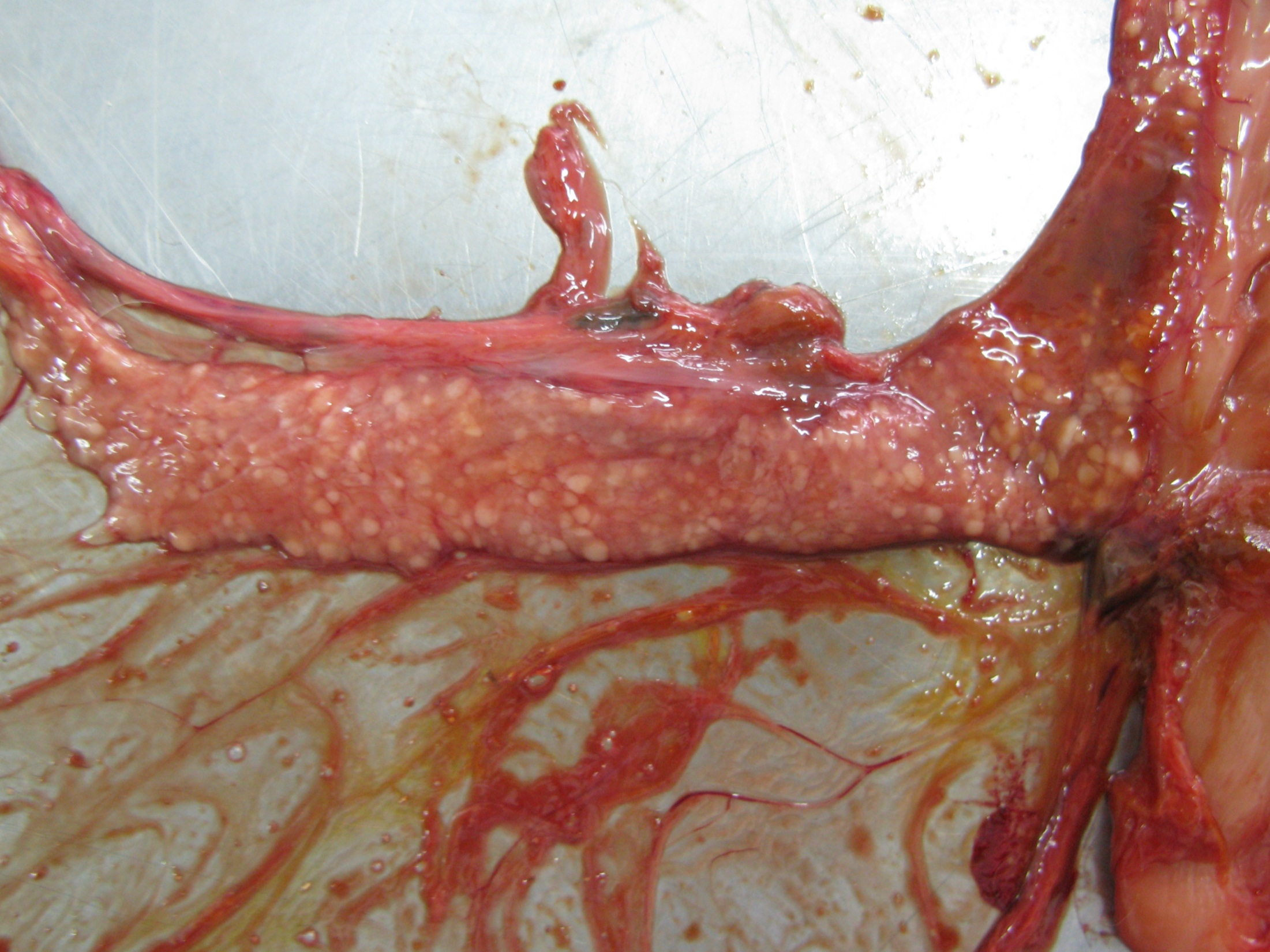
Cat
Pancreas, nodular hyperplasia of the exocrine parenchyma
Small slightly elevated pancreatic nodules
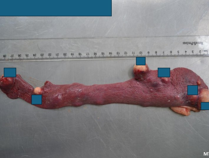
Spleen, Nodular hyperplasia (NH) and metastatic tumors – cholangiocarcinoma (CC)
In NH we don’t recommend splenectomy, observation and recheck every 6 months
NH have small nodules up to 3 mm in diameter
Ddx for NH: more common in cats (do FNA and US) - lymphoma in the spleen, mesenchymal tumor (fibrosarcoma), metastatic cancer from another organ
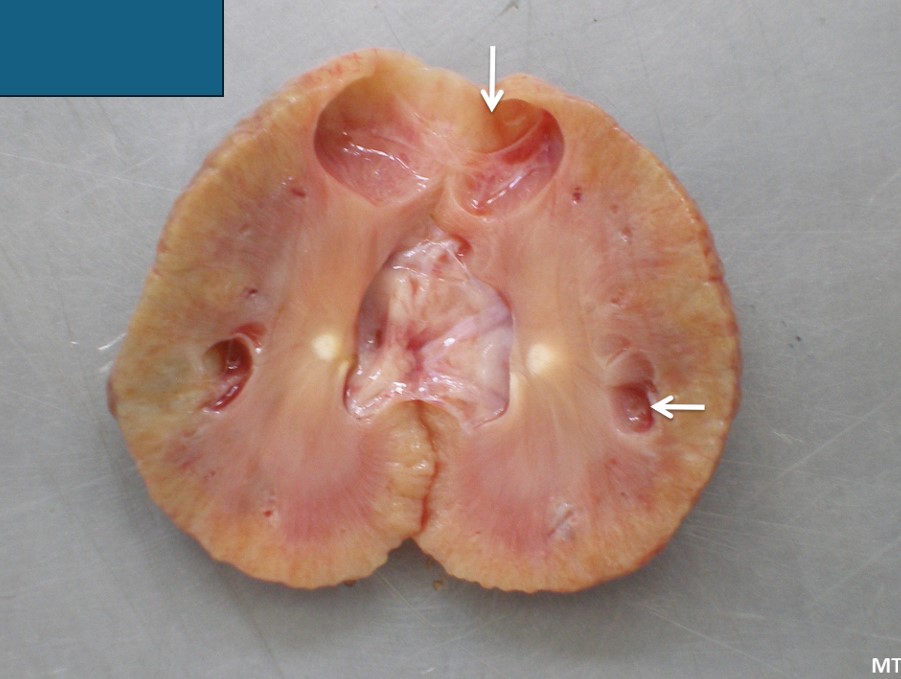
Cat
Renal cysts
Incidental finding
Kidney: irregularly shaped, reduced in volume, cortex with 2 cysts (incidental finding)
White nodules: chronic interstitial nephritis
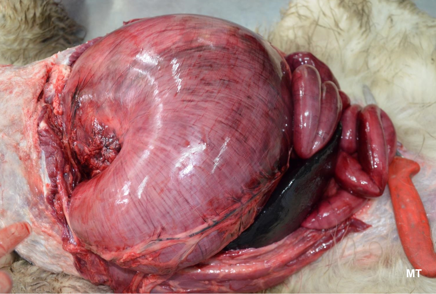
Dog, large breed, gastric dilation and volvulus
Vascular changes: edema, congestion
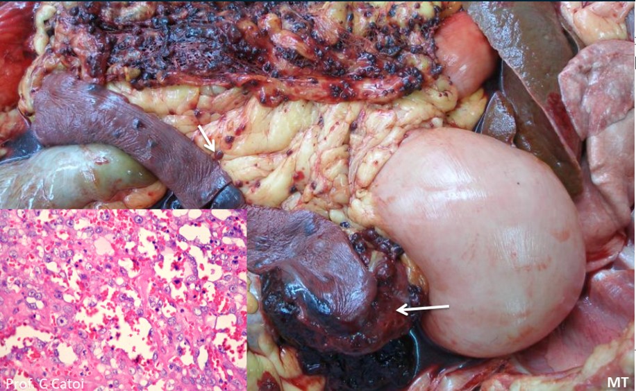
Dog, German Shepherd (large breed !!!)
Splenic hemangiosarcoma with multiple metastatic nodules in the omentum and mesentery
Multifocal dark red-purple nodules scattered across the spleen, omentum and mesentery
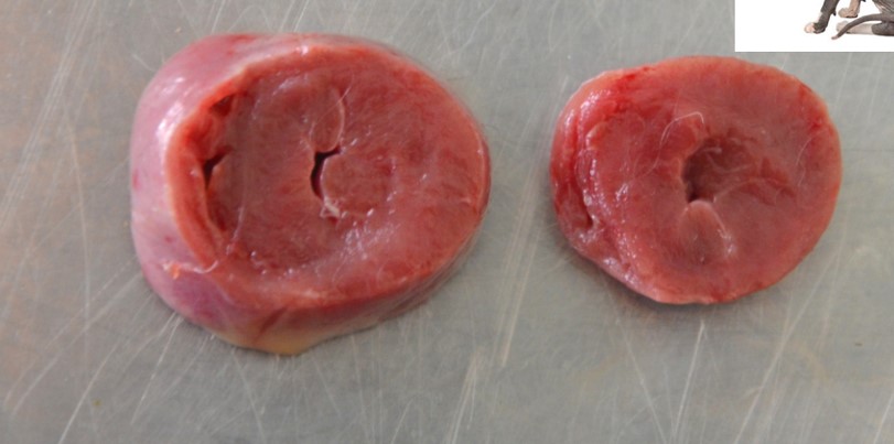
Cat, Sphinx, heart
Hypertrophic cardiomyopathy (HCM)
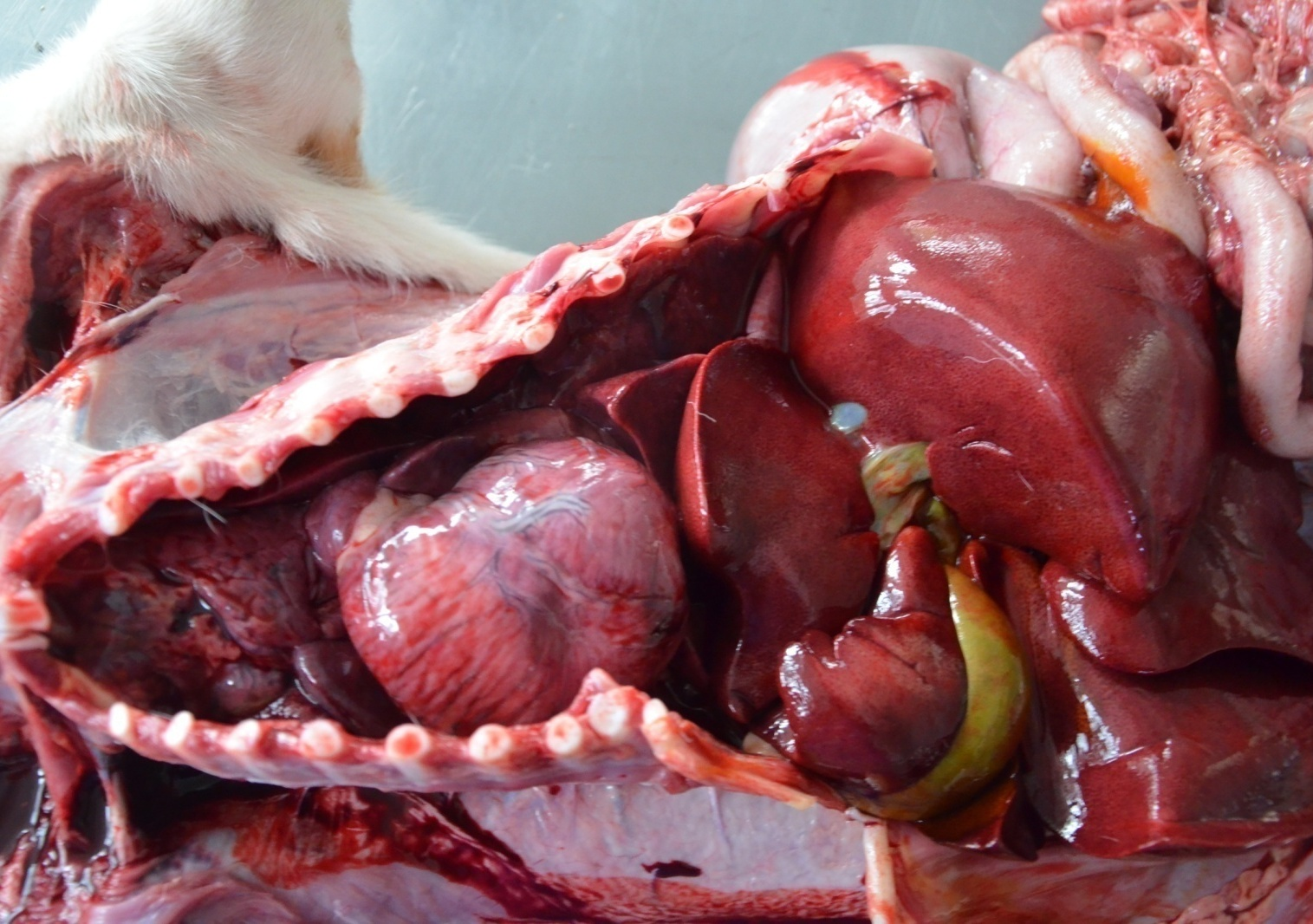
Young dog (2 months),
PDA
Cardiomegaly due to hypertrophy of the RV
Young dog (2 months),
PDA
Cardiomegaly due to hypertrophy of the RV
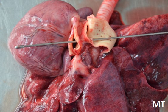
Young dog (2 months),
PDA
Cardiomegaly due to hypertrophy of the RV
Communication of aorta and pulmonary trunk
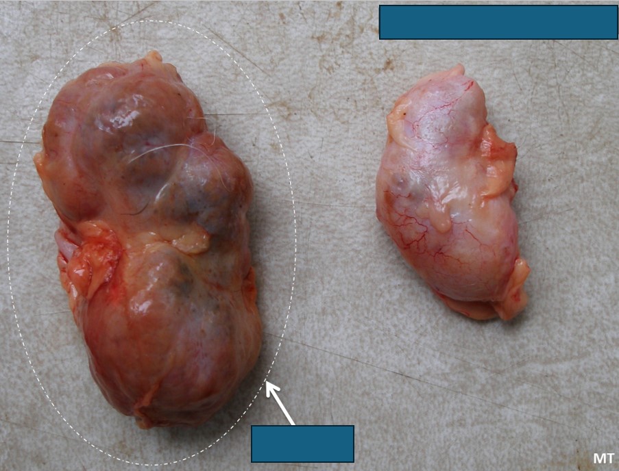
Old dog, kidneys, bilateral atrophy
End stage kidneys: irregularly shaped, severe bilateral atrophy and fibrosis
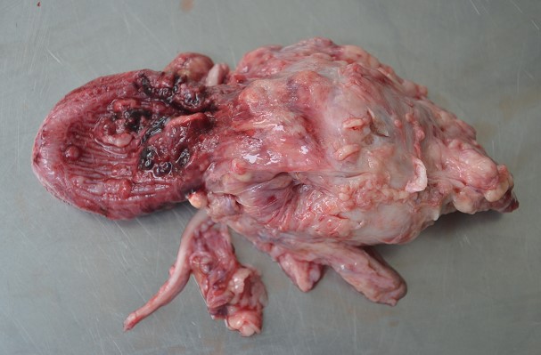
Male dog, prostate gland adenocarcinoma with pulmonary metastases
Prostate: large, multinodular, firm, irregular mass that replaced the normal parenchyma
Areas of hemorrhage and necrosis
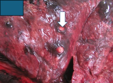
Male dog, prostate gland adenocarcinoma with pulmonary metastases
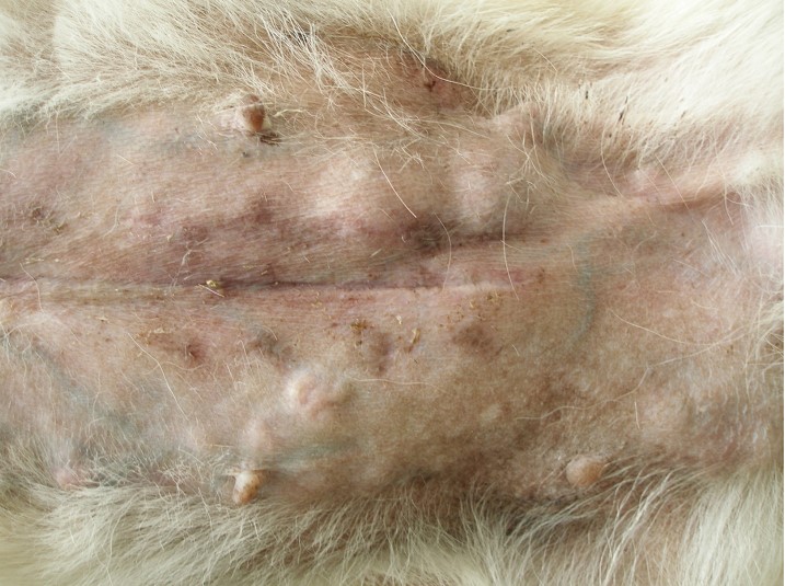
Dog, female, mammary gland adenocarcinoma
Dissemination of the neoplastic cells via
lymphatic vessels (histo picture!)
Pulmonary metastases
Skin: firm, cutaneous nodules along the mammary chain
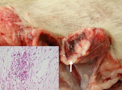
Dog, female, mammary gland adenocarcinoma
Dissemination of the neoplastic cells via
lymphatic vessels (histo picture!)
Pulmonary metastases
Lymphatic dissemination
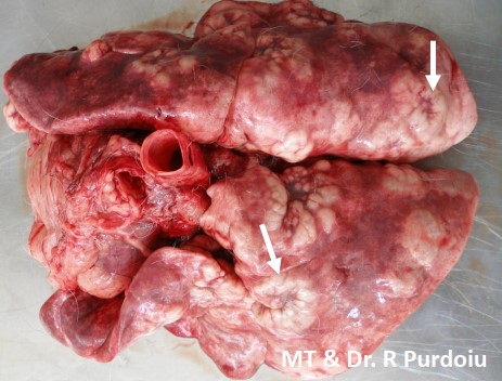
Dog, female, mammary gland adenocarcinoma
Dissemination of the neoplastic cells via
lymphatic vessels (histo picture!)
Pulmonary metastases
Pulmonary metastases: multifocal to coalescing, raised, pale tan nodules
>80% of pulmonary parenchyma was replaced
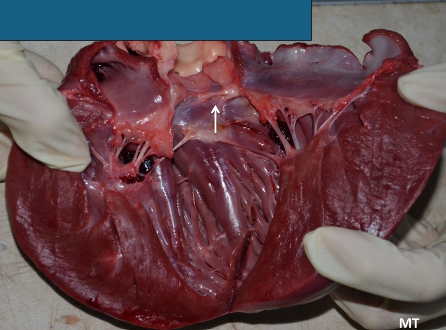
Dog, heart, left side, subvalvular aortic (subaortic) stenosis
A fibrous band is present beneath the aortic valve (arrow)
Sudden death!
Severe concentric hypertrophy of LV
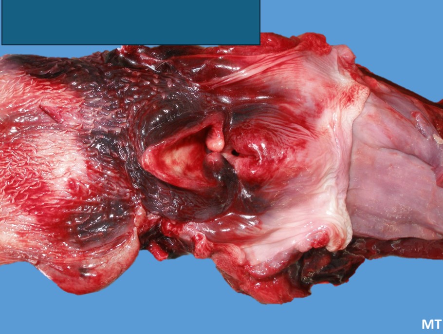
Dog
Laryngeal and pharyngeal hemorrhages and edema
Anticoagulant poisoning
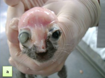
Head injury: brain laceration and unilateral exophthalmos
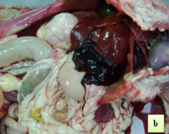
Internal bleeding in a cat due to hepatic rupture
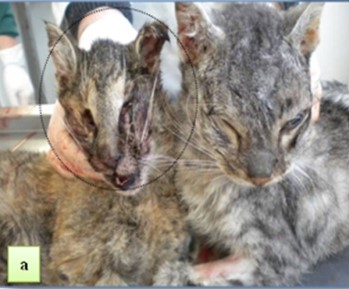
Neurogenic shock
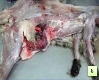
Eviscerations of abdominal wall
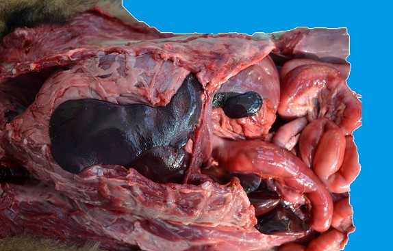
Dog, diaphragmatic hernia Horse, diaphragmatic hernia
Sudden death!
Displacement of the abdominal organs through a foramen in the diaphragm
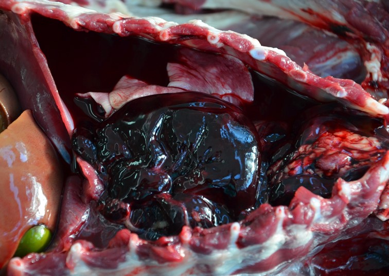
Dog, Sudden death!!!
Etiology: anticoagulant poisoning
Hemothorax: severe internal bleeding in the pleural cavity → hypovolemia, respiratory distress
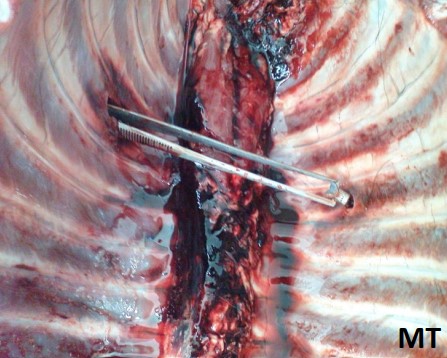
Dog, Sudden death!!!
Etiology: trauma
Hemothorax: rupture of the lungs
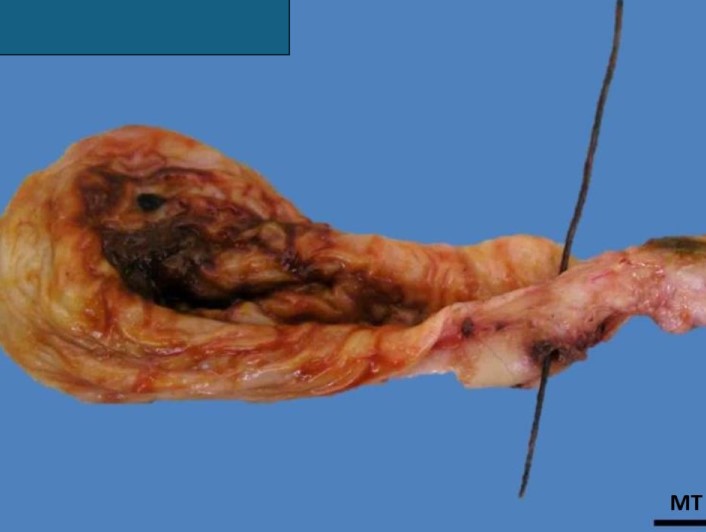
Hemorrhage and necrosis
Intravita as borders infiltrated with blood
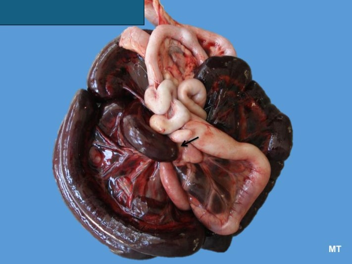
Dog, 3y old: sudden death!
Mesenteric hernia and venous infarction
Intestines: necrosis, hemorrhages, infarcts
Acute lesion
Secondary compression
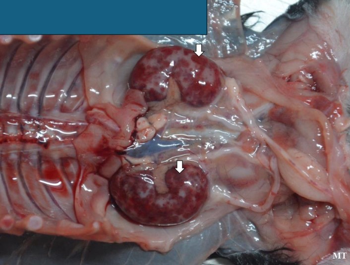
2-week-old newborn puppy, neonatal death
Hemorrhages in the renal cortex + cortical necrosis
Cause: systemic infection with canine herpesvirus
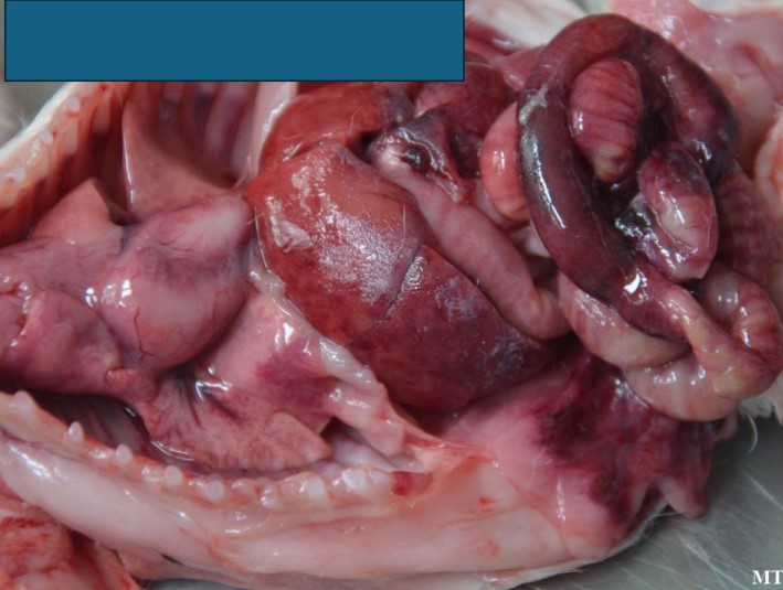
1-week-old newborn puppy, neonatal death
Acute fibrinous peritonitis, hemorrhagic enteritis
Cause: bacterial infection (Salmonella spp, E. coli)
Lungs: pulmonary congestion and edema
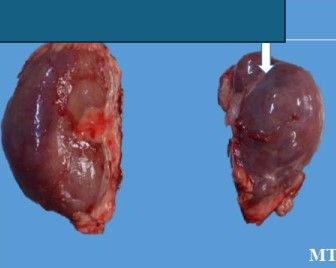
Puppy, kidneys
Unilateral hypoplasia and dysplasia
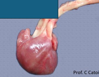
Puppy, heart
Complete transposition
of arteries (grade 4)
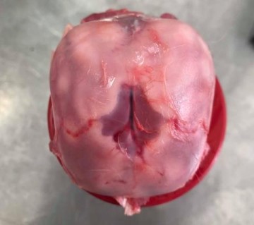
Puppy, Bichon Frisé
Internal hydrocephalus and atrophy of the nervous tissue
Brain: accumulation of large amounts of CSF within the ventricles → severely domed and
thinned cerebral cortex, ventricles visible through the translucent, stretched nervous tissue
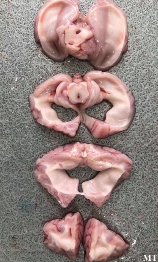
Puppy, Bichon Frisé
Internal hydrocephalus and atrophy of the nervous tissue
Severe dilation of the lateral ventricles and nervous tissue atrophy
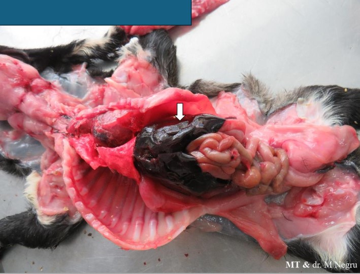
Puppy, neonatal death
Traumatic hepatic rupture and hemoperitoneum
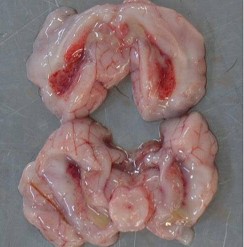
Puppy, neonatal death
Purulent choroiditis with accumulation of exudate in the lateral ventricles
Bacteriology: Pseudomonas spp. (green colonies) and E. coli
Cause: maternal factor - mastitis
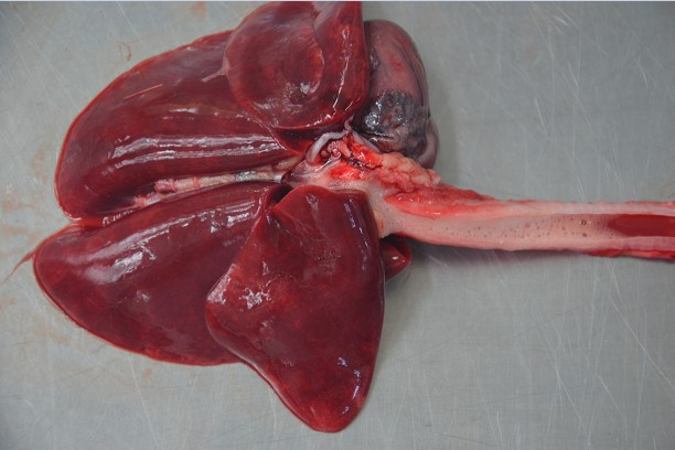
Cat, perianesthetic death (during general anesthesia)
Severe, acute, negative pressure (?) pulmonary edema
Negative pressure pulmonary edema (NPPE), the noncardiogenic
pulmonary edema, is caused by upper airway obstruction and
rapid negative intrapleural pressure increasing due to attempts of
inspiration against the obstruction. This occurs as a serious
complication of general anesthesia with tracheal intubation, and
more than half of these cases are related to postanesthetic
laryngospasm
Lungs: diffuse pulmonary congestion and edema, lungs appear wet and dark red
Trachea: significant amount ofcwhite-pinkish froth within the lumen
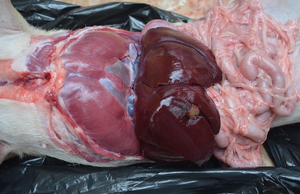
Dog, perianesthetic death (during general anesthesia)
Severe pneumothorax
Diaphragm: abnormal position, convexed towards abdominal cavity
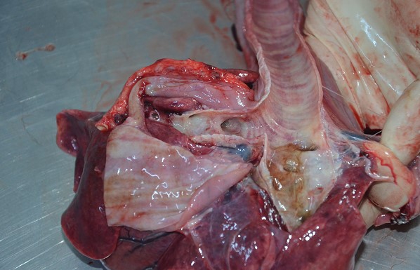
Dog, perianesthetic death (after general anesthesia)
Aspiration pneumonia
Lungs: multifocally dark red and congested
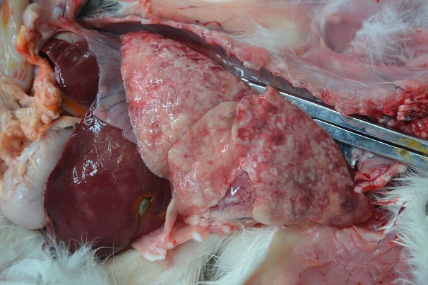
Cat, perianesthetic death (during general anesthesia)
History: Traumatic injuries due to a car accident
Postmortem: Severe, chronic, verminous pneumonia
Aelurostrongylus abstrusus
Lung: fail to collapse, multifocal, diffuse, slightly raised, whitish nodules replaced >80% of pulmonary parenchyma
Ddx in a young cat 2-3 months: fungal infection (cryptococcus, aspergillus), metastatic cancer
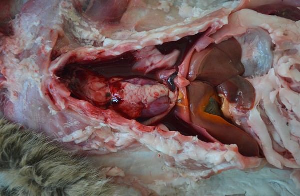
Cat, perianesthetic death
Severe, acute, traumatic hemothorax
The lesions are caused by resuscitation
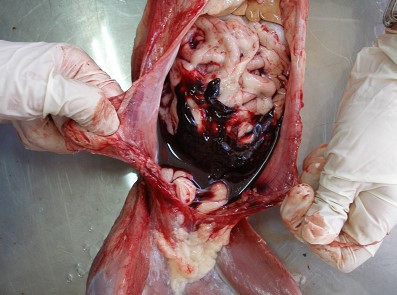
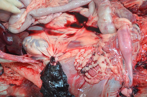
Dog, F, death after surgery (ovariohysterectomy)
Acute, severe, hemoperitoneum
Ovarian artery hemorrhage
Hemoperitoneum: abdominal cavity filled with a large volume of dark red, non-clotted and clotted blood
Dog, F, death after surgery (ovariohysterectomy)
Acute, severe, hemoperitoneum
Ovarian artery hemorrhage
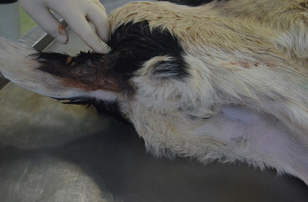
Dog, Parvoviral enteritis
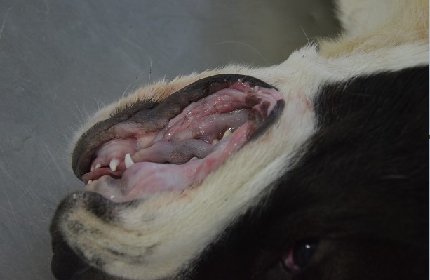
Dog, Parvoviral enteritis
Pale mucosa
GI content in oral cavity
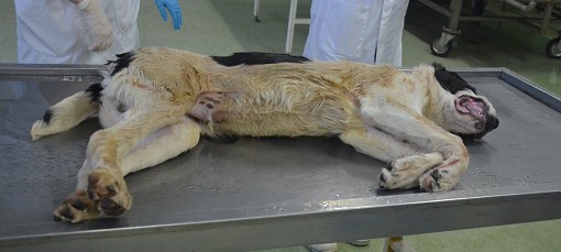
Dog, Parvoviral enteritis
Dehydration
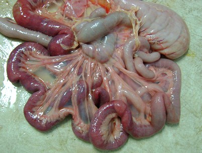
Dog, Parvoviral enteritis
Intestines: congestion, edema, necrosis, hemorrhage
Mesenteric LN: swollen
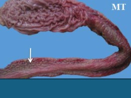
Dog, Parvoviral enteritis
Severe, diffuse, acute necrotizing enteritis
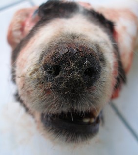
Dog, CDV infection (Morbillivirus)
Crusty nose
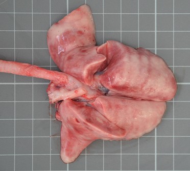
Dog, CDV infection
Diffuse bronchointerstitial pneumonia: the lungs are diffusely rubbery with rib imprints on the pleural surface
Lungs: diffuse bronchointerstitial pneumonia, lungs are diffusely rubbery
with rib imprints on the pleural surface
Lungs are expanded, gray, fail to collapse
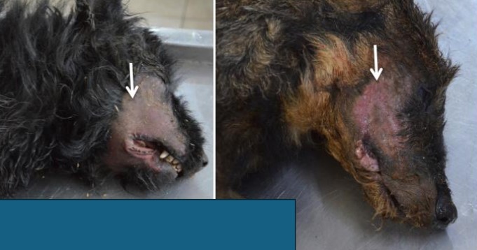
Dogs,
Facial skin abrasions (due to intense itching)
Pseudorabies (Aujeszky's disease)
Etiology: Suid herpesvirus 1 (SuHV-1)
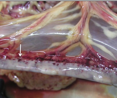
Dog, liver, CAV1 infection
Petechial hemorrhages on the intestinal serosa
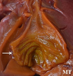
Dog, liver, CAV1 infection
Diffuse thickening (edema) of the gallbladder wall
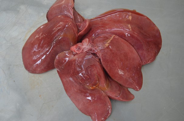
Dog, liver, CAV1 infection
Acute, severe necrotizing hepatitis
Hepatomegaly, pale, fragile, congested, edematous, necrosis
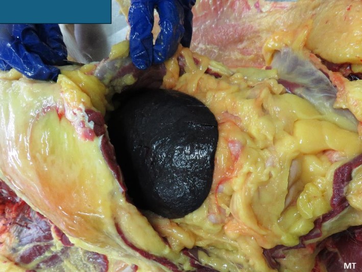
Dog, acute leptospirosis
Jaundice, splenic congestion
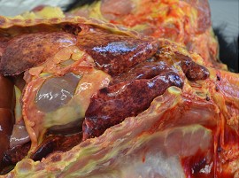
Dog
Cause: Leptospira spp.
Lungs: pulmonary edema, congestion, multifocal to coalescing dark red areas of hemorrhages
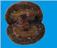
Dog, kidney
Hemorrhages (petechiae) in the renal cortex + cortical necrosis and jaundice
Cause: Leptospira spp.
Ddx of acute fever and jaundice: IMHA, babesia, Erlichia
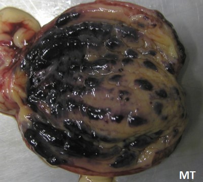
Urinary bladder hemorrhages
Cause: Leptospira spp.
Ddx of acute fever and jaundice: IMHA, babesia, Erlichia
Dog
Severe pulmonary hemorrhage, edema and jaundice
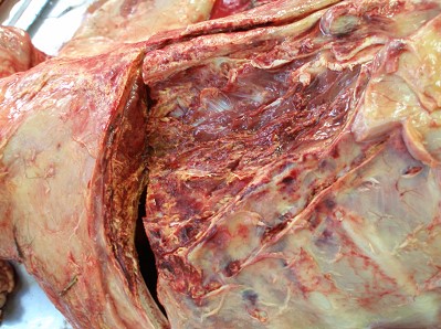
Necrotizing fasciitis (toxic shock syndrome)
Areas are discolored, yellow, pale, red due to hemorrhage, cutsection of the muscle reveal extensive necrosis of muscle tissue
Dog
Severe pulmonary hemorrhage, edema and jaundice
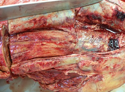
Necrotizing fasciitis (toxic shock syndrome)
Areas are discolored, yellow, pale, red due to hemorrhage, cutsection of the muscle reveal extensive necrosis of muscle tissue
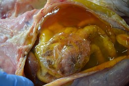
Feline infectious peritonitis (FIP), effusive form
Intraperitoneal gelatinous fluid admixed with numerous white-yellow fibrin strands
Etiology: mutated coronavirus
Serofibrinous peritonitis
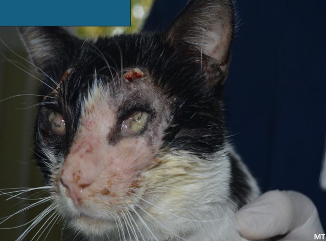
Cat, FHV1 infection
Bilateral, purulent keratoconjunctivitis
Multifocal, ulcerative dermatitis
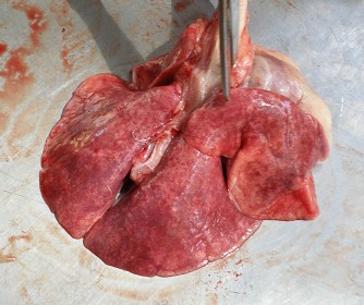
Cat, FHV1 infection
Acute bronchointerstitial pneumonia
Lungs: acute bronchointerstitial pneumonia, expanded, congested, edematous, fail to collapse, rubber texture
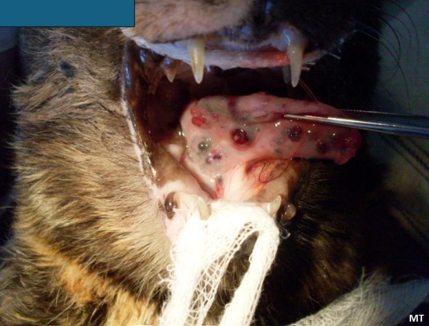
Cat, FCV infection
Multifocal ulcerative glossitis
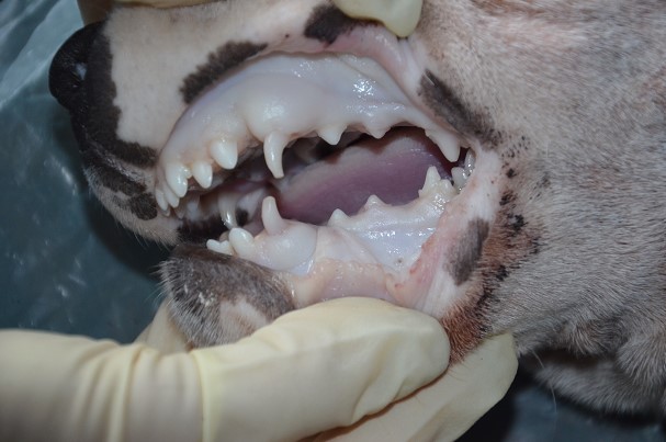
Dog, anticoagulant rodenticide (Brodifacoum) poisoning \
Oral Cavity: pale mucous membranes - severe anemia due to blood loss
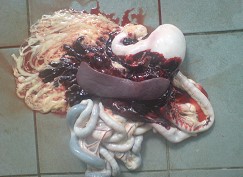
Dog, anticoagulant rodenticide (Brodifacoum) poisoning
Abdominal cavity: hemoperitoneum with large volumes of un-clotted blood surrounding the internal organs
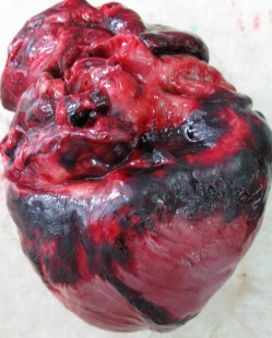
Dog, anticoagulant rodenticide (Brodifacoum) poisoning
Heart: epicardial hemorrhage
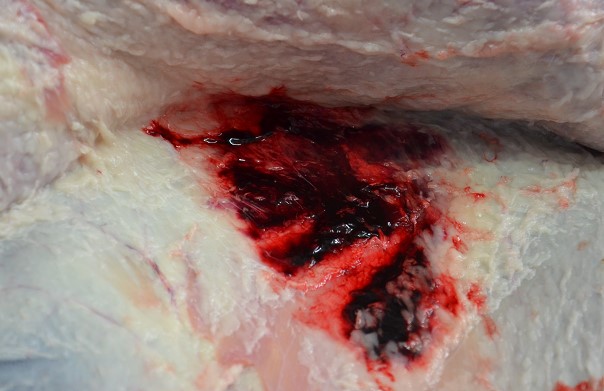
Dog, anticoagulant rodenticide (Brodifacoum) poisoning
SC tissue: large areas of ecchymosis and hematoma formation
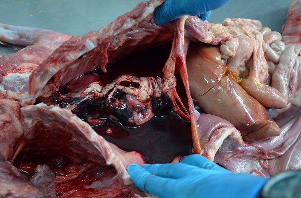
Dog, anticoagulant rodenticide (Brodifacoum) poisoning
Thoracic cavity: hemothorax
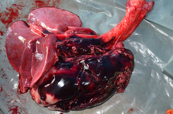
Dog, anticoagulant rodenticide (Brodifacoum) poisoning
Lungs: dark-red due to multifocal to coalescing pulmonary hemorrhage
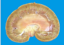
Dog, kidney
Acute, severe, diffuse, tubular necrosis, calcium oxalate crystals
Cause: ethylene glycol intoxication (calcium oxalate)
Kidney is pale-brown-yellow due to extensive necrosis
Striation on the cut section
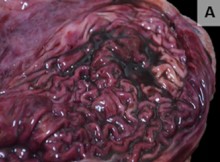
Metallic phosphide poisoning in a dog
Stomach: congestion of mucosa, erosions
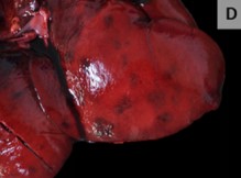
Metallic phosphide poisoning in a dog
Liver: congestion, friable, multifocal hemorrhage and necrosis
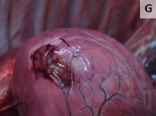
Metallic phosphide poisoning in a dog
Heart apex: myocardial necrosis and hemorrhage
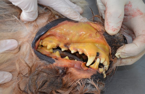
Dog, AIHA
Jaundice (icterus), splenomegaly
Hemoglobinuria (“port wine” urine)
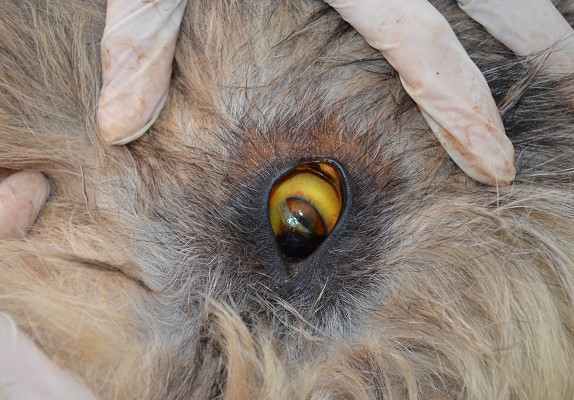
Dog, AIHA
Jaundice (icterus), splenomegaly
Hemoglobinuria (“port wine” urine)
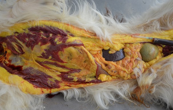
Dog, AIHA
Jaundice (icterus), splenomegaly
Hemoglobinuria (“port wine” urine)
deep red – black urine in the syringe (not blood)
Spleen: splenomegaly due to congestion is a consistent finding
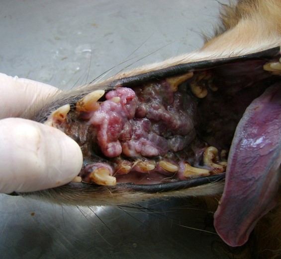
Dog, progressive weight loss
Oral cavity, infiltrative melanoma
Oral cavity: mass is large, irregularly shaped, infiltrate the nasal cavity and bones
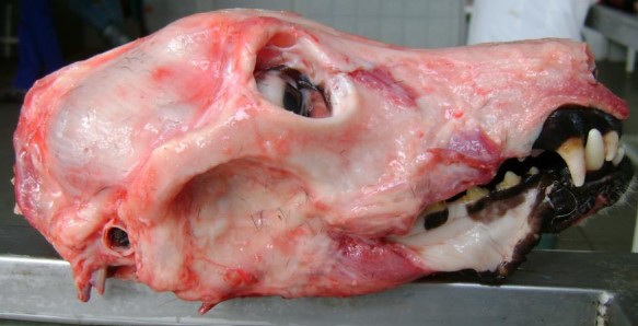
Dog, progressive weight loss
Cause: masticatory myositis
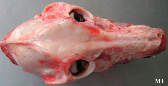
Dog, progressive weight loss
Cause: masticatory myositis
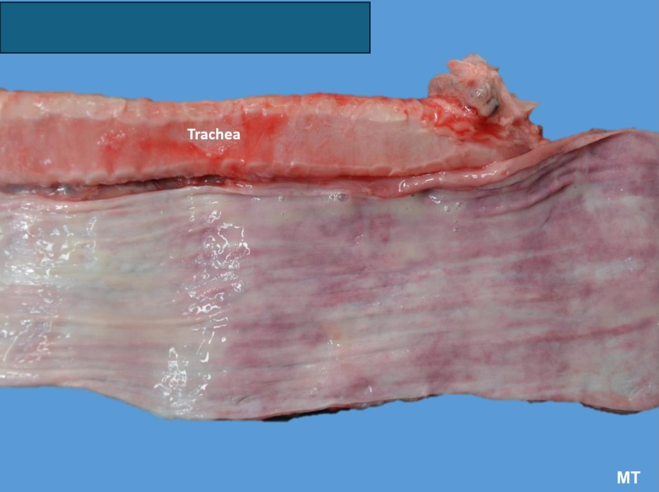
Dog, progressive weight loss
Cause: Congenital idiopathic megaesophagus (CIM)
Esophagus: esophageal dilation, congested mucosal surface due to
the chronic retention of food and saliva
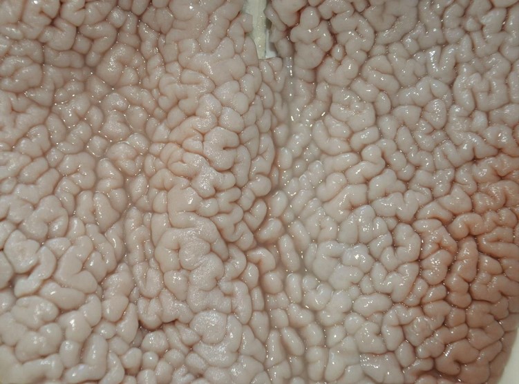
Dog, progressive weight loss
Hypertrophic gastropathy
Diffuse form
Stomach: pale thickened mucosa with “brain-like” appearance
Transverse folds (don’t confuse with rigor mortis – not transverse folds only longitudinal)
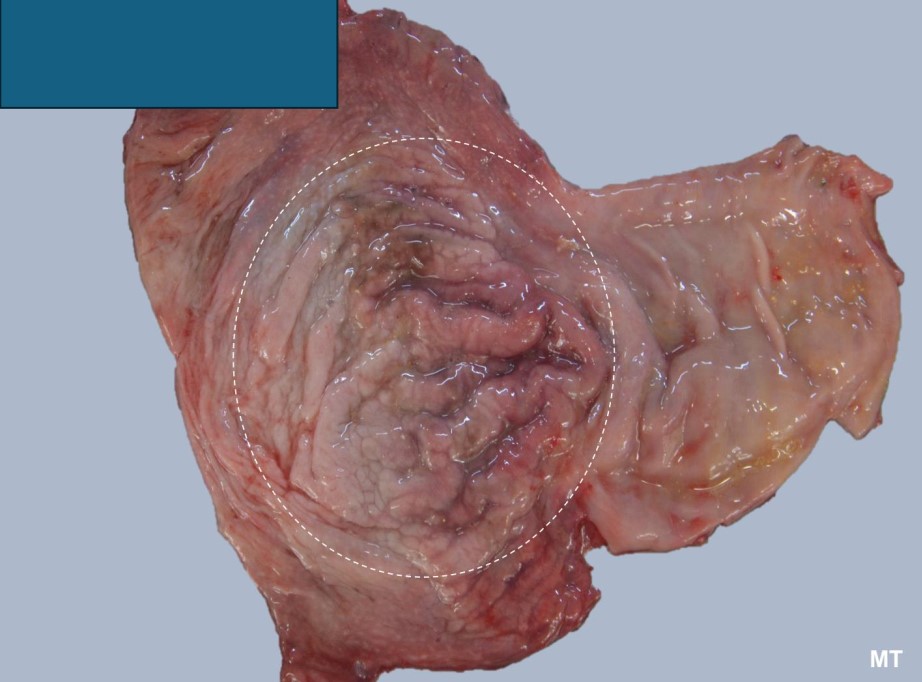
Dog, progressive weight loss
Stomach - body region
Giant hypertrophic gastropathy
Menetrier-like disease
Stomach: mass-like lesion, gastric
mucosa is thickened, elevated, exophytic,
full of giant folds
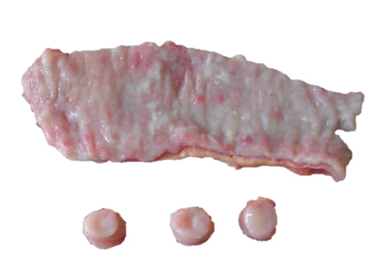
Dog, progressive weight loss
Lymphoplasmacytic enteritis and lymphangiectasia
Small intestines: thickened mucosa with cribriform like appearance, multifocal white-yellow, opaque spots representing lymphangiectasia
On cut section we see no lumen due to mucosa thickening
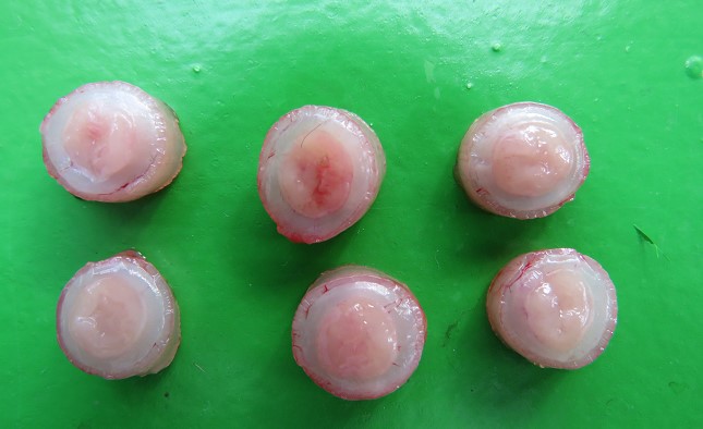
Cat, progressive weight loss Alimentary (intestinal) lymphoma + mesenteric lymphadenopathy
FeLV+
On cut section we see no lumen due to mucosa thickening
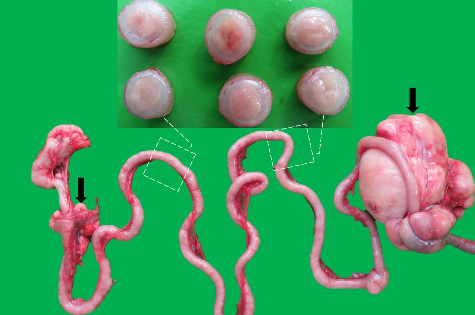
Cat, progressive weight loss Alimentary (intestinal) lymphoma + mesenteric lymphadenopathy
FeLV+
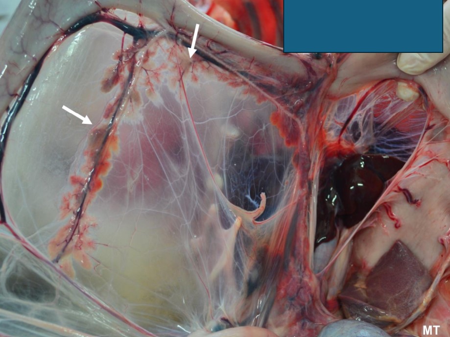
Dog, progressive weight loss
Pancreatic hypoplasia/ atrophy
Pancreatic insufficiency
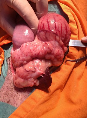
Dog, progressive weight loss
Pancreatic lipomatosis (fatty replacement)
Pancreatic insufficiency
Pancreas: irregularly shaped, diffusely thickened, pale-white-yellow
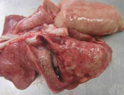
Cat, progressive weight loss
Verminous pneumonia
Aelurostrongylus abstrusus
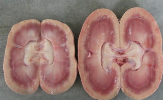
Dog, progressive weight loss
Metastatatic mineralization
Renal failure
Bilateral renal tubular calcification
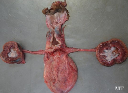
Dog, progressive weight loss
Chronic renal failure
Bilateral hydronephrosis
......ovariohysterectomy (!)
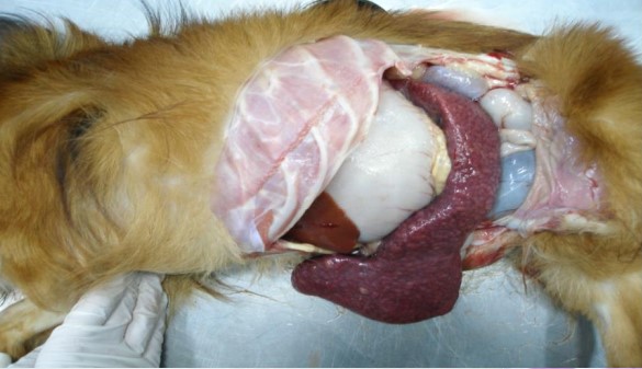
Dog, progressive weight loss
Malignant, multicentric lymphoma
- enlargement of spleen & lymph nodes
- cytological features
Spleen: irregularly shaped, splenomegaly due to congestion, hemorrhage, white coalescent nodules composed of uniform lymphocytes (large cell lymphoma)
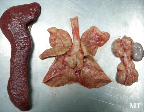
Dog, progressive weight loss
Malignant, multicentric lymphoma
- enlargement of spleen & lymph nodes
- cytological features
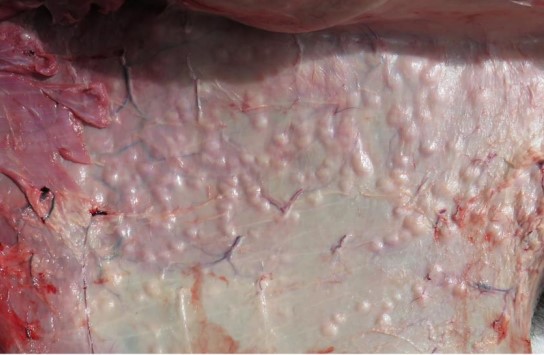
Dog, progressive weight loss
Skin lymphoma
Skin: diffuse multifocal to coalescing, raised, erythematous and crusting nodules