Knee joint, anterior and lateral leg
1/71
There's no tags or description
Looks like no tags are added yet.
Name | Mastery | Learn | Test | Matching | Spaced | Call with Kai |
|---|
No analytics yet
Send a link to your students to track their progress
72 Terms
Identify the major bony features of the patella, tibia, and fibula
Recognise the major features of the knee joint and understand their importance in the functioning of the lower limb.
Identify the popliteal fossa, recognising its boundaries and contents.
Recognise the major muscles of the anterior and lateral compartments of the leg and describe their actions.
Describe the routes of the tibial and common fibular nerves and their major muscular branches and recall the groups of muscles each of these supplies.
Describe the pattern of blood supply and venous drainage to the leg and recognise the major vessels involved.
What does the knee joint transition between?
thigh and leg
What are the two articulating surfaces of the knee joint?
Tibiofemoral surface and Patellofemoral surface
Why is the knee joint termed multiaxial?
performs several movement eg flexion, extension, lateral rotation, medial rotation
Describe the bones articulating at the knee joint
femur with tibia and patella (known as knee cap) lies anterior
What is the biggest bone of the leg?
tibia
Which bone of the leg is not involved in the knee joint?
fibula - lateral side of leg, very thin and lower so not involved in knee joint
From which view is the patella not visible? What other landmark distinguishes anterior from posterior view of the knee joint?
posterior
anterior view only has one shiny surface // posterior has 2 shiny surfaces/condyles
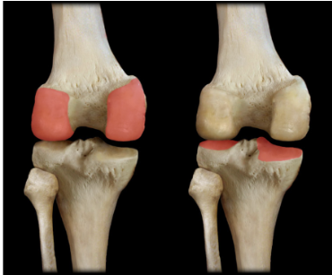
What is a way to situate ourselves when looking at the knee joint?
patella is anterior and fibula is lateral
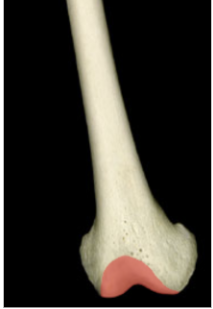
Is this anterior or posterior view of the femur?
posterior because only one condyle
Which movements of the multiaxial knee joint are not complete?
lateral and medial rotations - mainly allow joint stability
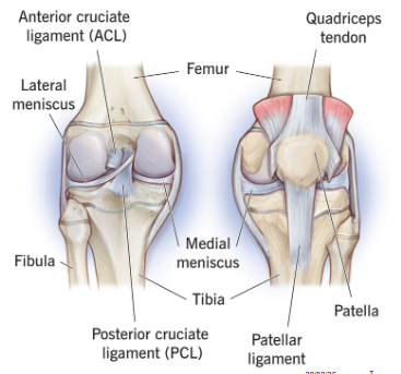
What are the extracapsular ligaments of the knee joint?
Patellar ligament (tendon)
Medial collateral ligament
Lateral collateral ligament
Why is the patellar ligament bordering on a tendon?
comes from the quadriceps tendon
What are the intracapsular ligaments of the knee joint?
Anterior cruciate ligament (ACL)
Posterior cruciate ligament (PCL)
Menisci (medial and lateral meniscus)
What does the lateral collateral ligament connect?
femur to tibia and fibula
What does the medial collateral ligament connect?
femur to tibia
Where does cruciate come from? Why are ACL and PCL commonly torn?
cross
always used and are internal so torn easily and heal badly
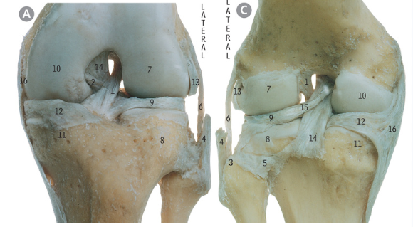
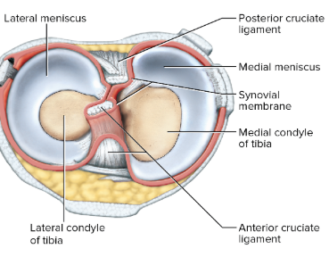
Label the image : 16, 6, 1, 14, 9, 12, 15
16 is medial collateral ligament, 6 is lateral collateral ligament, 1 is ACL, 14 is PCL, 9 is lateral meniscus (on top of fibula), 12 is medial meniscus, 15 is the fibres connecting PCL to lateral meniscus
Where does the sciatic nerve go as we leave the thigh?
Runs on posterior thigh right into the back of the knee
Where do the femoral nerve, artery and vein go as we leave the thigh?
From femoral triangle, they run on anteromedial aspect of the thigh
Pass through adductor hiatus in the adductor canal and emerge on posterior thigh just above the back of the knee
What does the femoral nerve become as we leave the thigh?
saphenous nerve
What does the femoral artery become as we leave the thigh?
popliteal artery
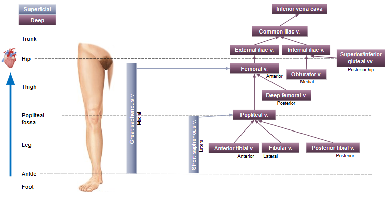
What drainage does the femoral vein receive from below the thigh?
popliteal vein
Define the popliteal fossa
‘Diamond’ space on posterior aspect of knee join that is a passage for nerves + blood vessels between thigh and leg
Which structures pass though the popliteal fossa?
3 nerves, 3 main vessels, lymph nodes
What are the boundaries of the popliteal fossa?
4 borders + floor + roof
Which 3 muscles make up the popliteal fossa?
semimembranosus and semitendinosus + biceps femoris + gastrocnemius (lateral and medial heads)
What are the 4 borders of the popliteal fossa?
Superomedial – semimembranosus & semitendinosus mm.
Superolateral – biceps femoris m.
Inferomedial – gastrocnemius m. (medial head)
Inferolateral – gastrocnemius m. (lateral head)
What makes up the roof of the popliteal fossa?
fascia, skin
What makes up the floor of the popliteal fossa?
knee capsule, distal femur, proximal tibia and popliteus muscle
What are the contents of the popliteal fossa ?
Sciatic nerve
→ Tibial nerve
→ Common fibular nerve
Popliteal vein
← Small (short) saphenous vein
Popliteal artery
→ 5 genicular arteries
Popliteal lymph nodes
What does the sciatic nerve divide into as it exits the popliteal fossa?
tibial nerve and common fibular nerve
What does the common fibular nerve divide into entering the leg and what do they each innervate?
→ Deep fibular nerve (anterior leg)
→ Superficial fibular nerve (lateral leg)
Which nerve and artery enter the leg from the knee joint?
common fibular nerve and anterior tibial artery, tibiofibular trunk
What does the common fibular nerve divide into as it enters the leg?
deep fibular nerve and superficial fibular nerve
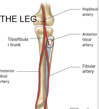
What does the tibiofibular trunk divide into at it enters the leg?
posterior tibial artery and fibular artery
Which nerve and artery enter the knee joint from the thigh?
sciatic nerve and popliteal artery
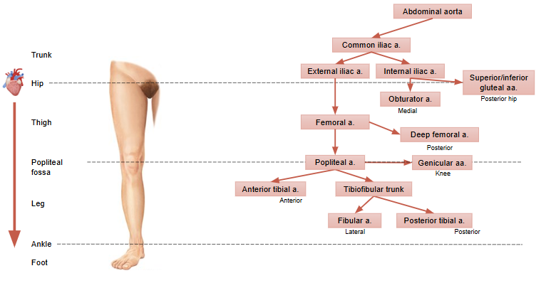
What does the popliteal artery divide into entering the leg and which compartment of the leg do they supply?
→ Anterior tibial artery (anterior leg)
→ Tibiofibular trunk
→ Posterior tibial artery (posterior leg)
→ Fibular artery (lateral leg)
What compartments does the leg have vs the thigh?
anterior, posterior and lateral // posterior, anterior, medial
Describe the branches of the tibiofibular artery down the leg?
first branch is anterior tibial artery which supplies the anterior leg
next 2 branches are posterior tibial artery (posterior) and fibular artery (lateral)

What are the 2 bones of the leg?
tibia and fibula
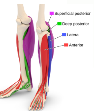
What are the 3 fascial/muscle compartments of the leg?
anterior, lateral, posterior (superficial and deep)
What is the term for the big toe?
hallux
What are movements of the leg?
Dorsiflexion
Plantarflexion
Eversion
Inversion
Flexion/extension of toes
Describe eversion
plants of foot go away from body
Describe inversion
plants of foot go in towards body
Describe toe extension
toe in towards leg
Describe dorsiflexion
feet go in towards leg
Describe plantarflexion
feet pointed eg ballet dancer
Which movements allow walking on uneven surfaces eg stones?
eversion and inversion
What are the muscles of the anterior compartment of the leg?
extensor hallucis longus, extensor digitorum longus, tibialis anterior, fibularis tertius
Action of tibialis anterior of anterior leg
dorsiflexion of ankle and inversion of foot
Action of fibularis tertius
Dorsiflexion of ankle, eversion of foot
What is the common and different movements of extensor hallucis longus vs extensor digitorum longus of the anterior compartment of the leg?
both do dorsiflexion of the ankle , hallucis only extends the big toe // digitorum extends toes 2-5
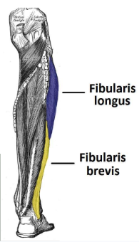
What are the muscles of the lateral part of the leg?
fibularis longus and brevis

How does the irrigation of the anterior vs lateral leg differ?
deep fibular artery // superficial fibular artery
How does blood supply to the anterior vs lateral leg differ?
anterior tibial artery // fibular artery
Action of fibularis longus
Eversion and plantarflexion of foot
Action of fibularis brevis
Eversion and plantarflexion of foot

Irrigation of anterior compartment of the leg
anterior tibial artery
Blood supply to anterior compartment
deep fibular nerve
How many muscles make up the anterior vs lateral compartments of the leg?
4 vs 2
Describe blood supply through the knee joint and to the leg
Describe blood drainage through the knee joint and from the leg
Describe the structure of the knee joint, both bones and ligaments
articulates 3 bones: femur and patella at the patellofemoral surface, and femur and tibia at the tibiofemoral surface
reinforced by extracapsular (patellar ligament/tendon, medial collateral ligament, lateral collateral ligament)
and intracapsular (anterior cruciate ligament (ACL), posterior cruciate ligament (PCL), medial and lateral meniscus) ligaments.
Summarise the anterior compartment of the leg
consists of muscles that dorsiflex the foot: tibialis anterior (also inverts), fibularis tertius (also everts), extensor hallucis longus (also extends hallux) and extensor digitorum longus (also extends toes 2-5)
innervated by the deep fibular nerve and are supplied by the anterior tibial artery.
Summarise the lateral compartment of the leg
consists of muscles that evert and plantarflex the foot: fibularis longus and fibularis brevis
innervated by the superficial fibular nerve and are supplied by the fibular artery