lec 9 - drosophila limb development
1/25
There's no tags or description
Looks like no tags are added yet.
Name | Mastery | Learn | Test | Matching | Spaced | Call with Kai |
|---|
No analytics yet
Send a link to your students to track their progress
26 Terms
The core question: how does a tiny flat disc of ~30 cells grow into a perfectly patterned 3D wing (50,000 cells, 4 days later)?
morphogen gradients along two perpendicular axes
what is an imaginal disc
small sacs of epithelial cells
float in larval body through metamorphosis (when most larval tissue is destroyed), and then unfold into adult structures
how do wing discs unfold into adult structures
wing disc eversion
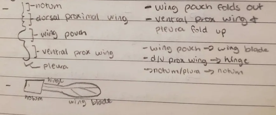
what are the parts of the wing disc
noctum
dorsal proximal wing
wing pouch
ventral proximal wing
pleura
describe the process of wing disc eversion
how are imaginal discs patterned
orthogonal morphogen gradients (AP/DV)
cells read orthogonal morphogen gradients like coordinate planes
the AP gradient is patterned by
Hh, Engrailed (Eg), and Dpp
what happens when Shh is placed ectopically in the limb bud
chick: mirror image wing
fly: extra wing
human: polydactyly
the DV gradient is patterned by
Wg/Wnt, Apterous (dorsal TF)
____ defines the posterior segments
Eg
how does Eg defining the posterior segment tranlate into the imaginal discs
when cells invaginate into imaginal discs, they inherit the posterior expression of Eg
how do we know Eg patterns posteriorly
reporter genes
replace Eg gene with LacZ genes → LacZ is under the control of Eg enhancers
when washed with soluble colorless X-gal, LacZ converts it to blue precipitate
posterior wing is blue - therefore Eg is posterior
What is Dpp
BMP protein
fly TGF-B homolog
Hh transcriptional target
morphogen
what is the process of AP axis patterning
posterior Eg activates Hh production but restricts Ci
Hh diffuses over the compartment (AP) boundary into the anterior section - where there is Ci
Hh pathway is activated → Patched receptor production is activated → Patched expresses at the AP/compartment boundary
Hh activates Dpp, a morphogen - spreads outwardly from the compartment boundary
Dpp pathway
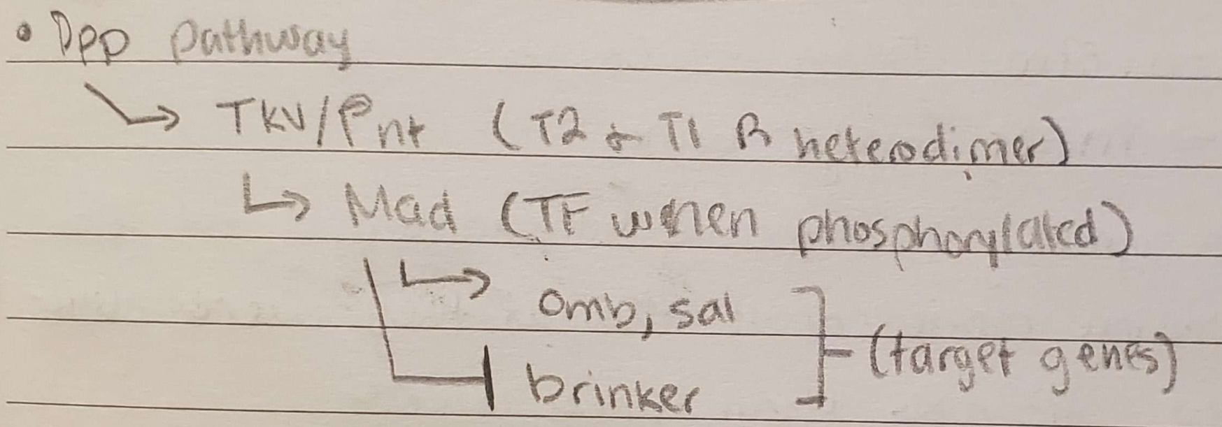
TKV/Pnt
Type 2 and Type 1 Dpp receptor heterodimer
Mad
Dpp TF when phosphorylated
Omb, Sal
Dpp target genes (Dpp activates)
brinker
Dpp target gene (Dpp inhibits)
How do we know Dpp is a morphogen?
antibodies for p-Mad show Dpp diffusion
when Dpp is tagged with GFP, can visualize the diffusion - area of protein is much wider than the region Dpp is produced in
high [Dpp] → Omb and Sal activation, but low [Dpp] → only Omb activation
french flag model - changes in [Dpp] → changes in gene expression = morphogen
when GDP-Dpp is bound to the AP axis cell membrane by anti-GFP nanobodies, brinker (only active when Dpp is not present) expression is able to move closer to the AP axis than it normally does— implies that Dpp diffused outwardly and induced a certain cell fate
![<ol><li><p>antibodies for p-Mad show Dpp diffusion</p></li><li><p>when Dpp is tagged with GFP, can visualize the diffusion - area of protein is much wider than the region Dpp is produced in</p></li><li><p>high [Dpp] → Omb and Sal activation, but low [Dpp] → only Omb activation</p><ol><li><p>french flag model - changes in [Dpp] → changes in gene expression = morphogen</p></li></ol></li><li><p>when GDP-Dpp is bound to the AP axis cell membrane by anti-GFP nanobodies, brinker (only active when Dpp is not present) expression is able to move closer to the AP axis than it normally does— implies that Dpp diffused outwardly and induced a certain cell fate</p></li></ol><p></p>](https://assets.knowt.com/user-attachments/aa419bd7-39d7-4d25-a5cd-eb55f9d813d6.png)
Clone 1: Eg- cells cloned and transplanted into posterior region
Eg- → increased Ci but decreased Hh → Hh from surrounding cells diffuse into cloned cell area → Dpp signaling only in cloned area
Clone 2: Eg+ cells are cloned and transplanted in anterior region
Eg+ → increased Hh but decreased Ci → Hh diffuses outwardly → increased Dpp signaling around the cloned cell region
Clone 3: Patched- cells are cloned and transplanted in anterior region
Patched- → Smo- → increased Hh signaling → increased Dpp signaling in and around cloned cell area
Clone 2/3 (Hh/Dpp signaling in anterior wing disc) - what is the phenotype and why
phenotype: mirror image wing
why: because the Hh signaling pathway was activated so far from the compartment boundary, nearby cells believed that they were close to the compartment boundary and thus began patterning as such.
how is the DV axis patterned
Apterous expressed dorsally - Wg signaling restricted
Wg expresses at DV boundary
How do we know that Wg is diffusion/a morphogen
expression domain much tighter than protein location