Lab 1 - The Human Brain and Cranial Nerves
1/57
There's no tags or description
Looks like no tags are added yet.
Name | Mastery | Learn | Test | Matching | Spaced | Call with Kai |
|---|
No analytics yet
Send a link to your students to track their progress
58 Terms
Brain protection structures
Cranial bones, meninges cerebrospinal fluid, and blood brain barrier
Cranial meninges layers
Dura mater, arachnoid mater, pia mater
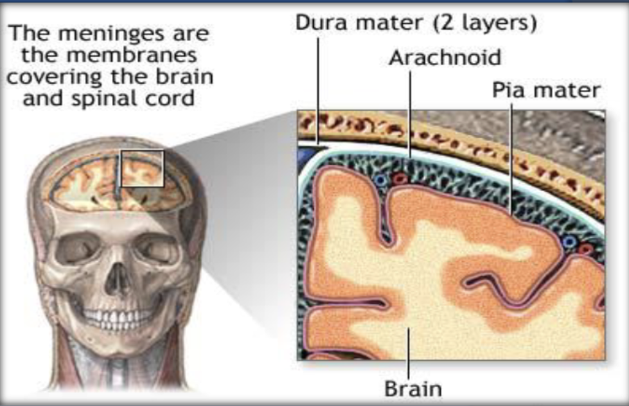
Dura mater
Tough outer meningeal layer attached to skull with endosteal and meningeal layers

Arachnoid mater
Middle web like layer with subarachnoid space containing CSF and arachnoid villi for CSF reabsorption
Pia mater
Thin inner layer directly on brain surface containing blood vessels
Subarachnoid space
Space between arachnoid and pia filled with CSF
Cerebrospinal fluid CSF
Fluid that cushions brain maintains chemical environment and circulates through ventricles and meninges
Choroid plexus
Structure in ventricles that produces CSF
Ventricles
Four brain chambers that produce and circulate CSF

Four brain regions
Cerebrum, diencephalon, brainstem, and cerebellum
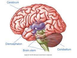
Cerebrum
Largest brain region responsible for higher thinking sensory perception and voluntary movement
Cerebral cortex
Outer gray matter layer responsible for higher intellect and functional areas
Gyrus
Ridge or fold on brain surface
Sulcus
Shallow groove on brain surface
Fissure
Deep groove separating brain regions
Longitudinal fissure
Separates left and right hemispheres
Corpus callosum
White matter tract connecting cerebral hemispheres
Frontal lobe
Involved in reasoning judgment motor function and personality
Parietal lobe
Processes sensory information like touch temperature and pain
Temporal lobe
Responsible for hearing memory and language
Occipital lobe
Primary visual processing center
Insula
Deep lobe involved in taste and internal awareness
Central sulcus
Separates frontal and parietal lobes
Lateral sulcus
Separates temporal lobe from frontal and parietal lobes
Parieto occipital sulcus
Separates parietal and occipital lobes
Primary motor cortex
Controls voluntary movements located in precentral gyrus
Primary sensory cortex
Receives sensory input located in postcentral gyrus
Broca area
Motor speech production area in frontal lobe
Wernicke area
Language comprehension area in temporal lobe
Visual cortex
Processes vision in occipital lobe
Auditory cortex
Processes sound in temporal lobe
Olfactory cortex
Responsible for smell in temporal lobe
Gustatory cortex
Responsible for taste in parietal lobe
Diencephalon
Region containing thalamus and hypothalamus
Thalamus
Relay station that sends sensory information to cerebral cortex
Hypothalamus
Maintains homeostasis regulates temperature hunger thirst hormones and autonomic functions
Pituitary gland
Master endocrine gland controlled by hypothalamus
Pineal gland
Produces melatonin and regulates sleep cycles
Brainstem components
Midbrain pons medulla oblongata
Midbrain
Relays motor and sensory signals and contains cranial nerve origins
Pons
Relay station sending sensory information to thalamus and cerebellum
Medulla oblongata
Controls vital functions like breathing heart rate and digestion
Decussation
Crossing of motor fibers in medulla causing opposite side control
Cerebellum
Coordinates movement balance posture and motor learning
Arbor vitae
Tree like white matter in cerebellum
Cranial nerves
Twelve pairs of nerves serving head and neck sensory and motor functions
Olfactory nerve I
Smell sensory
Optic nerve II
Vision sensory
Oculomotor nerve III
Eye movement and pupil constriction motor
Trochlear nerve IV
Eye movement downward and inward motor
Trigeminal nerve V
Facial sensation and chewing mixed
Abducens nerve VI
Lateral eye movement motor
Facial nerve VII
Facial expression and taste anterior tongue mixed
Vestibulocochlear nerve VIII
Hearing and balance sensory
Glossopharyngeal nerve IX
Taste posterior tongue swallowing mixed
Vagus nerve X
Parasympathetic control of organs in thorax and abdomen mixed
Accessory nerve XI
Neck and shoulder movement motor
Hypoglossal nerve XII
Tongue movement motor