APR Module 1: Body Orientation
1/214
There's no tags or description
Looks like no tags are added yet.
Name | Mastery | Learn | Test | Matching | Spaced | Call with Kai |
|---|
No analytics yet
Send a link to your students to track their progress
215 Terms
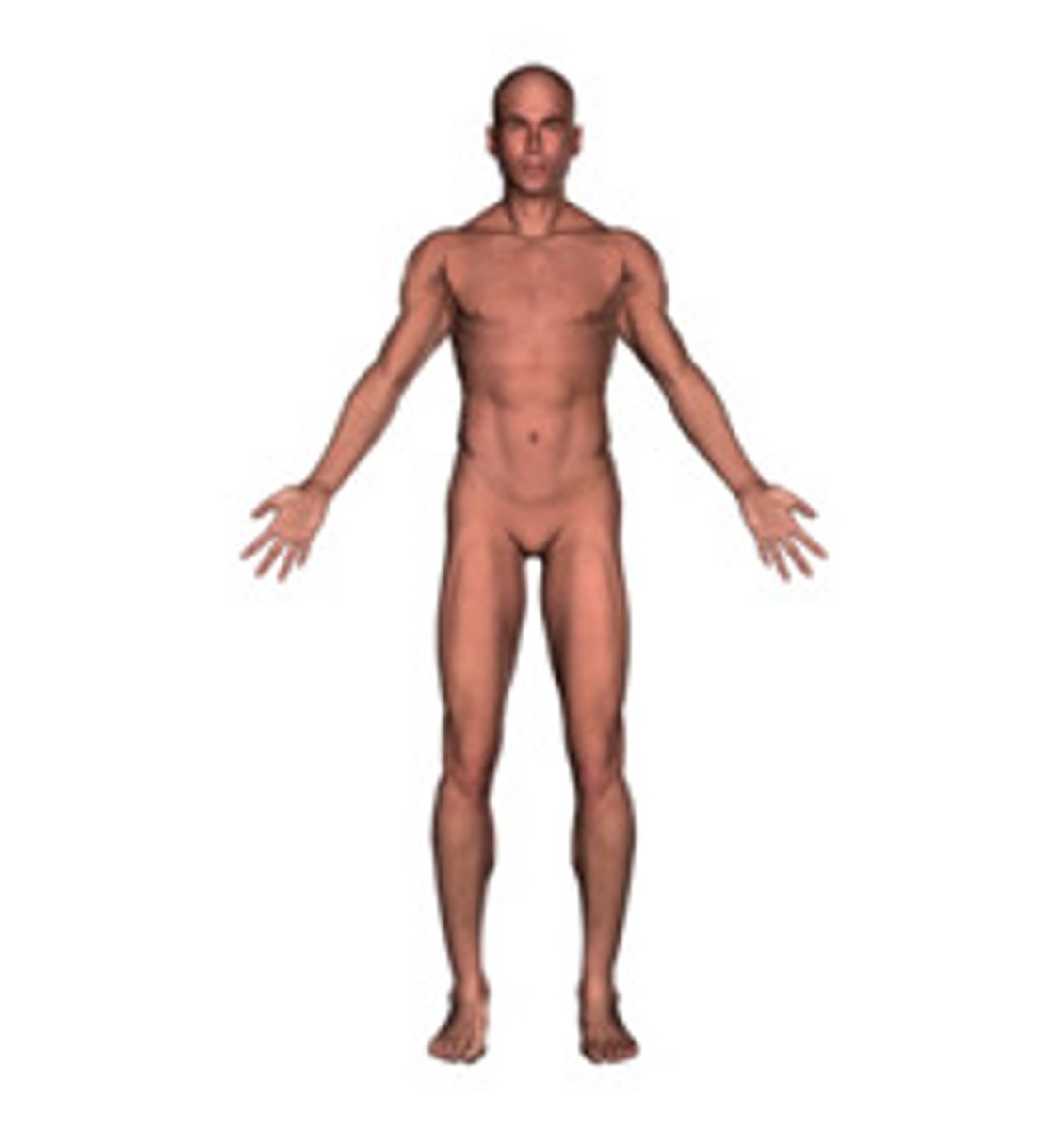
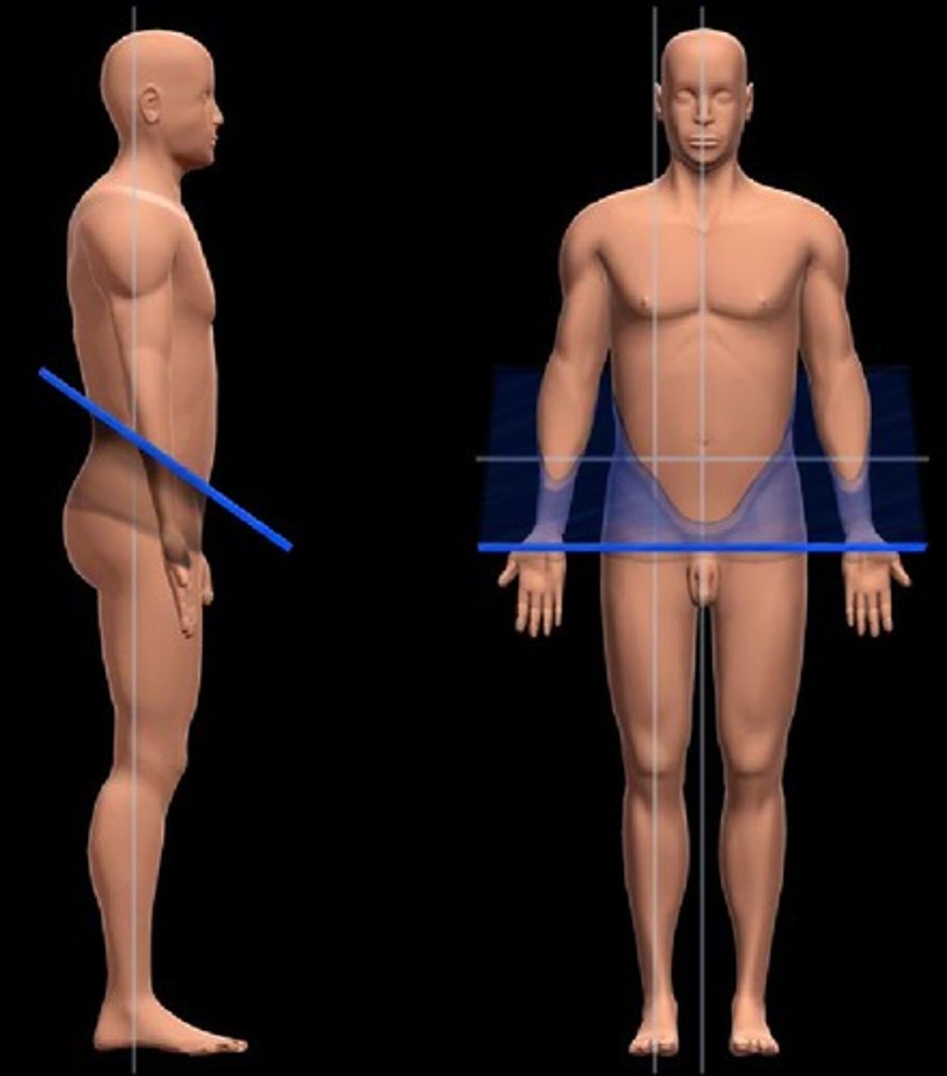
anatomical position
An individual in anatomical position is standing erect with arms at sides, palms facing forward with fingers pointing downward, feet parallel to each other and flat on the floor, and eyes directed forward
prone
Position of the body when lying face down
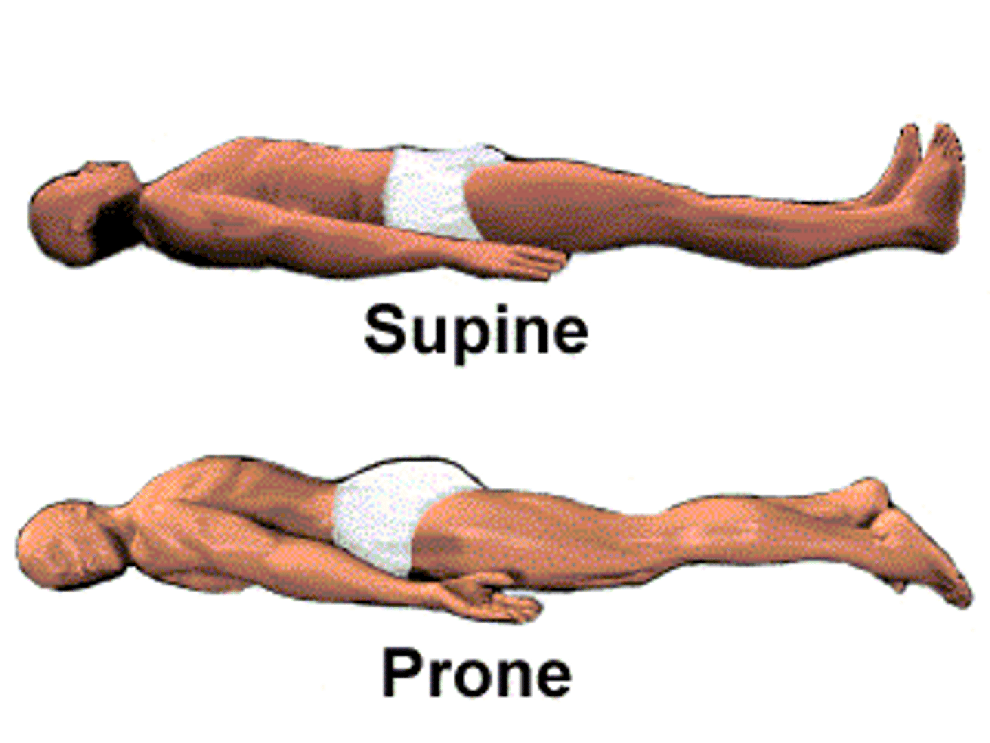
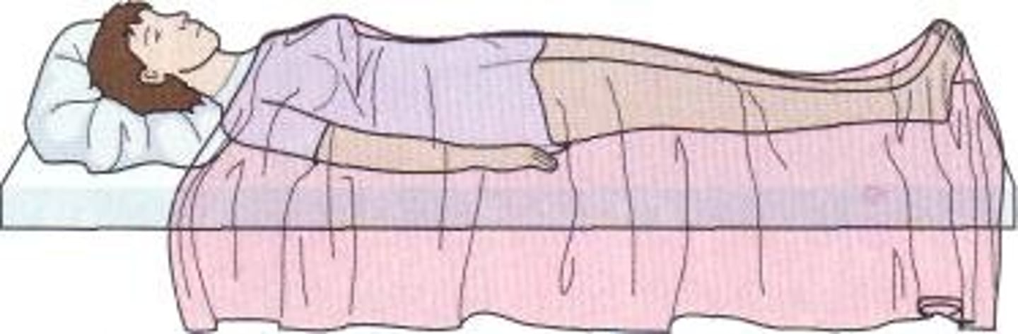
supine
Position of the body when lying face up
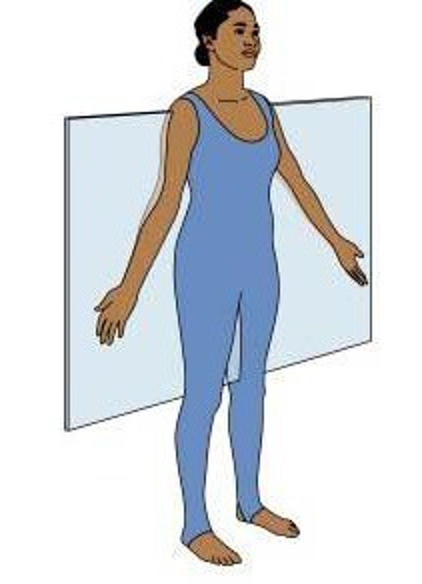
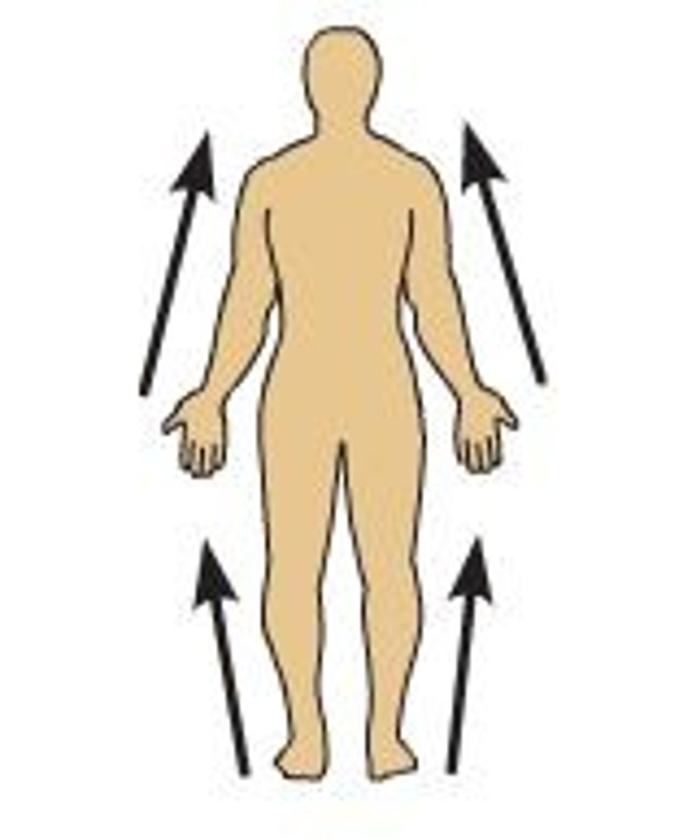
coronal plane
A plane that passes side-to-side through the body, dividing it into anterior and posterior portions (also called frontal plane)
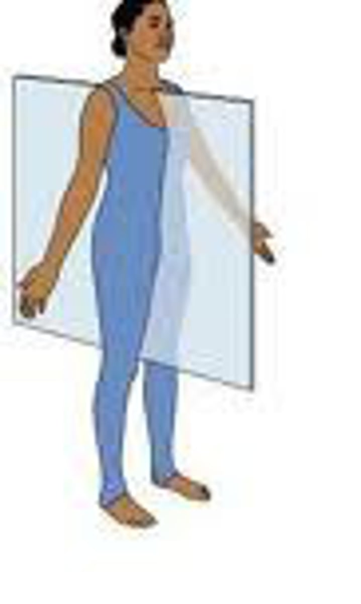
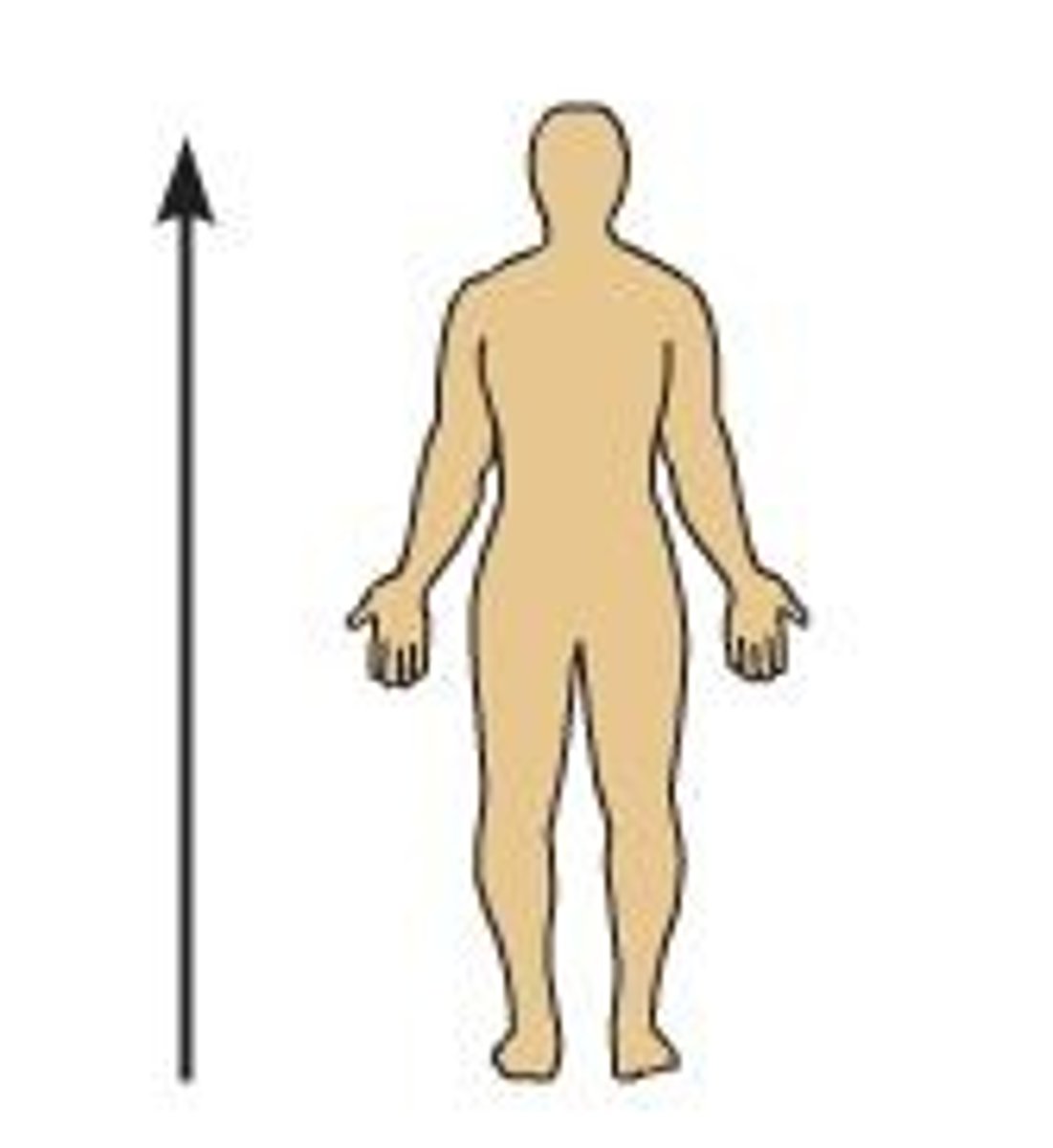
midsagittal plane
A plane that passes from front to back through the midline of the body, dividing it into right and left equal halves (median plane; same as sagittal)

oblique plane
A slanted plane (i.e., not horizontal or vertical) that passes through the body
sagittal plane
A plane that passes from front to back through the body, dividing it into right and left portions
Description:
A vertical plane that passes parallel to the long axis of the body, dividing it into right and left portions
Comment:
Sometimes called parasagittal plane
Median (midsagittal) plane passes through midline of body and divides it into equal right and left halves
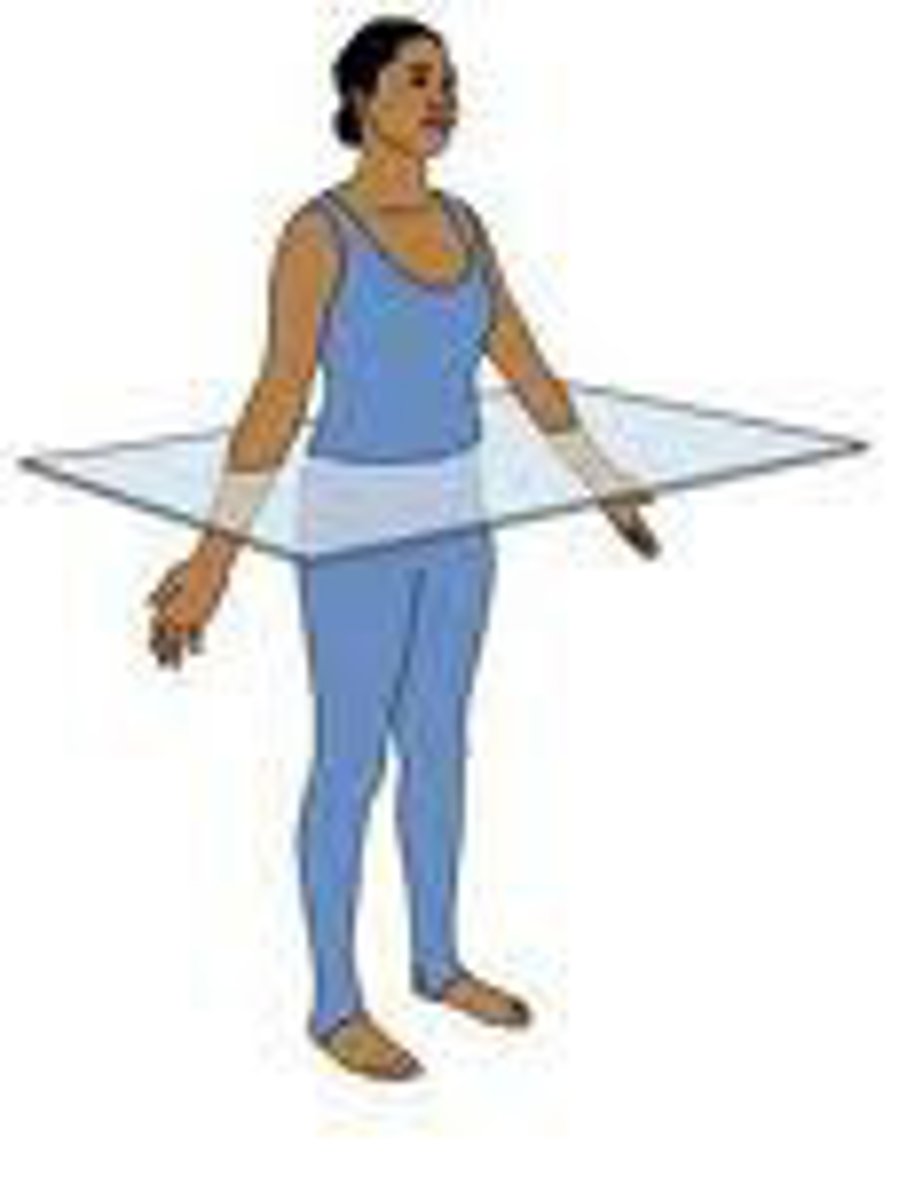
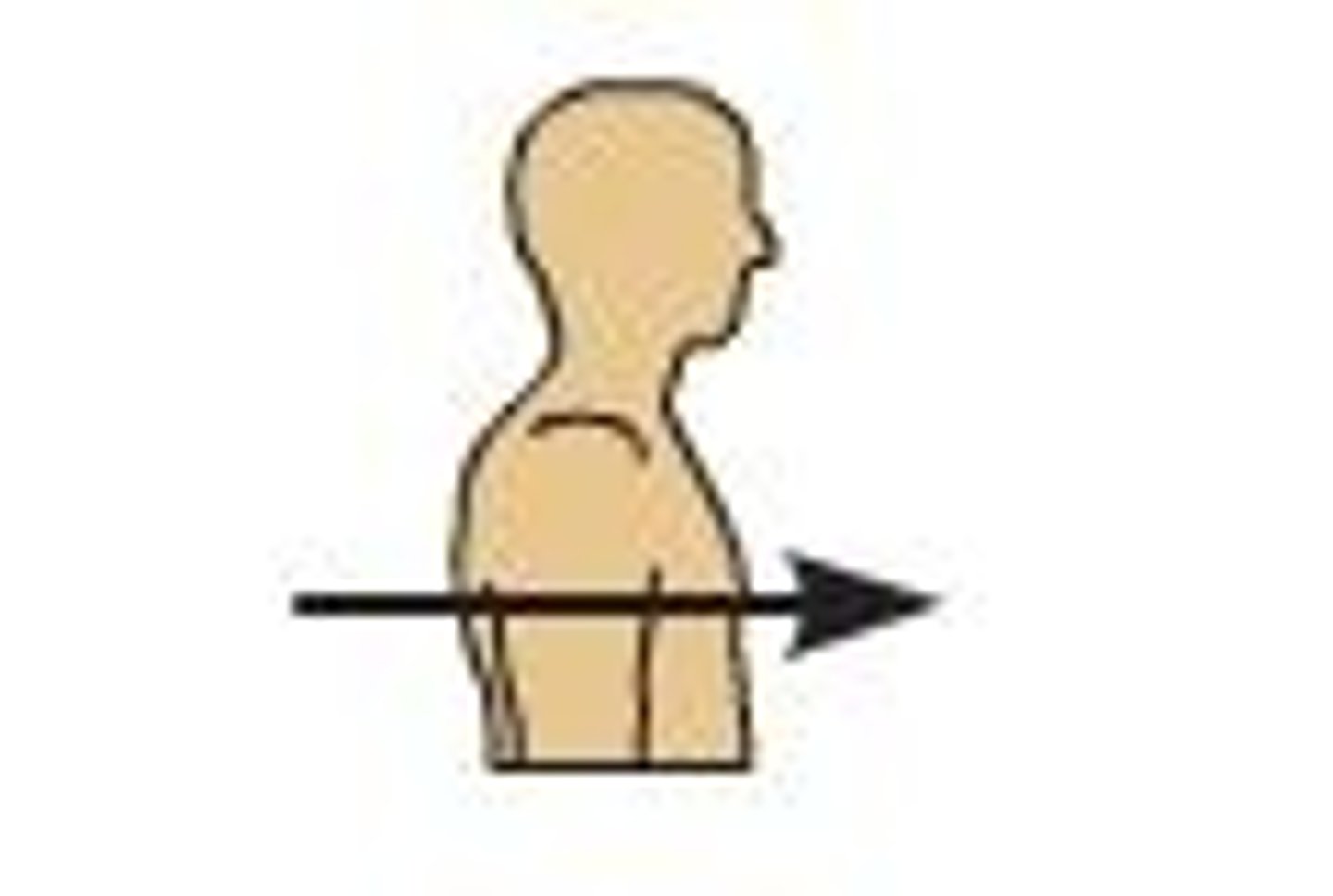
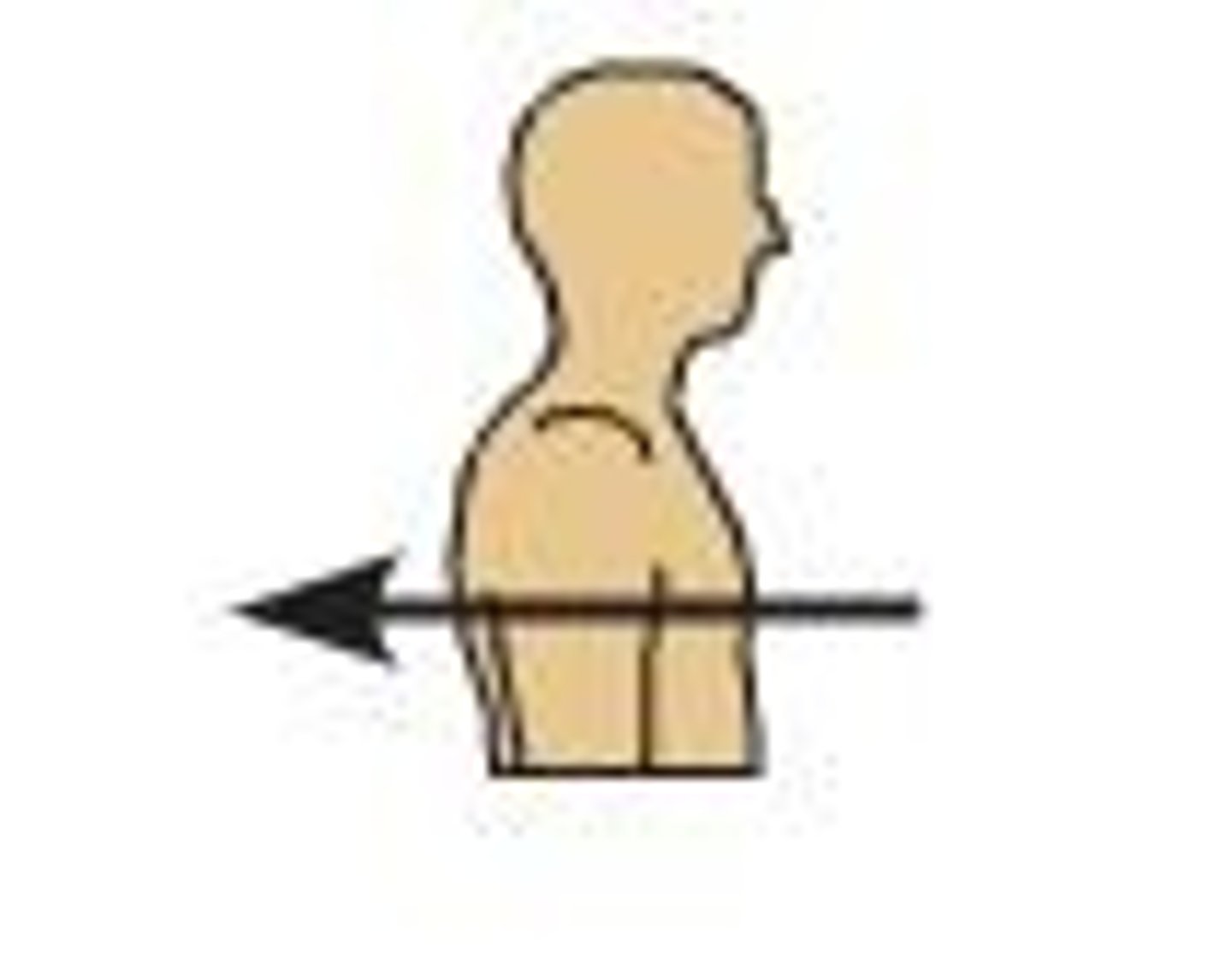
transverse plane
A plane that passes perpendicular to the long axis of the body, dividing it into superior and inferior portions (horizontal/cross-section)
Description:
A horizontal plane that passes perpendicular to the long axis of the body, dividing it into superior and inferior portions
Comment:
Also called horizontal plane or cross-section
Transverse planes can also be named for specific landmarks that they pass through, e.g. subcostal plane, transumbilical plane, and intertubercular plane
anterior
-Description:
Toward the front of the body (e.g., the sternum is anterior to the heart)
Opposite of posterior
-Comment:
Ventral, sometimes used synonymously with anterior, relates to the belly
deep
-Away from the surface of the body (e.g., in kidney, the medulla is deep to the cortex)
-Opposite of superficial
distal
-Farther from trunk or origin of a structure (e.g., the wrist is distal to the elbow)
-Opposite of proximal
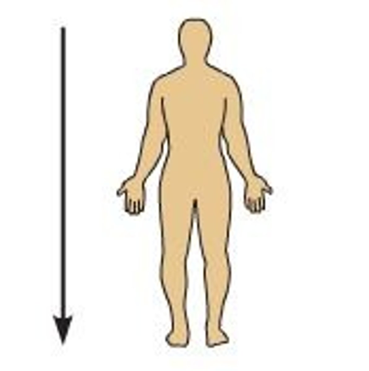
inferior
-Description:
Downward or below (e.g., the diaphragm is inferior to the heart)
Opposite of superior
-Comment:
In humans, synonymous with caudal (toward the tail)
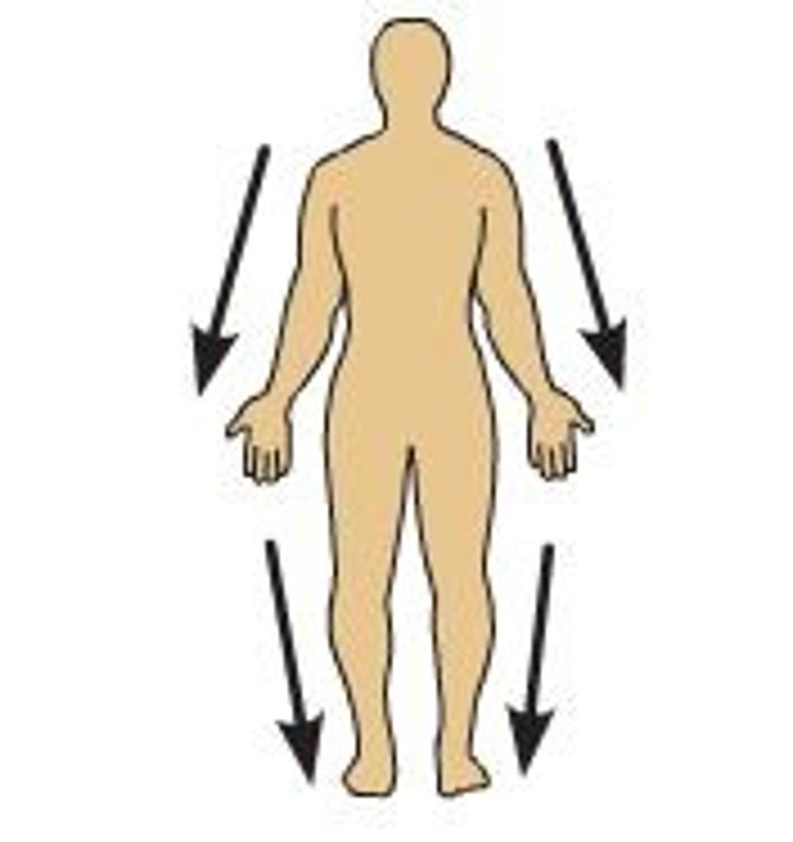
lateral
-Away from the midline of the body (e.g., the lungs are lateral to the heart)
-Opposite of medial
medial
-Toward the midline of the body (e.g., the heart is medial to the lungs)
-Opposite of lateral
posterior
-Toward the back of the body or relating to the back (e.g., the heart is posterior to the sternum)
Opposite of anterior
-Comment:
Dorsal, sometimes used synonymously with posterior, relates to the back (L. dorsum = back of the body)
proximal
-Closer to trunk or origin of a structure (e.g., the elbow is proximal to the wrist)
-Opposite of distal
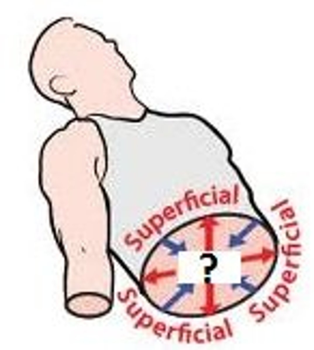
superficial
Toward the surface of the body (e.g., in kidney, the cortex is superficial to the medulla)
Opposite of deep

superior
Description:
Upward or above (e.g., the heart is superior to the diaphragm)
Opposite of inferior
Comment:
Cranial, sometimes used synonymously with superior, relates to the head (L. cranium = head of the body)
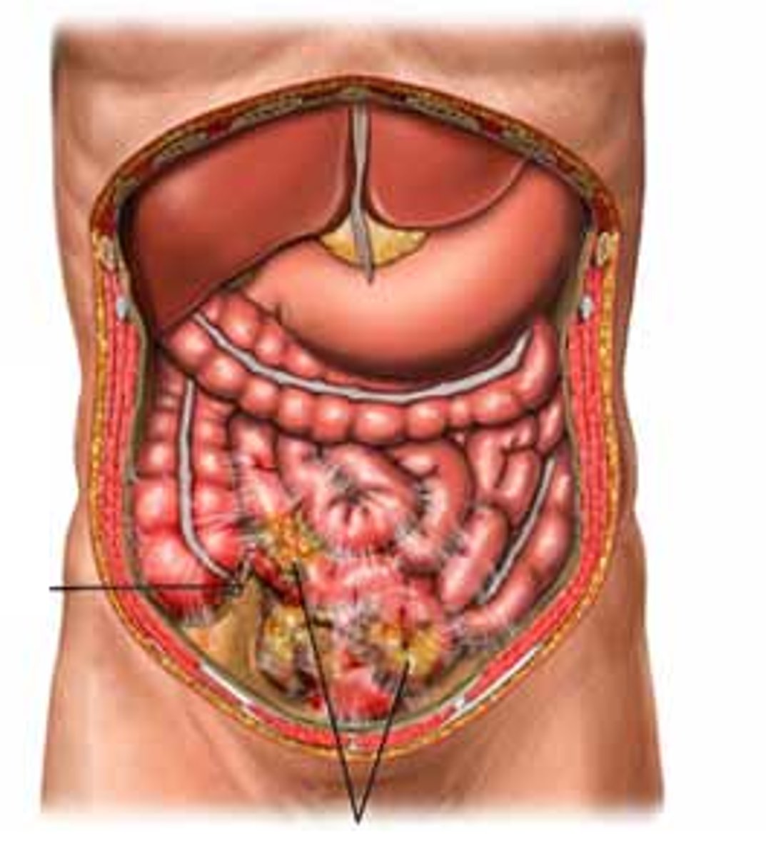
abdominal cavity
-Location:
Abdominal region
-Description:
Bounded by abdominal walls, thoracic diaphragm (superior), and pelvic brim (inferior)
Major organs include: stomach, intestines, liver, gallbladder, spleen, pancreas, kidneys and ureters, suprarenal glands, aorta, inferior vena cava, and lumbar nerve plexus
abdominopelvic region
-Location:
Trunk, inferior to thoracic region
-Description:
Topographic (surface) subdivision of trunk
ankle region
-Location:
Foot
-Description:
Subdivision of foot
Skin, muscles and tendons, nerves, and vessels around distal ends of tibia and fibula, and talus

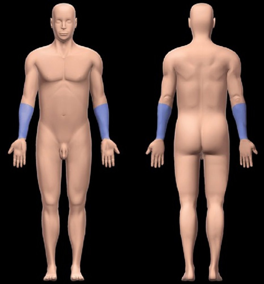
antebrachial region
-Location:
Upper limb (distal)
Between elbow and wrist joints
-Description:
Subdivision of upper limb
Skin, muscles, nerves, and vessels around radius and ulna
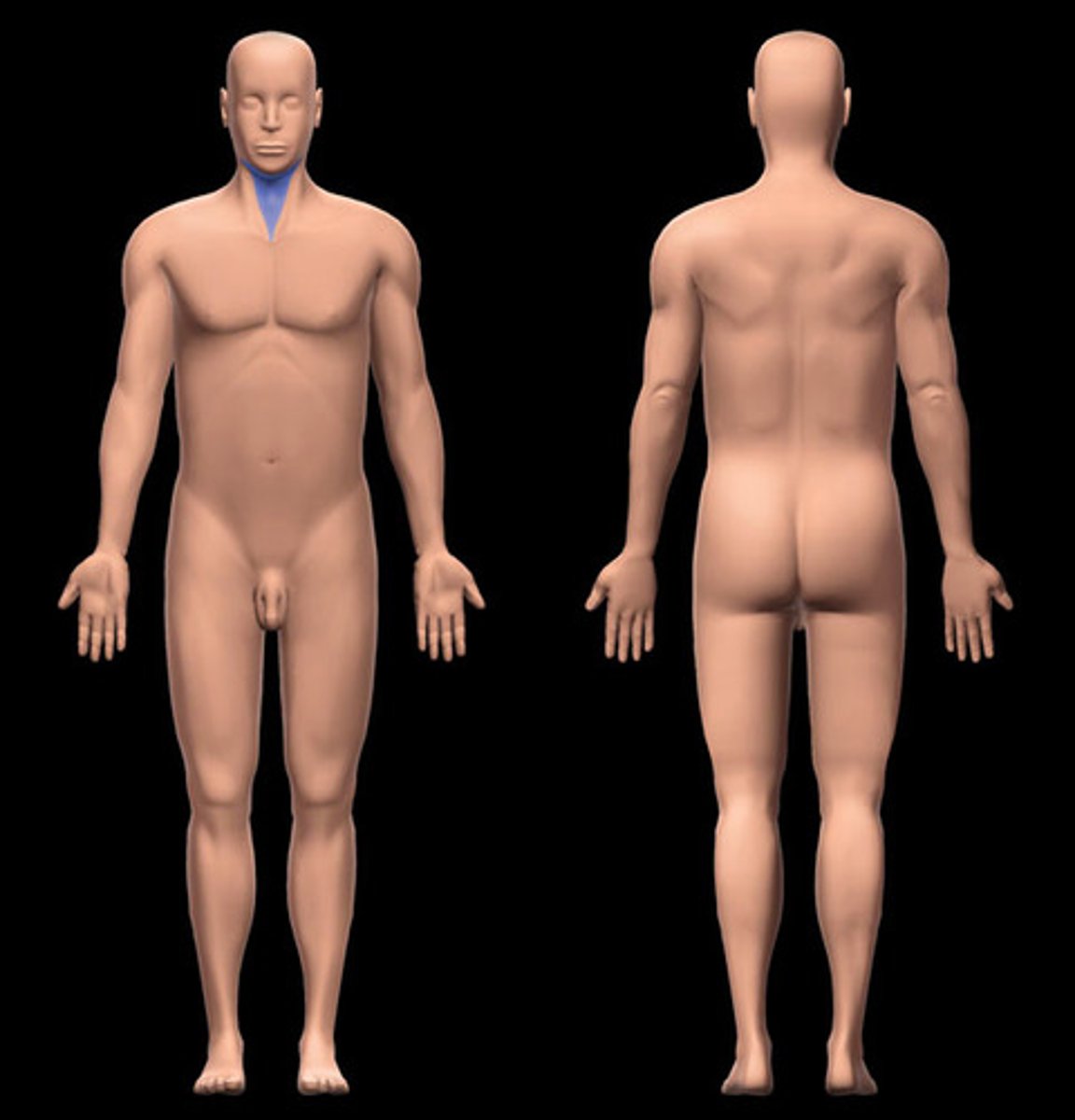
anterior cervical region
-Location:
Neck
Anterior to sternocleidomastoid region
-Description:
Anterior subdivision of neck
Anterior boundary: midline of neck
Posterior boundary: anterior edge of sternocleidomastoid
Superior boundary: inferior edge of mandible
Apex: jugular notch
Roof: subcutaneous tissue (with platysma muscle) and investing layer of deep cervical fascia
Floor: pharynx, larynx, and thyroid gland
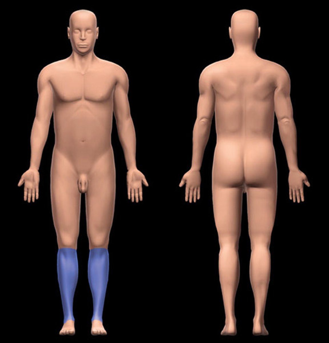
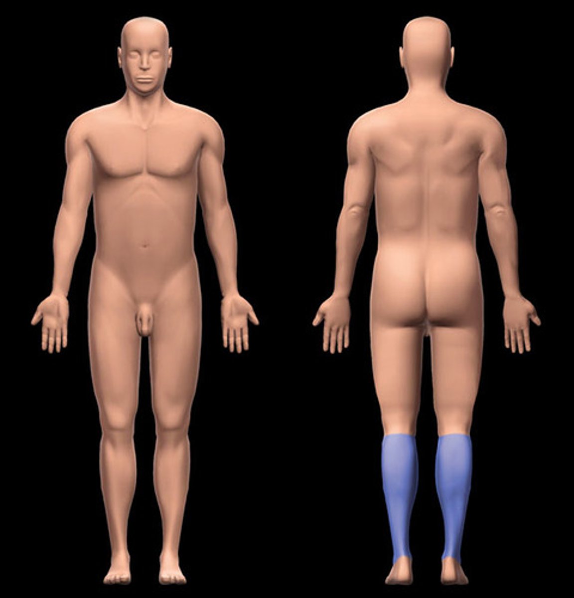
anterior leg region
-Location:
Leg (anterior)
-Description:
Subdivision of leg
Includes anterior and lateral muscular compartments
Anterior border of shaft of tibia (shin) is subcutaneous
Skin, muscles, tendons, nerves, and vessels anterior to intermuscular septum of leg and interosseous membrane of leg
auricular region
-Location:
Head (lateral cranial region)
-Description:
Region of external ear

axillary region
Location:
Thoracic region (inferior to shoulder joint)
Between upper arm and lateral thoracic wall
Description:
Subdivision of thoracic region that includes axilla
Axilla is a pyramidal space
Anterior boundary: pectoralis muscles and clavipectoral fascia
Posterior boundary: scapula and subscapularis, teres major, and latissimus dorsi muscles
Medial boundary: serratus anterior and upper lateral thoracic wall (ribs 1-4 and intercostal muscles)
Lateral boundary: humerus (intertubercular sulcus)
Base: skin, subcutaneous tissue, and axillary (deep) fascia
Apex: passage between neck and upper limb (cervicoaxillary canal) formed by rib 1, clavicle, and superior border of scapula
Contents of axilla: axillary artery, vein, and lymph nodes; infraclavicular part of brachial plexus; and areolar tissue
back
-Location:
Trunk (posterior)
Between neck region and tip of coccyx
-Description:
Formed by skin and subcutaneous tissue, muscles, vertebral column (inferior to cervical region), ribs (thoracic region), and spinal cord and meninges
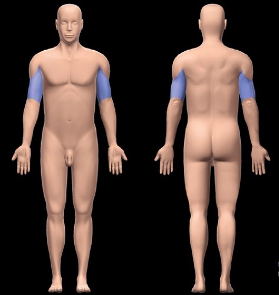
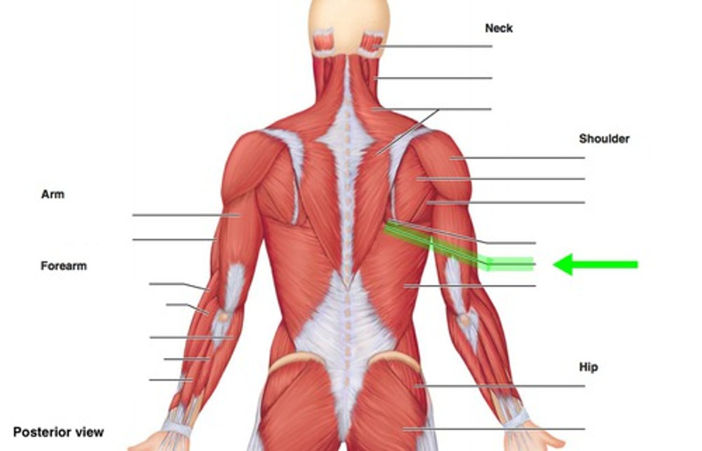
brachial region
-Location:
Upper limb (proximal)
Between glenohumeral (shoulder) and elbow joints
-Description:
Subdivision of upper limb
Skin, muscles, nerves, and vessels around humerus
buccal region
-Location:
Head (lateral face)
-Description:
Region of cheek
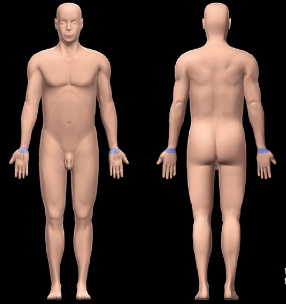
carpal region
-Location:
Hand
-Description:
Proximal subdivision of hand
Skin, muscles, nerves, and vessels around carpal bones and distal ends of radius and ulna
cranial region
-Location:
Head (superior part)
-Description:
Region of cranial cavity (i.e., surrounding brain)
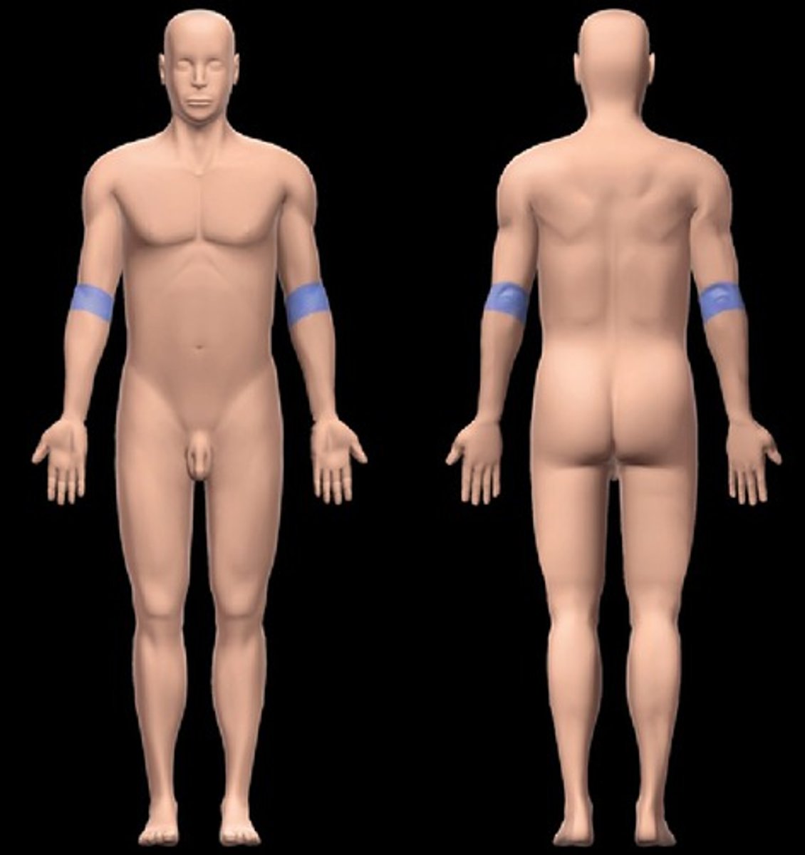
cubital region
-Location:
Upper limb
Anterior and posterior aspects of elbow
-Description:
Subdivision of upper limb
Skin, muscles, nerves, and vessels around distal end of humerus and proximal ends of radius and ulna
Includes cubital fossa, a shallow triangular depression on anterior aspect of elbow
Important structures related to cubital fossa include: median cubital vein, brachial artery and its primary branches (e.g., radial and ulnar arteries), median and radial nerves, and biceps brachii tendon
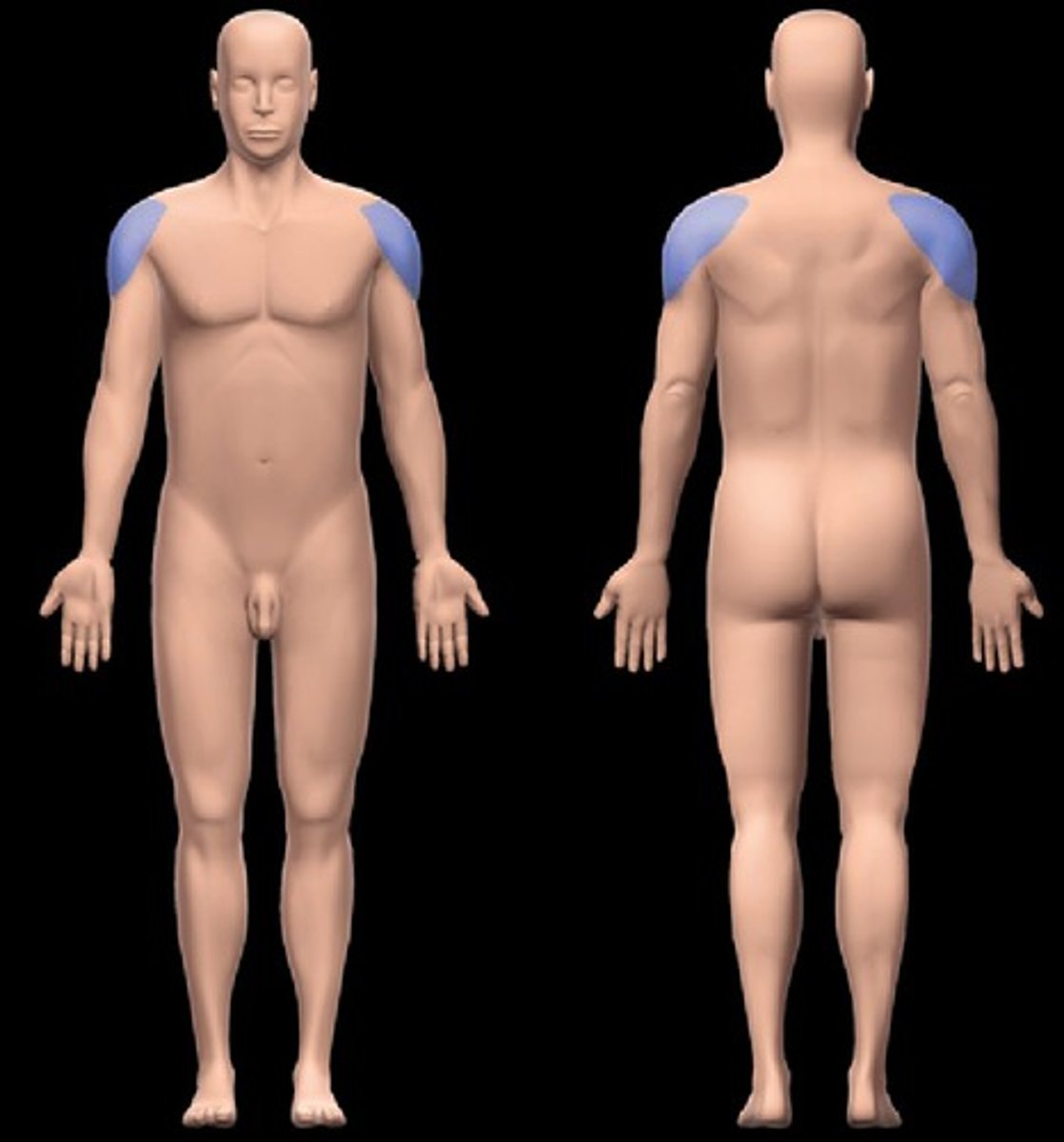
deltoid region
-Location:
Upper limb (proximal end)
-Description:
Subdivision of upper limb over deltoid muscle
Skin, muscles, nerves, and vessels around glenohumeral joint
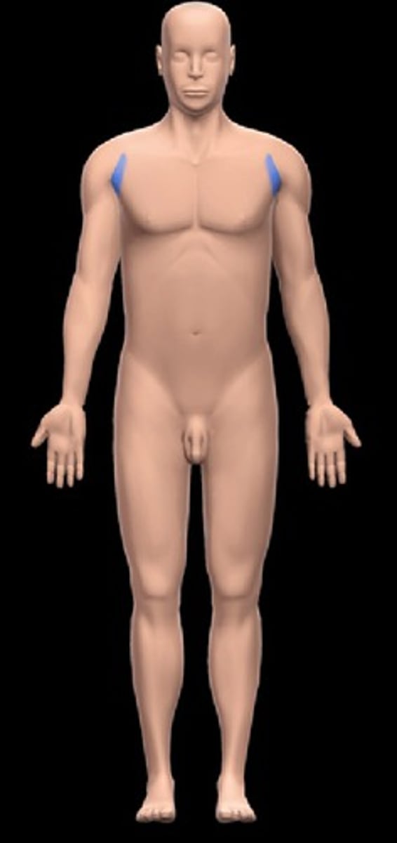
deltopectoral triangle
-Location:
Anterior shoulder
-Description:
Narrow, triangular groove bordered by clavicle, deltoid, and pectoralis major muscles

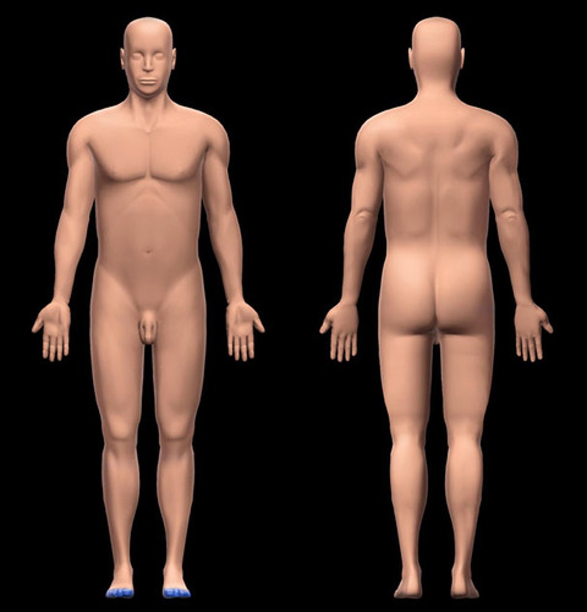
digits of foot
-Location:
Foot (distal end)
-Description:
Distal subdivision of foot
Includes: great (digit 1 or hallux), second (digit 2), third (digit 3), fourth (digit 4), and fifth or little (digit 5) toes
Skin, muscle tendons, nerves, and vessels around phalanges of toes
digits of hand
-Location:
Hand (distal end)
-Description:
Distal subdivision of hand
Includes: thumb (digit 1 or pollex), and index (digit 2), middle (digit 3), ring (digit 4), and little (digit 5) fingers
Skin, muscle tendons, nerves, and vessels around phalanges of fingers

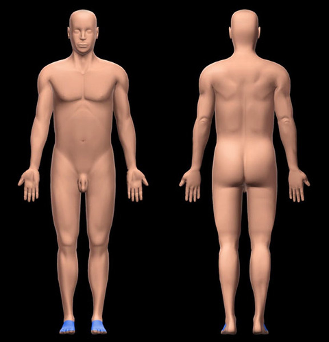
dorsum of foot
-Location:
Foot (dorsal)
-Description:
Dorsal aspect of foot (i.e., directed superiorly in anatomical position)
Skin, muscle tendons, nerves, and vessels on dorsal aspect of foot
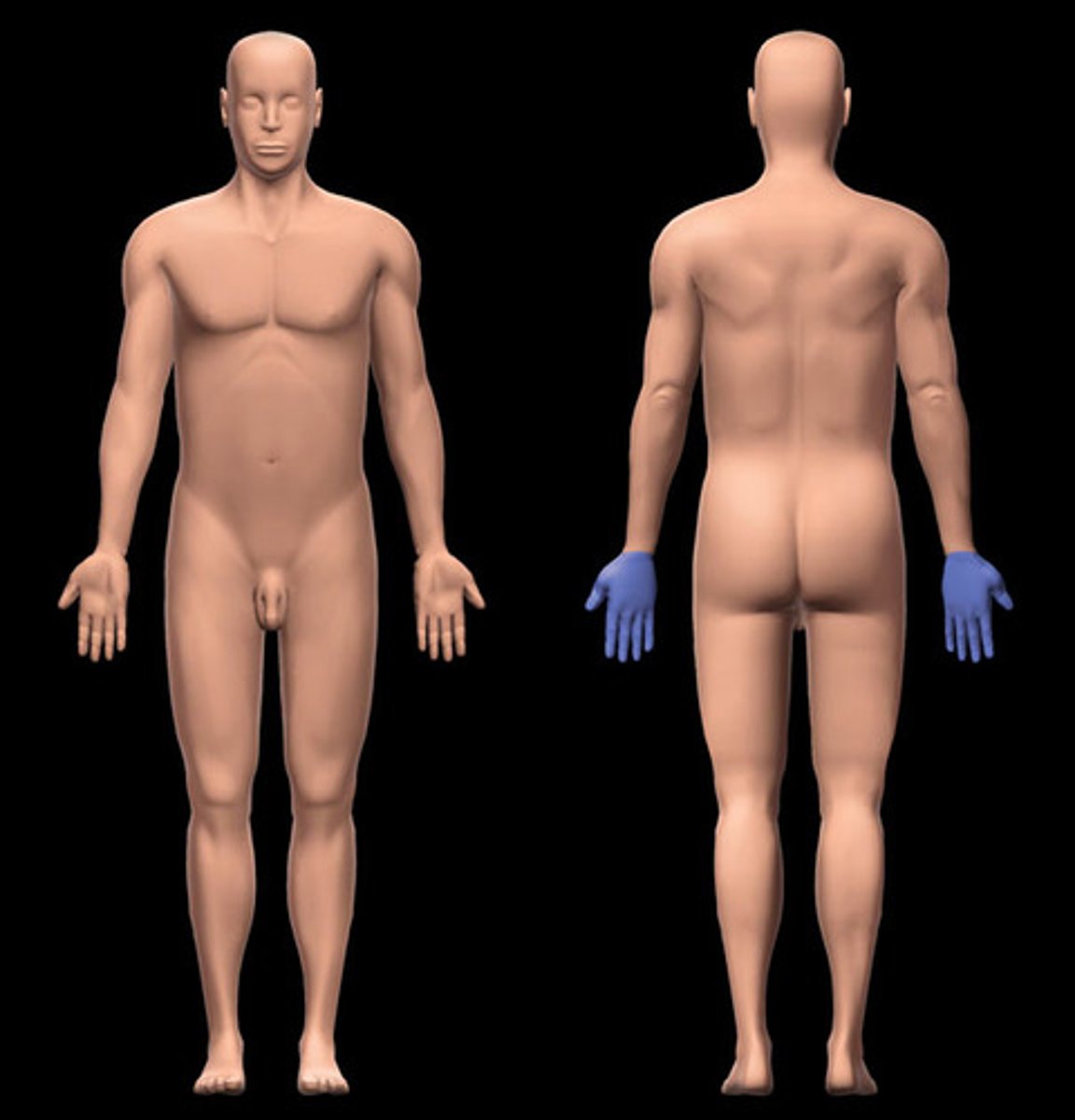
dorsum of hand
-Location:
Hand (proximal)
-Description:
Subdivision of hand
Subdivisions include: carpal and metacarpal regions, and digits
Skin, muscles and tendons, nerves, and vessels on posterior aspect of carpal bones, metacarpals, and phalanges
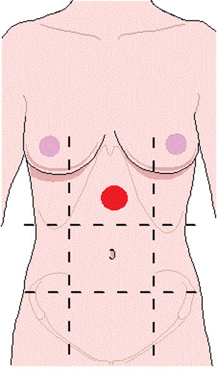
epigastric region
-Location:
Abdominal wall (anterior)
-Description:
One of nine regions of abdominal cavity
Upper median region (flanked by right and left hypochondriac regions)
Contents include suprarenal glands and parts of stomach, large intestine, liver and gallbladder, and pancreas
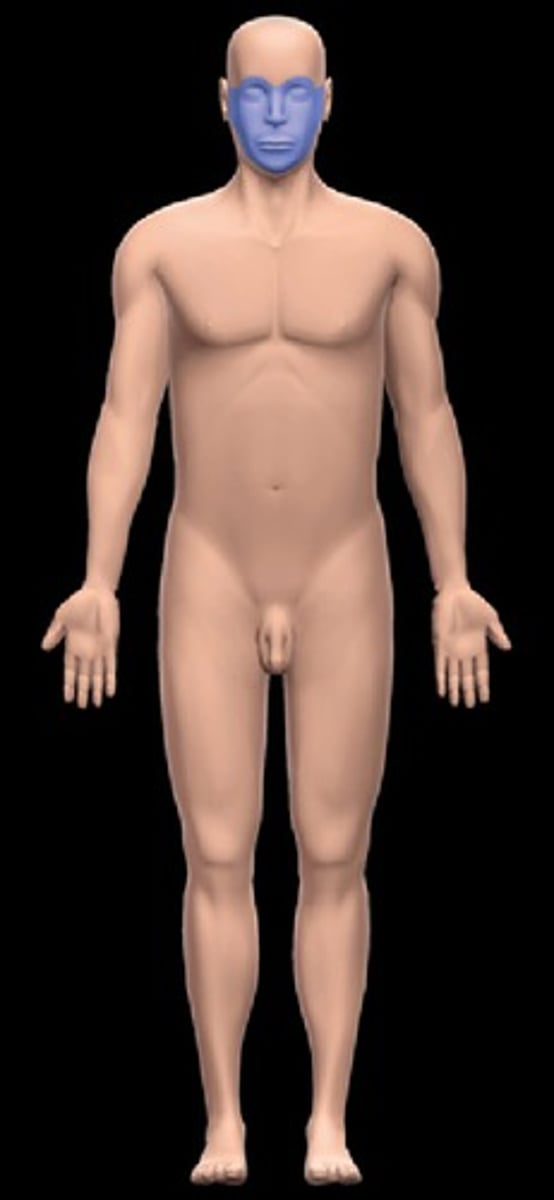
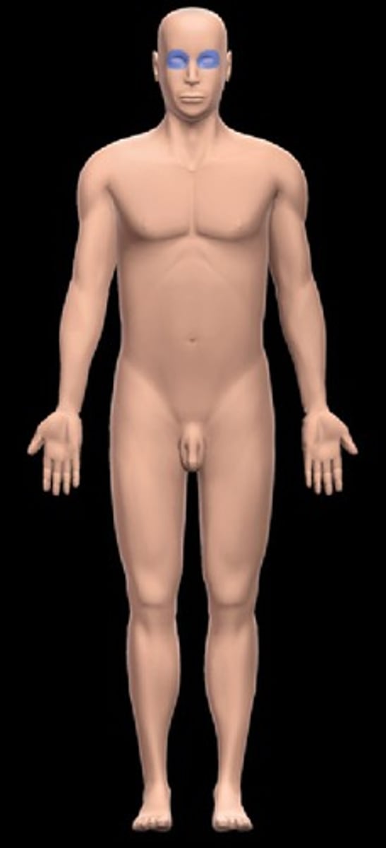
facial region
-Location:
Head (anterior inferior part)
-Description:
Region of face, including: eyes, nose, mouth, cheeks, and chin
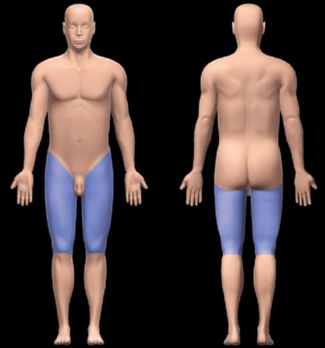
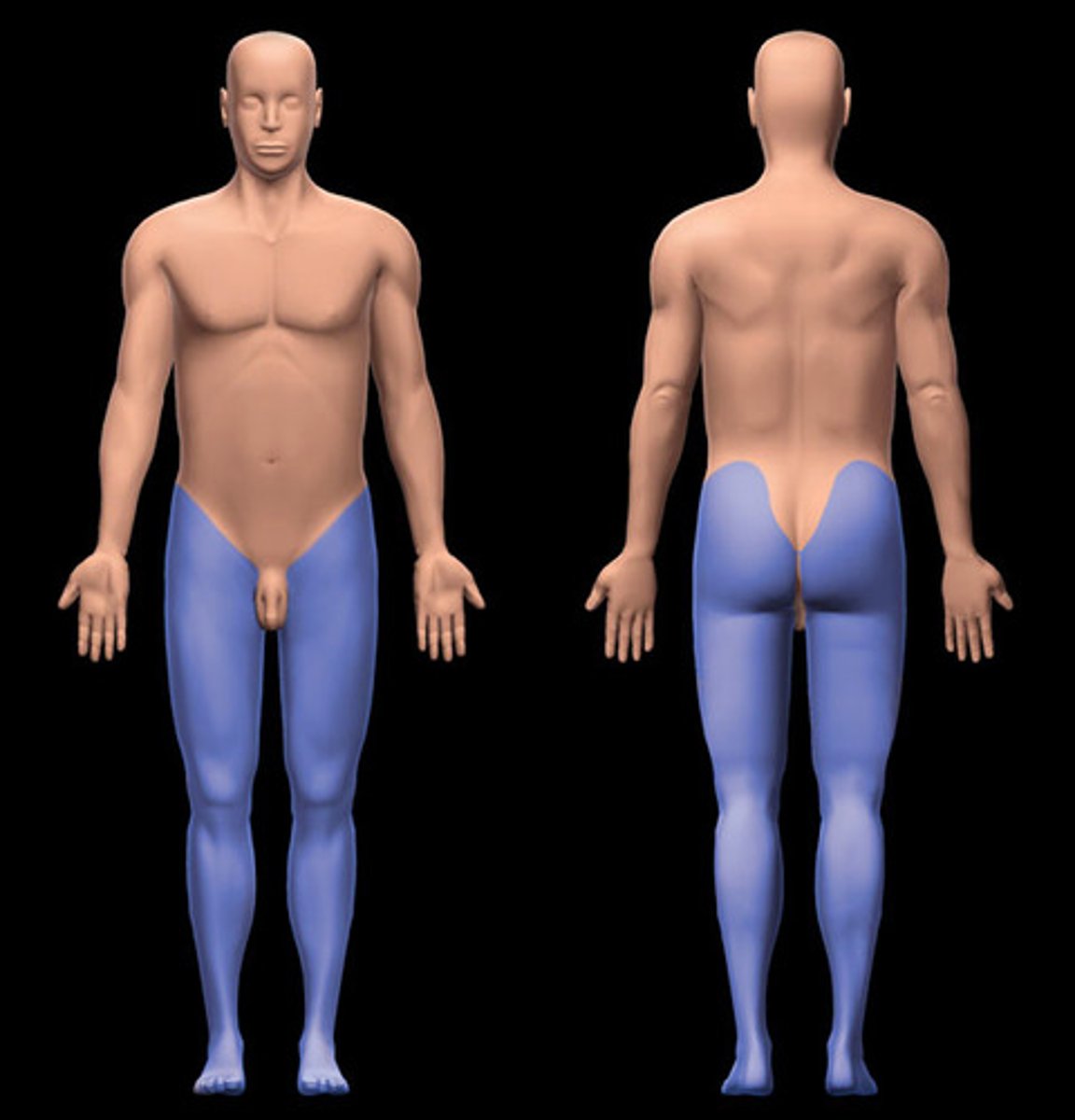
femoral region
-Location:
Lower limb (proximal)
-Description:
Subdivision of lower limb
Skin, muscle tendons, nerves, and vessels around femur
foot region
--Location:
Lower limb (distal end)
-Description:
Subdivision of lower limb
Subdivisions include: ankle, heel, dorsum, sole, metatarsal, and digits (toes)
Anatomical and functional subdivisions: hindfoot (talus and calcaneus), midfoot (navicular, cuboid, and cuneiforms), and forefoot (metatarsals and phalanges of toes)
Includes skin, muscles, tendons, nerves, and vessels around tarsal bones, metatarsals, and phalanges of toes

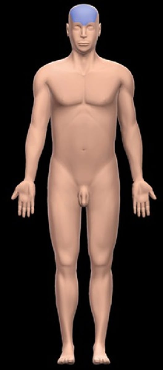
frontal region
-Location:
Head (anterior superior part of cranial region)
-Description:
Region of forehead
Related to frontal bone
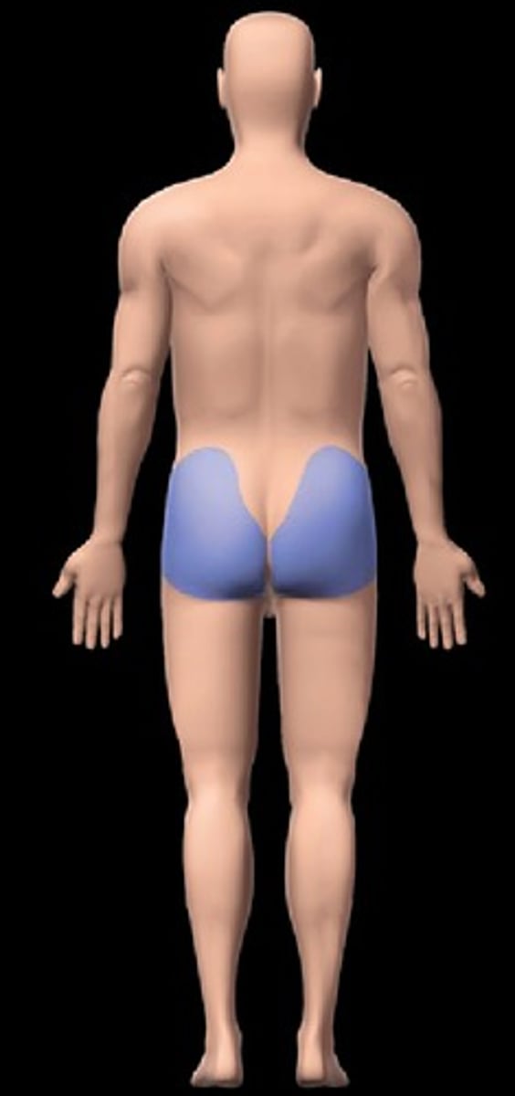
gluteal region
Location:
Lower limb (proximal end)
Description:
Subdivision of lower limb
Skin, muscle tendons, nerves, and vessels associated with posterior aspect of hip joint
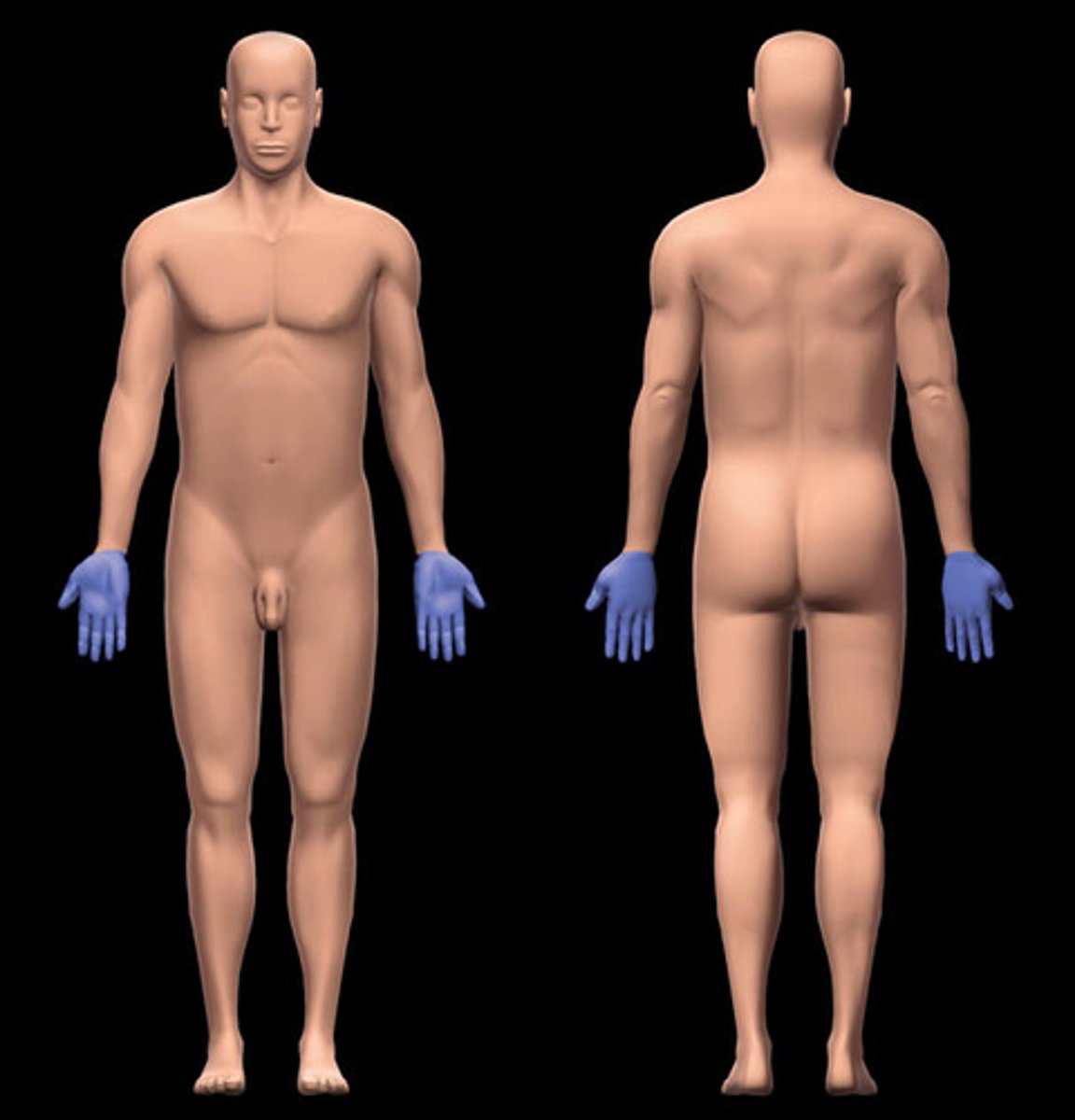
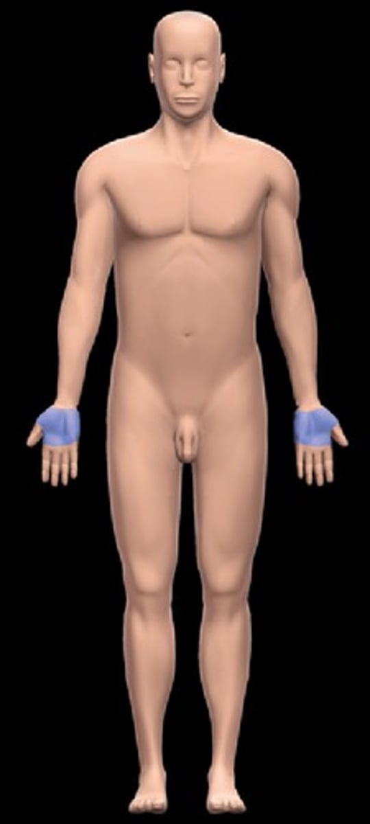
hand region
Location:
Upper limb (distal end)
Description:
Subdivision of upper limb
Subdivisions include: carpal (wrist), palmar, dorsum, and digits (fingers)
Includes skin, muscles, tendons, nerves, and vessels around carpal bones, metacarpals, and phalanges of fingers
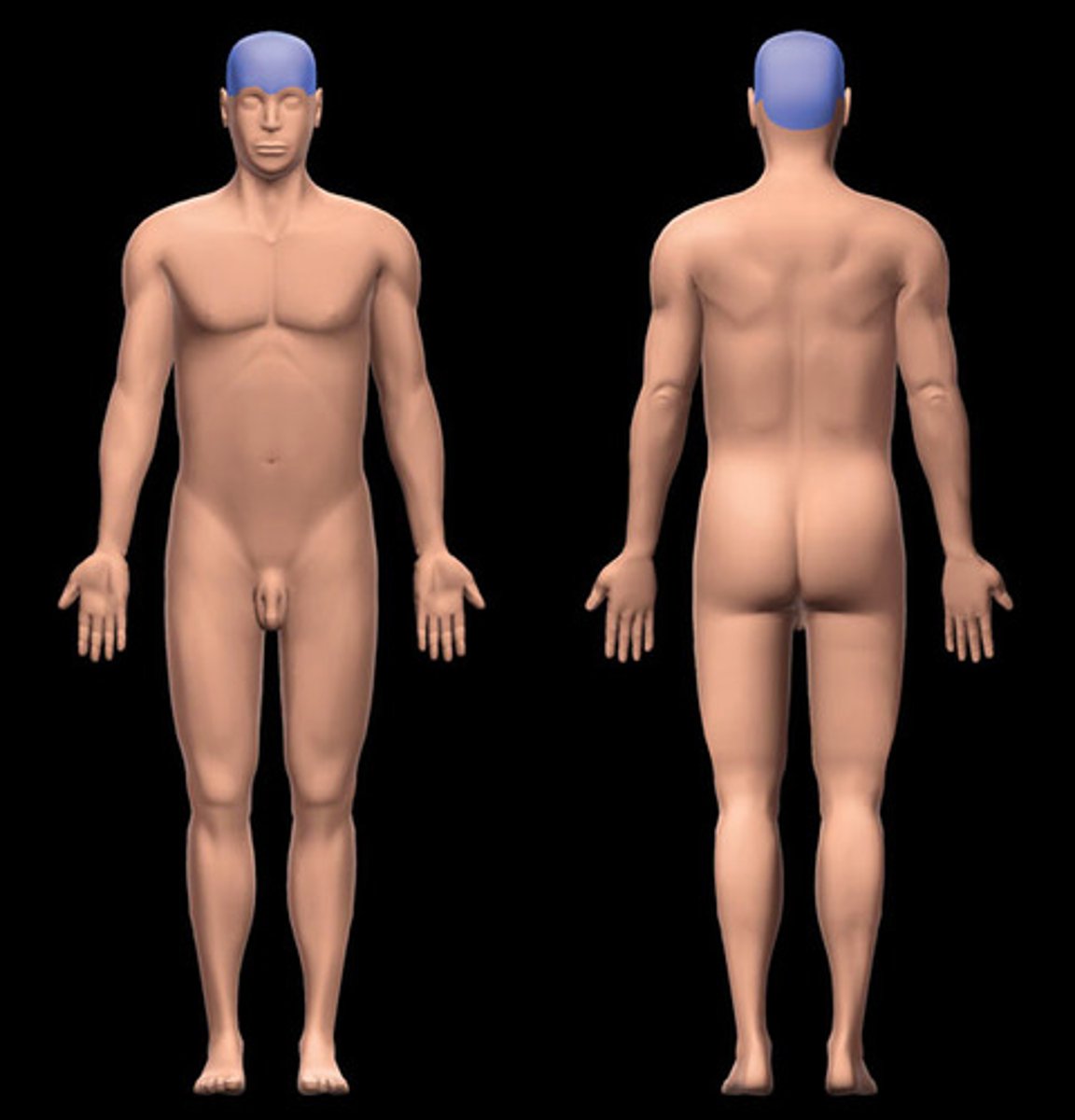
head
Location:
Superior to neck region
Description:
Skeleton includes skull and associated bones (hyoid and auditory ossicles)
Soft tissues and bones form cranial cavity, oral cavity, nasal cavities and paranasal sinuses, orbits, and ears
Contains brain and organs of special sense (vision, hearing and equilibrium, smell, and taste)
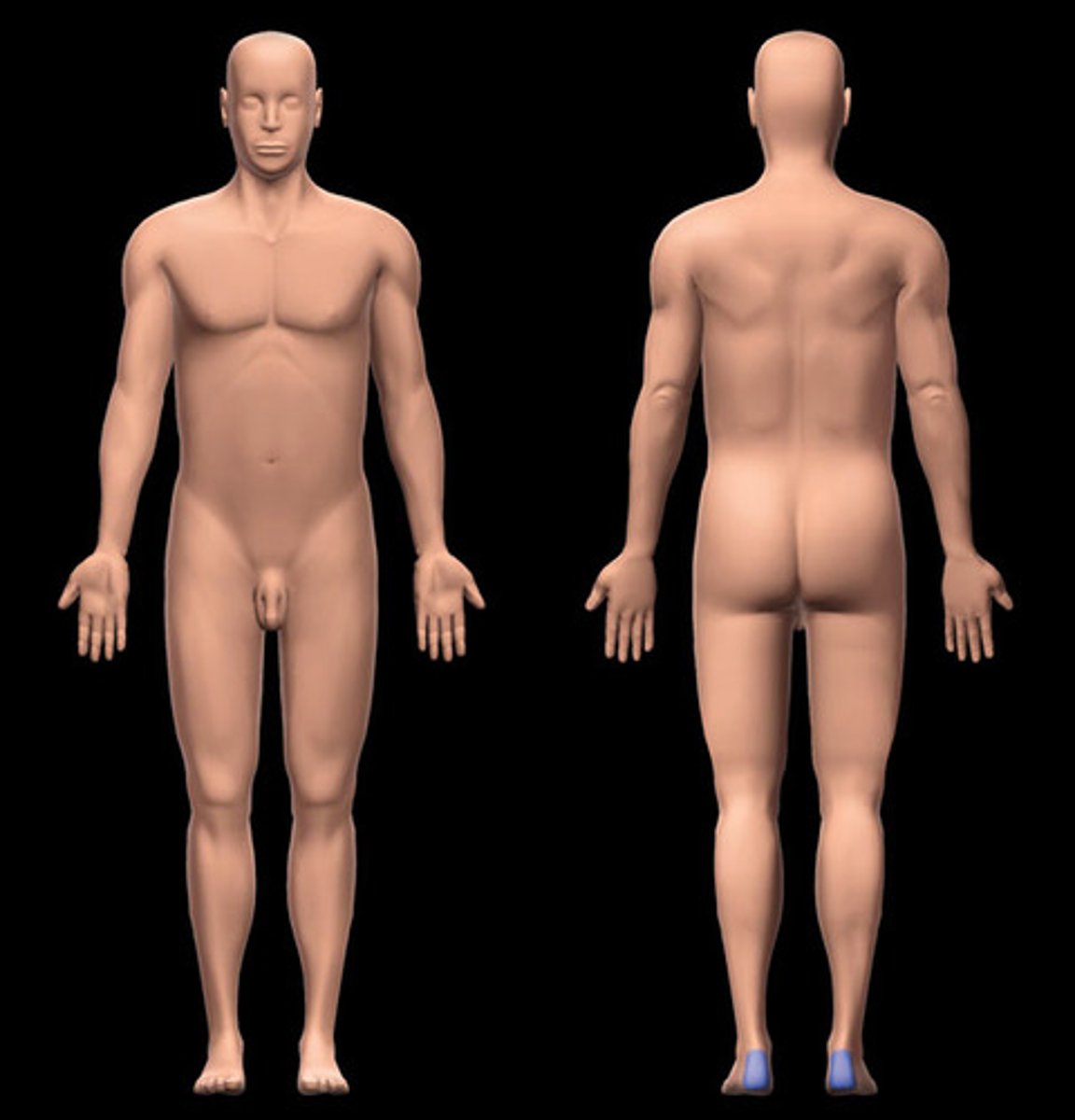
heel region
Location:
Foot (proximal)
Description:
Subdivision of foot
Skin, muscle tendons, nerves, and vessels around hindfoot (talus and calcaneus)
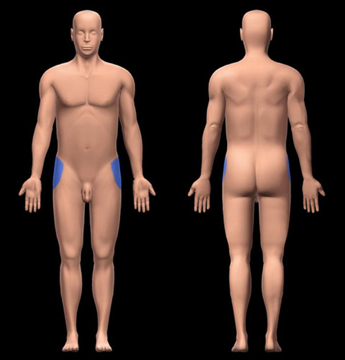
hip region
Location:
Lower limb (lateral proximal)
Description:
Lateral subdivision of lower limb from iliac crest ("waist") to proximal thigh
Skin, muscle tendons, nerves, and vessels around ilium, ischium, and proximal end of femur (especially head, neck, and greater trochanter)
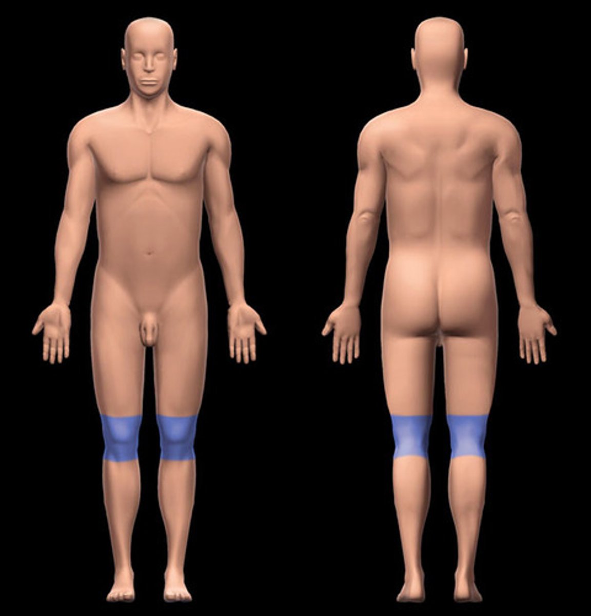
knee region
Location:
Lower limb (middle)
Description:
Subdivision of lower limb
Skin, muscles, nerves, and vessels around knee joint
Includes popliteal fossa on posterior aspect
lateral cervical region
Location:
Neck
Between posterior cervical and sternocleidomastoid (SCM) regions
Description:
Lateral subdivision of neck
Anterior boundary: posterior edge of SCM
Posterior boundary: anterior edge of trapezius
Inferior boundary: middle third of clavicle
Apex: superior nuchal line at junction of SCM and trapezius
Roof: subcutaneous tissue (with platysma muscle) and investing layer of deep cervical fascia
Floor: cervical muscles covered by prevertebral layer of deep cervical fascia

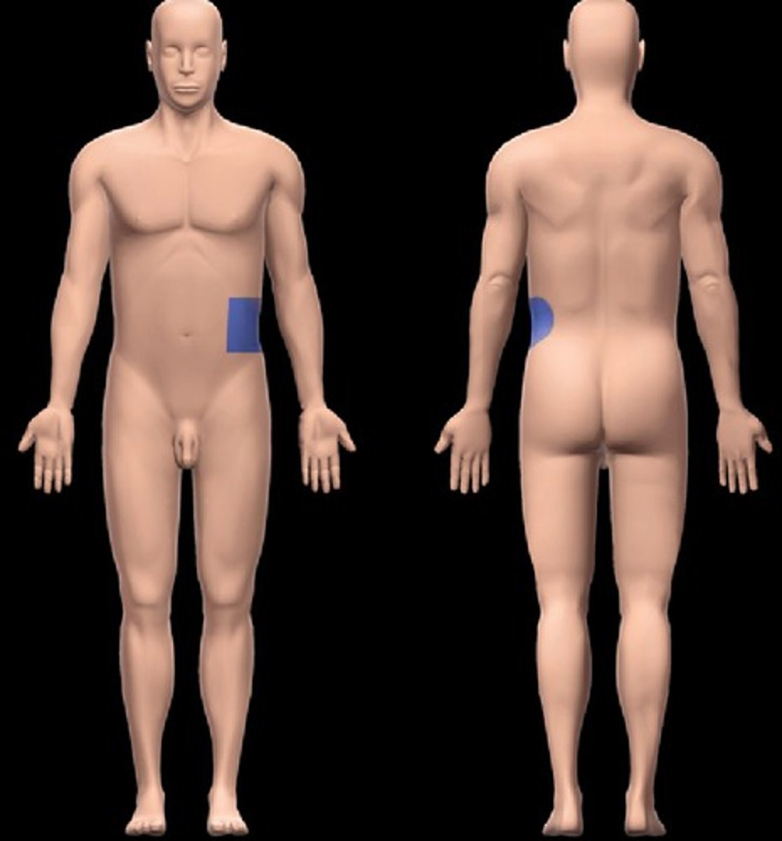
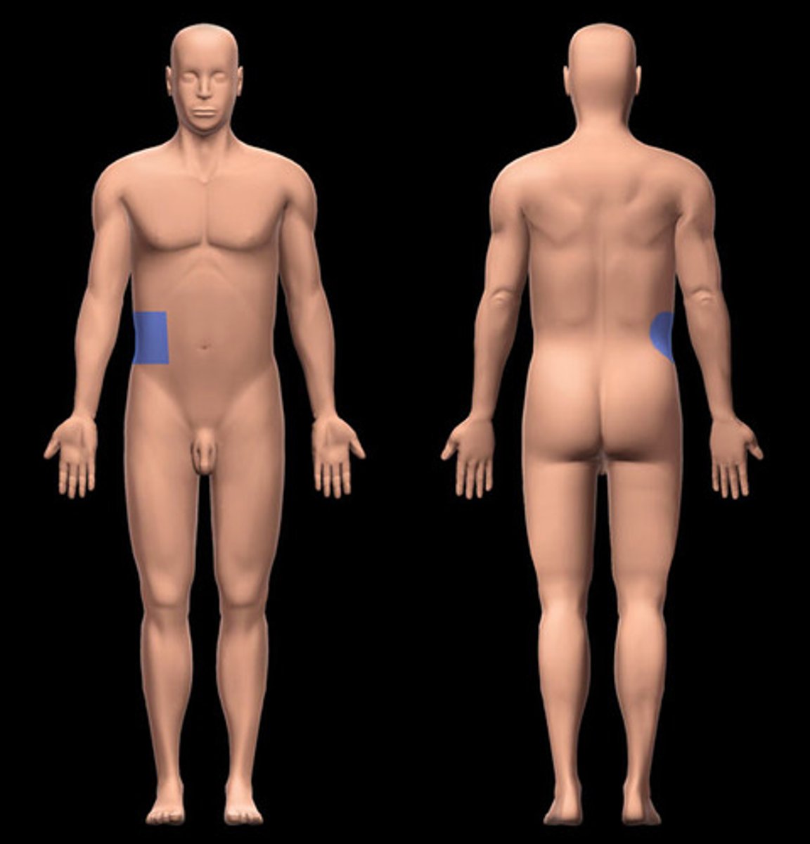
left flank region
Location:
Abdominal wall (anterior)
Description:
One of nine regions of abdominal cavity
Left lateral region
Contents include parts of small and large intestines, and left kidney
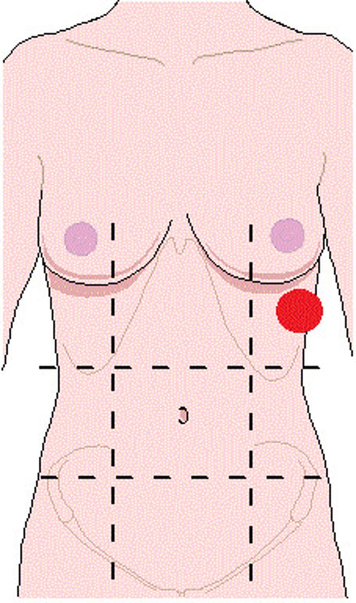
left hypochondriac region
Location:
Abdominal wall (anterior)
Description:
One of nine regions of abdominal cavity
Left upper lateral region
Contents include spleen and parts of stomach, large intestine, pancreas (tail), and left kidney
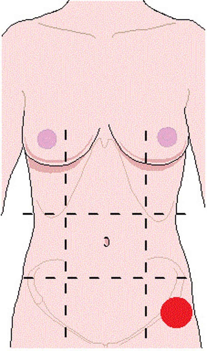
left inguinal region
Location:
Abdominal wall (anterior)
Description:
One of nine regions of abdominal cavity
Left lower lateral region
Contents include parts of small and large intestines
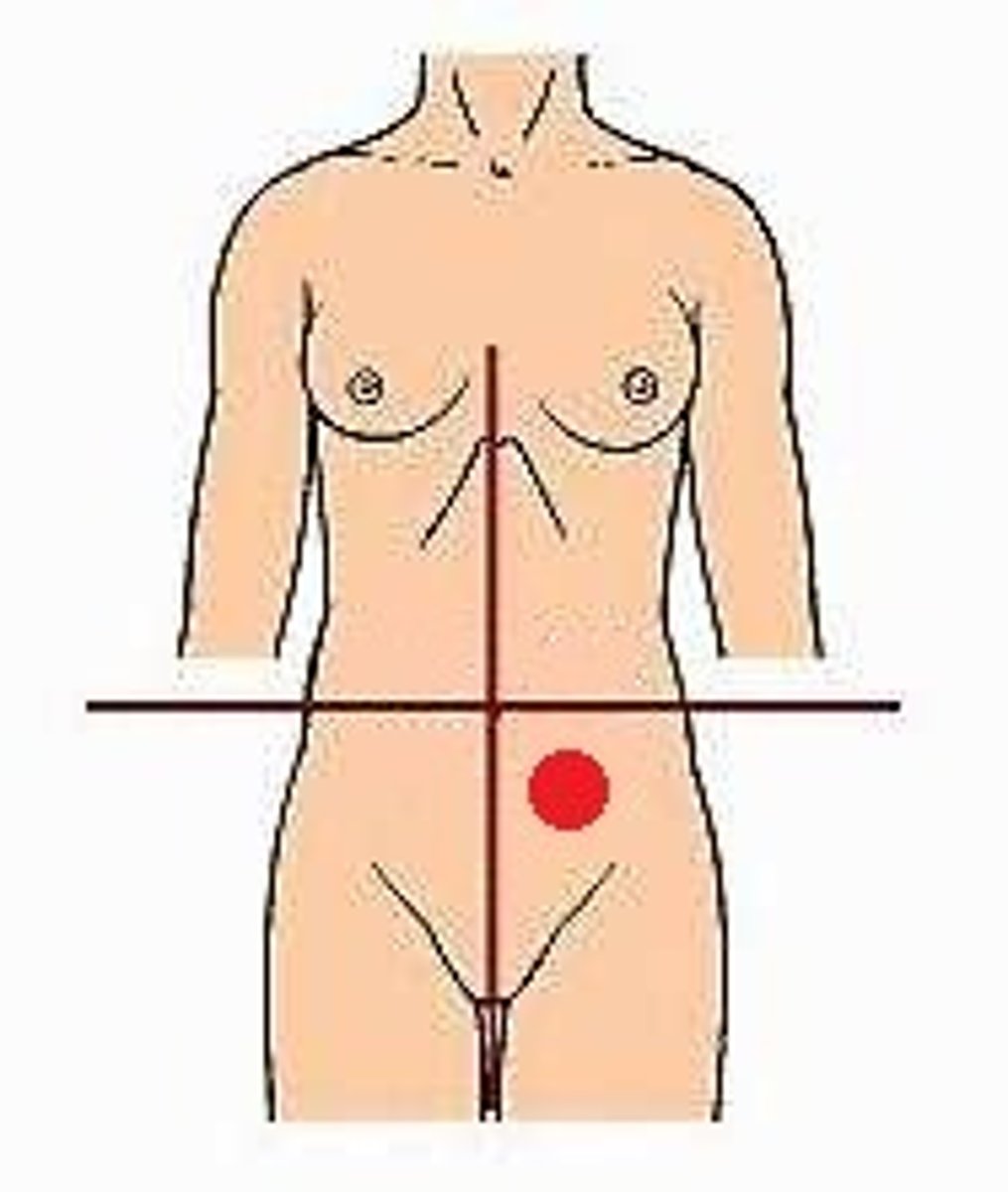
left lower quadrant
Location:
Left of midline, inferior to transverse plane through umbilicus
Description:
Lower left lateral area of abdominopelvic cavity
Contents include parts of small intestine, large intestine, urinary bladder (when distended), and left uterine tube and ovary
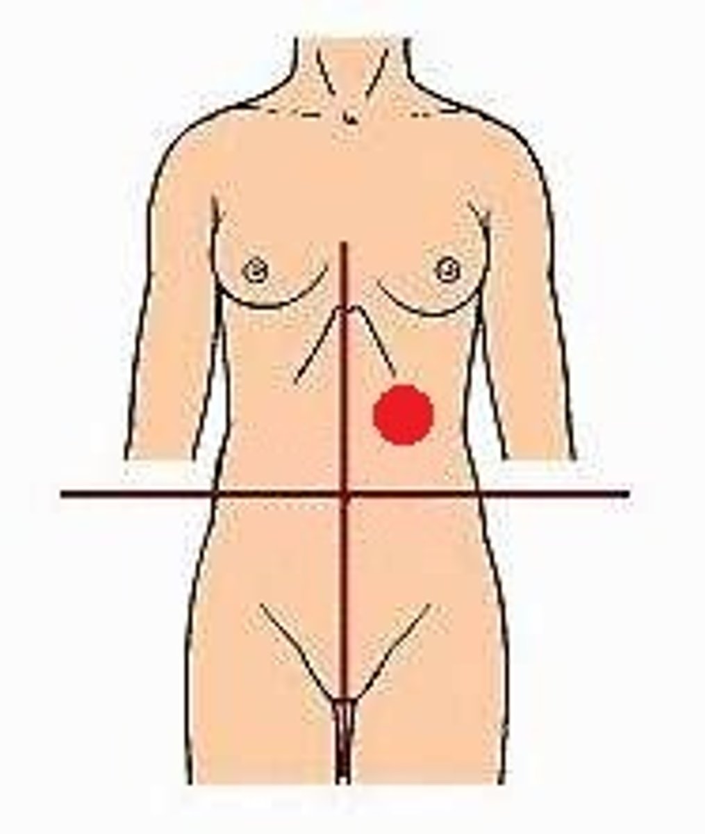
left upper quadrant
Location:
Left of midline, superior to transverse plane through umbilicus
Description:
Upper left lateral area of abdominopelvic cavity
Contents include spleen, left kidney and suprarenal gland, and parts of liver, stomach, pancreas, and small and large intestines
lower limb
Location:
Limb that, in anatomical position, extends inferiorly from trunk
Description:
Subdivisions: gluteal region (buttock), femoral region (thigh), leg, and foot (including ankle)
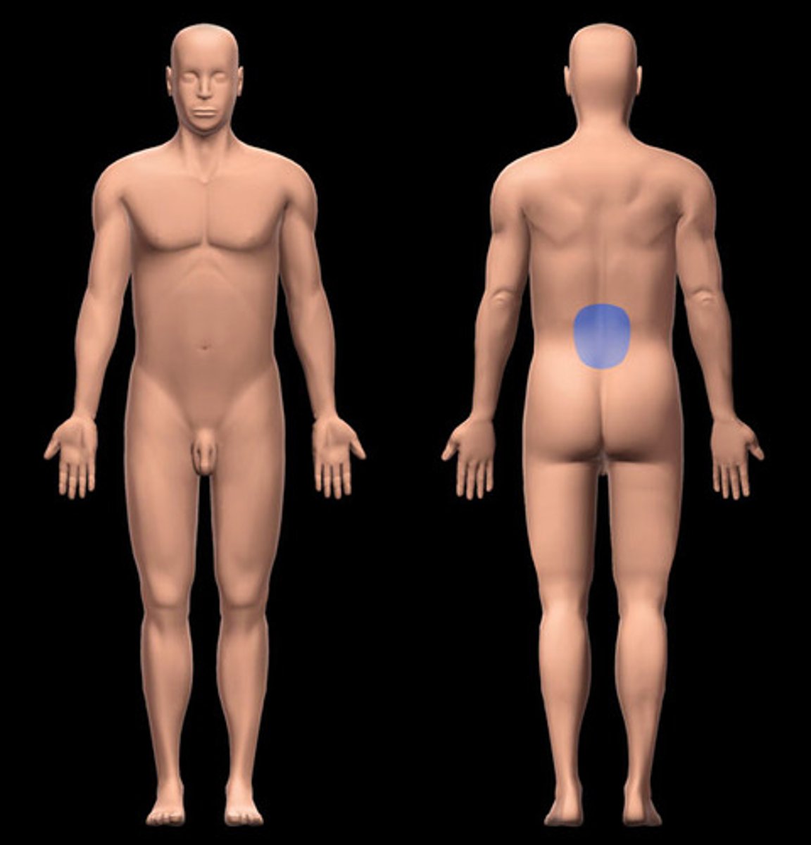
lumbar region
Location:
Back (inferior)
Description:
Subdivision of back
Includes lumbar vertebrae and attached muscles
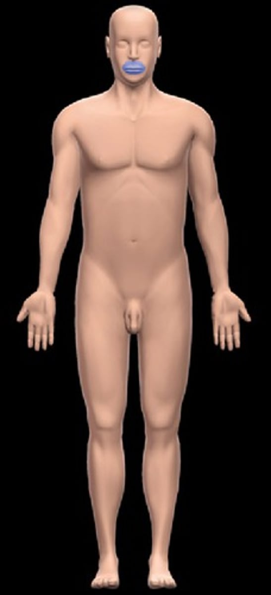
mental region
Location:
Head (anterior inferior part of facial region)
Description:
Region of chin
Related to anterior aspect of mandible

nasal region
Location:
Head (anterior)
Description:
Part of facial region that includes nose

neck
Location:
Between head (superiorly) and thorax and shoulders (inferiorly)
Description:
Connects head with upper limb and trunk
Skeleton formed by cervical vertebrae
Contains larynx and trachea, cervical part of spinal cord, parts pharynx and esophagus, and thyroid and parathyroid glands
Subdivided into anterior cervical, lateral cervical, posterior cervical, and sternocleidomastoid regions
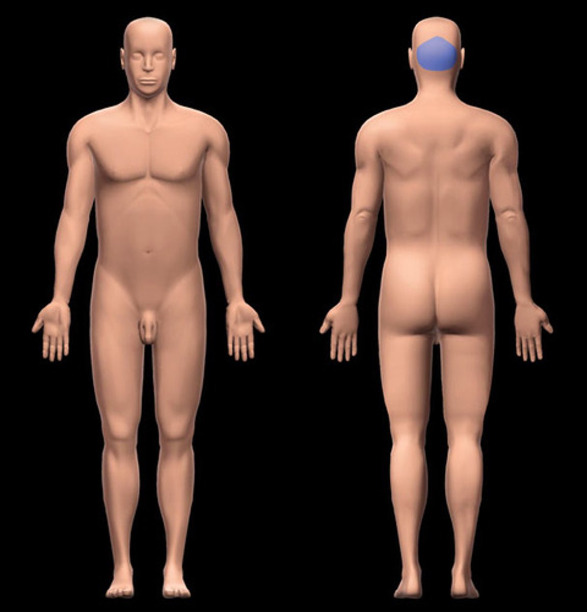
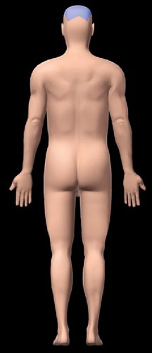
occipital region
Location:
Head (posterior)
Description:
Part of cranial cavity related to occipital bone
oral region
Location:
Head (anterior inferior facial region)
Description:
Region of mouth
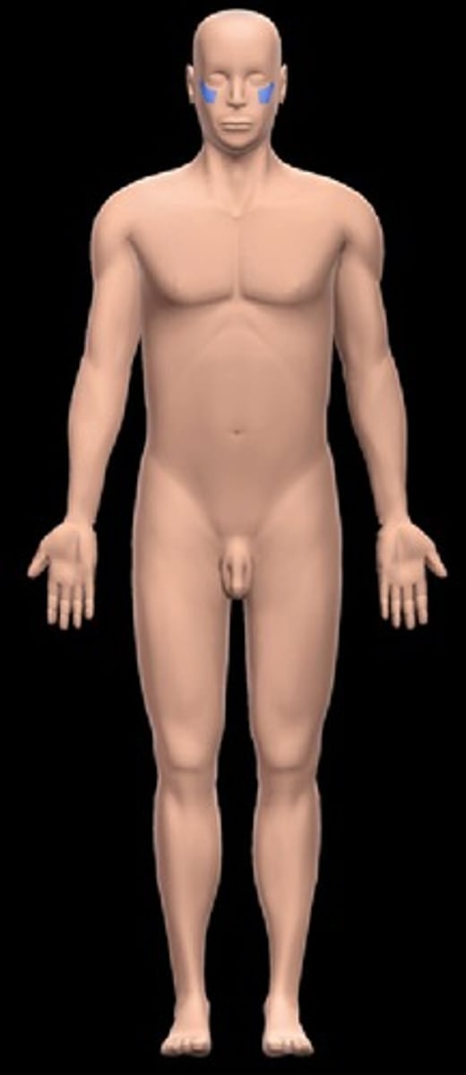
orbital region
Location:
Head (anterior)
Description:
Region of eye
Related to the orbits, the bony cavity that contain the eyes and accessory structures
palmar region
Location:
Hand (proximal)
Description:
Subdivision of hand
Subdivisions include: carpal and metacarpal regions, and thenar and hypothenar regions
Skin, muscles and tendons, nerves, and vessels on anterior aspect of carpal bones and metacarpals
parietal region
Location:
Head (lateral superior)
Description:
Part of cranial cavity related to parietal bone
parotid region
Location:
-Head (lateral)
-Inferior to auricular region
Description:
-Part of facial region related to parotid salivary gland and ramus of mandible

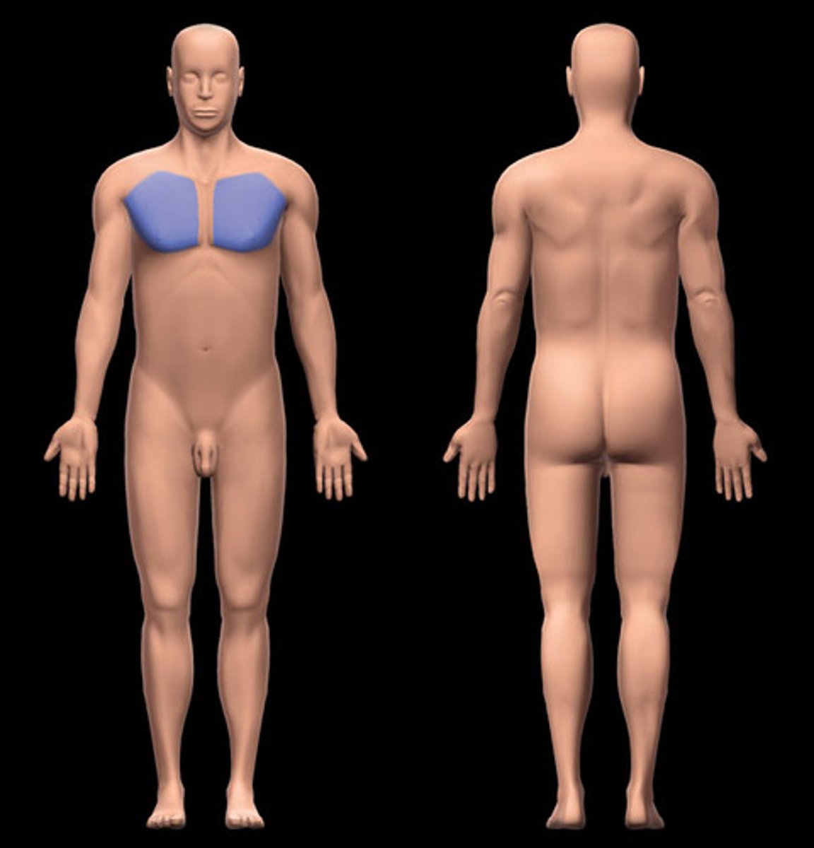
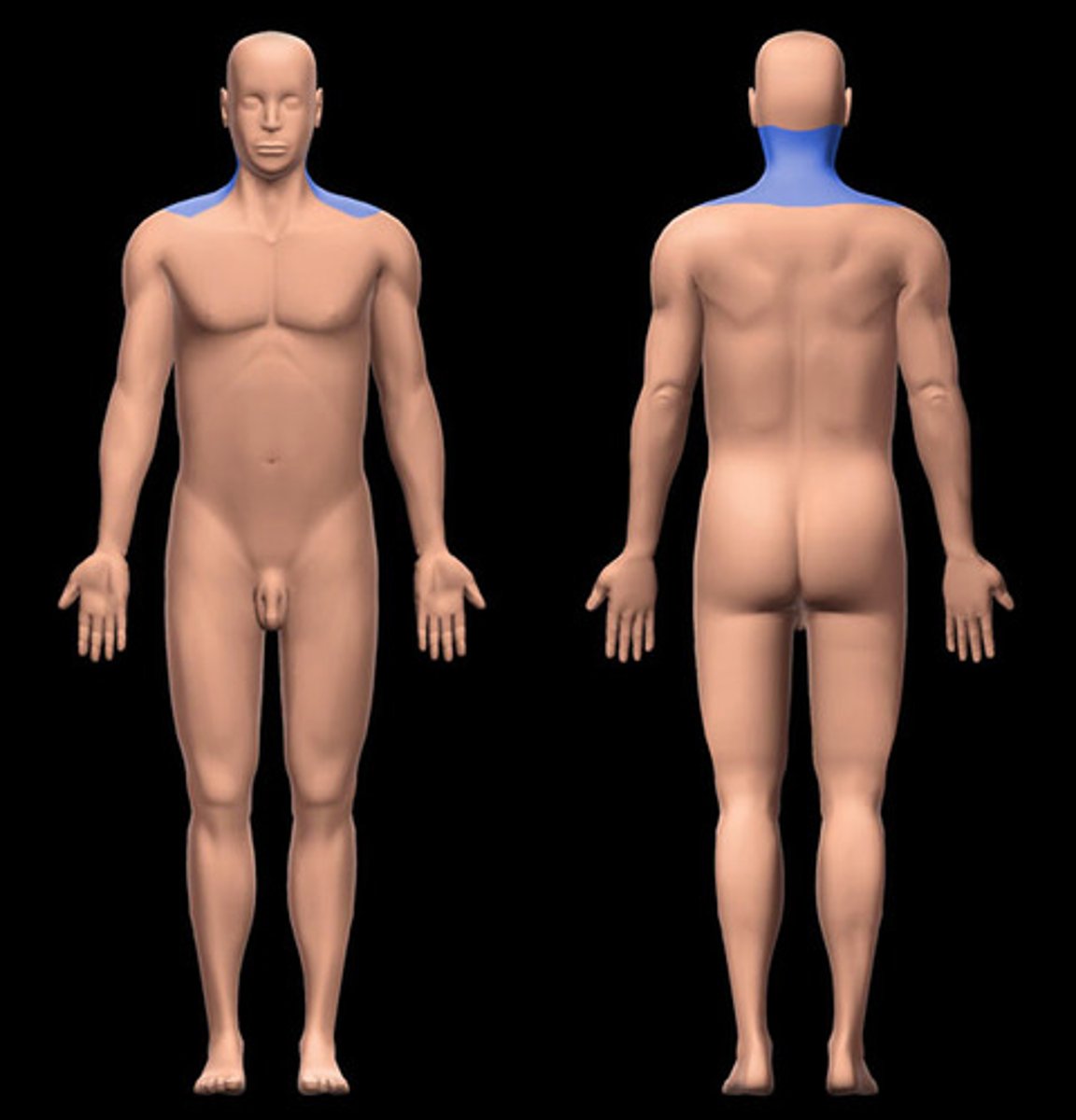
pectoral region
-Location:
Thoracic region (anterior)
-Description:
Subdividion of thoracic region over pectoralis major muscle
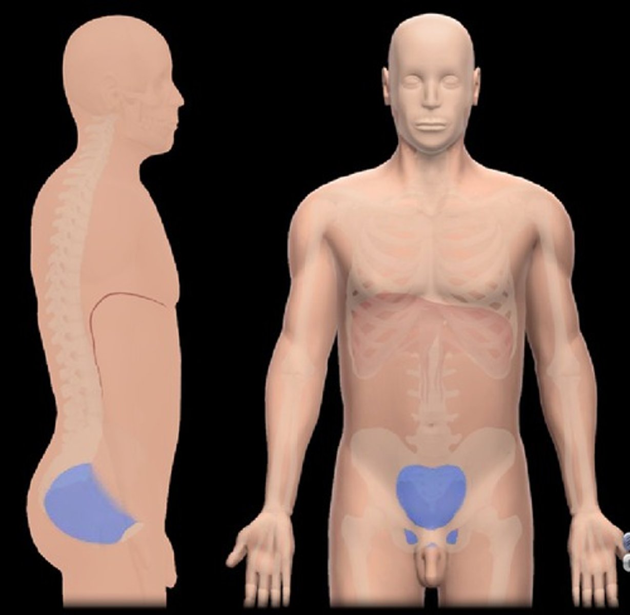
pelvic cavity
Location:
-Pelvic region
Description:
-Bounded by pelvic inlet (superiorly) and pelvic outlet (inferiorly)
-Major organs include: urinary bladder, loops of small intestine, inferior part of sigmoid colon, rectum, and reproductive organs (ovaries, uterus, vagina in female; prostate and seminal glands in male)
-Continuous superiorly with abdominal cavity
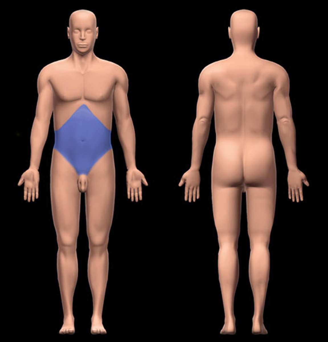
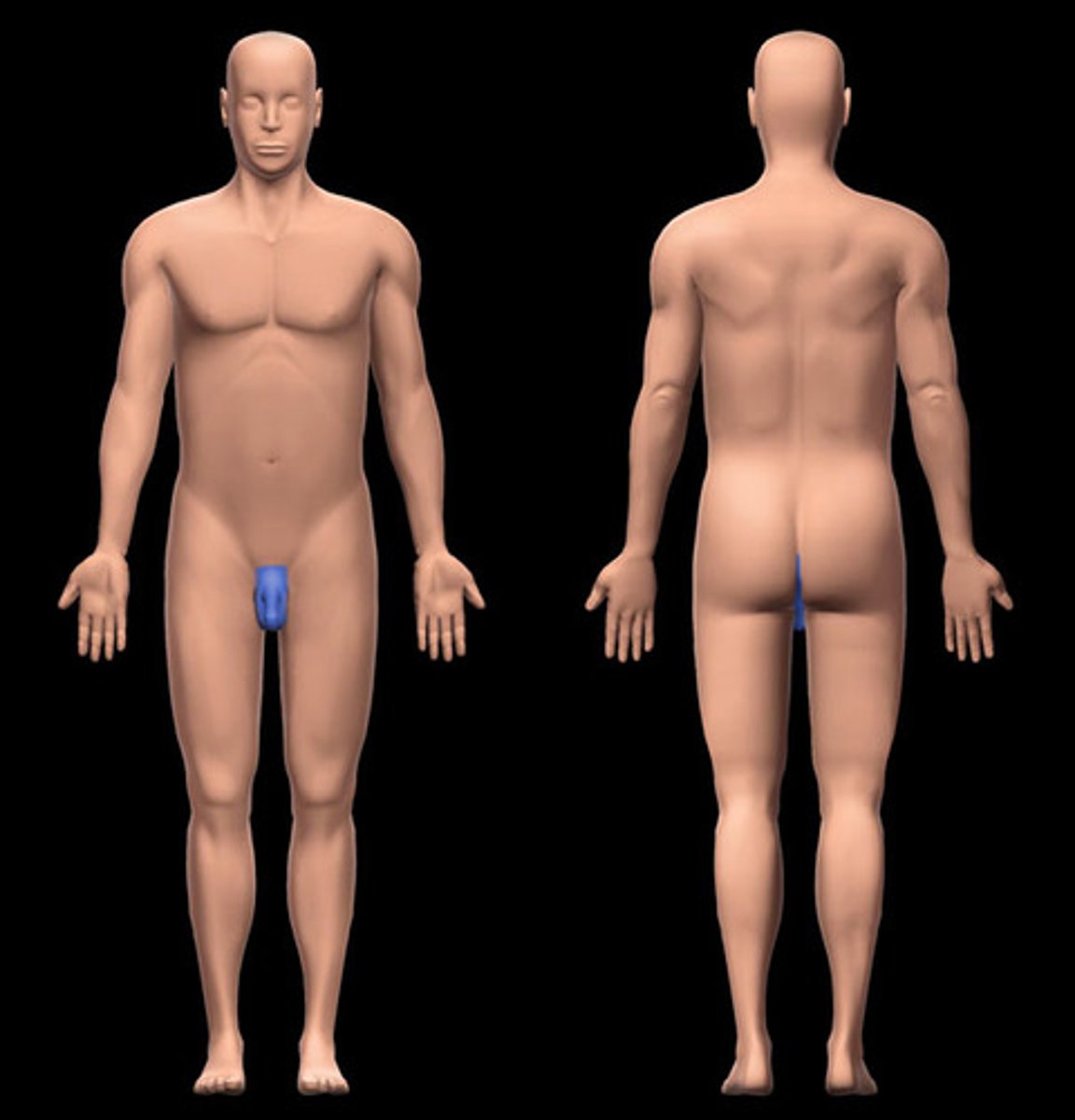
perineal region
Location:
-Between proximal thighs, from coccyx to pubic symphysis
Inferior to pelvic diaphragm
Description:
-Subdivision of trunk
-Forms diamond-shaped area when thighs abducted
-Anterior boundary: mons pubis
-Lateral boundary: medial surface of thigh
-Posterior boundary: gluteal folds and superior end of intergluteal (natal) cleft
-subdivided into anal and urogenital triangles
-Contents of anal triangle: anus
-Contents of urogenital triangle: clitoris, external urethral and vaginal orifices (female); penis, scrotum and its contents (male)
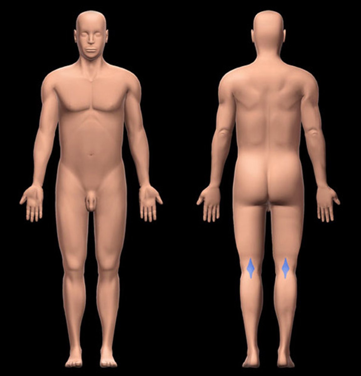
popliteal fossa
-Location:
Knee region (posterior)
-Description:
Subdivision of posterior knee region
Skin, muscles, nerves, and vessels associated with diamond-shaped region on posterior aspect of knee region
Bounded by biceps femoris, semimembranosus and semitendinosus, gastrocnemius, skin and popliteal fascia (roof), and posterior capsule of knee joint (floor)
Important structures include: small saphenous vein, popliteal artery and vein, tibial and common fibular nerves, posterior cutaneous nerve of thigh, and popliteal lymph nodes
posterior cervical region
Location:
Neck (posterior aspect)
Description:
Subdivision of neck
Deep to trapezius
Contents include: greater occipital n., occipital a., and suboccipital muscles
posterior leg region
Location:
Leg (posterior)
Description:
Subdivision of leg
Includes posterior muscular compartment
Skin, muscle tendons, nerves, and vessels posterior to posterior intermuscular septum of leg and interosseous membrane of leg
presternal region
Location:
Thoracic region (anterior midline)
Description:
Subdivision of thoracic region over sternum

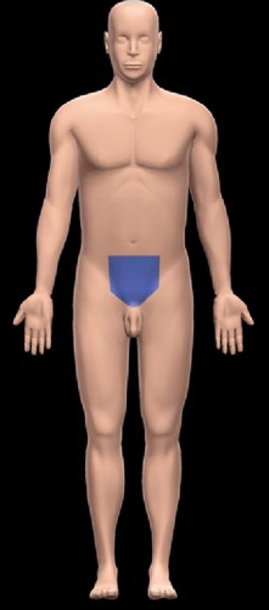
pubic region
Location:
Abdominal wall (anterior)
Description:
One of nine regions of abdominal cavity
Lower median region (flanked by right and left inguinal regions)
Contents include urinary bladder (when distended), and parts of small and large intestines
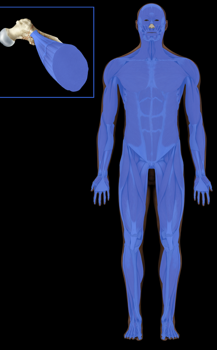
Muscular system
Organs and tissues:
Muscle
Tendon
Description:
Three types of muscle tissue: skeletal (striated), cardiac, and smooth
Skeletal muscles divided into axial and appendicular
Cardiac muscle found in walls of heart
Smooth muscle found in walls of hollow organs (e.g., gastroinestinal tract, blood vessels); in iris and ciliary body of eye
Function:
Movement of body
Maintenance of posture
Communication (especially muscles of facial expression)
Body functions, including respiration, circulation, digestion, defecation, urination, childbirth
Temperature regulation
Comment:
Skeletal muscle under voluntary control
Cardiac and smooth muscle under involuntary control
right flank region
Location:
Abdominal wall (anterior)
Description:
One of nine regions of abdominal cavity
Right lateral region
Contents include parts of small and large intestine, and right kidney
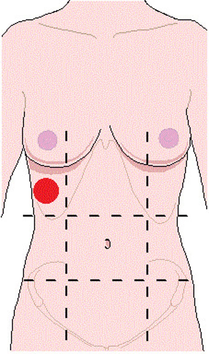
right hypochondriac region
Location:
Abdominal wall (anterior)
Description:
One of nine regions of abdominal cavity
Right upper lateral region
Contents include parts of large intestine, liver and gallbladder, and right kidney
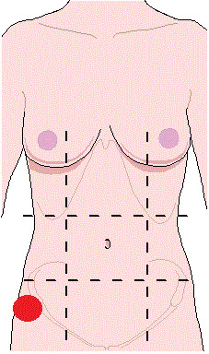
right inguinal region
Location:
Abdominal wall (anterior)
Description:
One of nine regions of abdominal cavity
Right lower lateral region
Contents include parts of small and large intestine (including cecum and vermiform appendix)
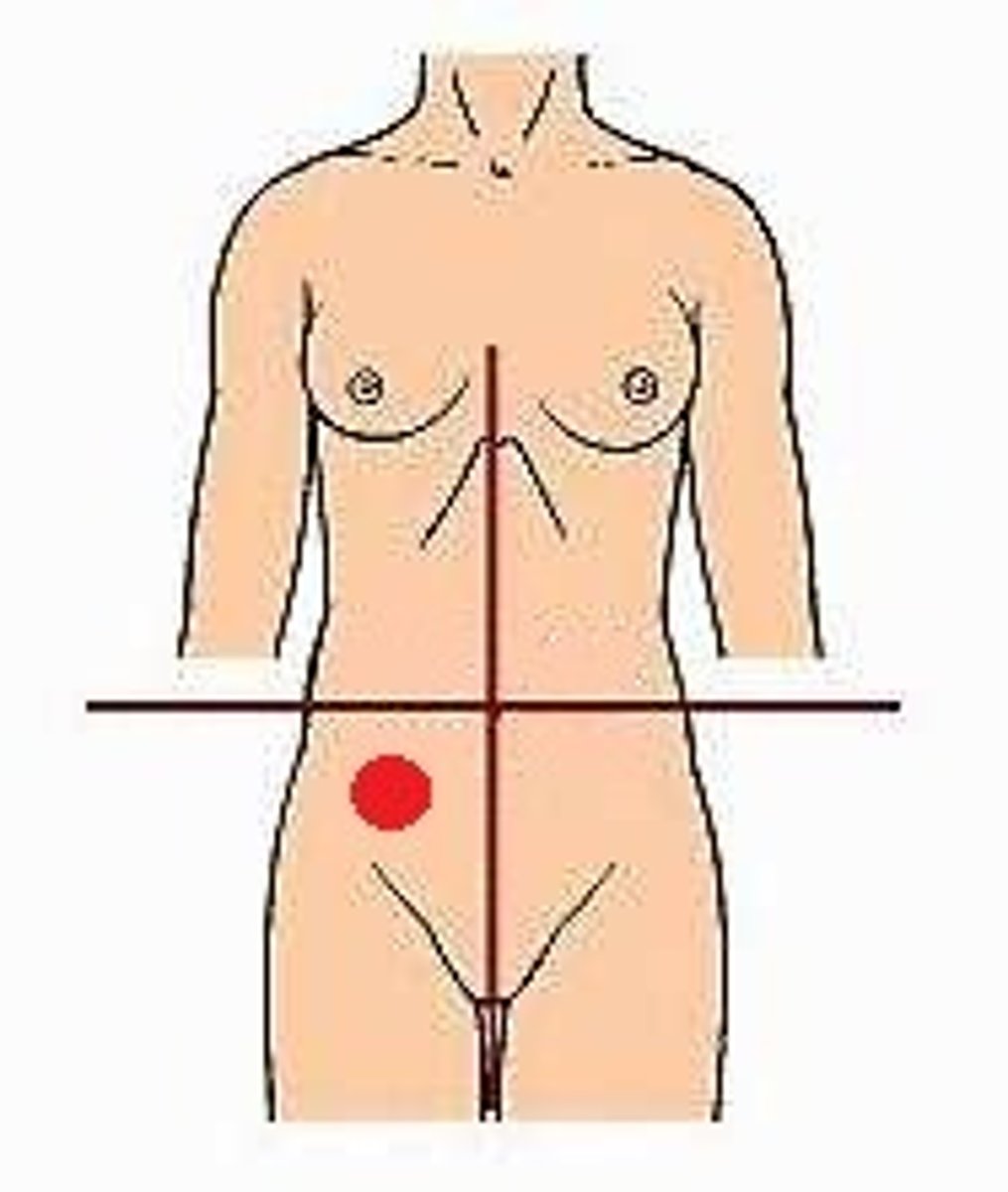
right lower quadrant
Location:
Right of midline, inferior to transverse plane through umbilicus
Description:
Lower right lateral area of abdominopelvic cavity
Contents include parts of small intestine, large intestine (including cecum and vermiform appendix), urinary bladder (when distended), and right uterine tube and ovary
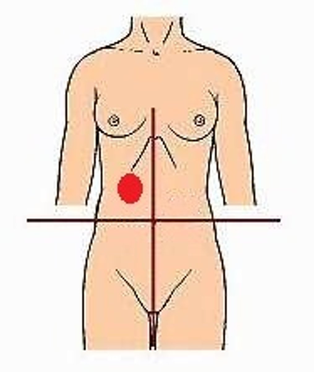
right upper quadrant
Location:
Right of midline, superior to transverse plane through umbilicus
Description:
Upper right lateral area of abdominopelvic cavity
Contents include right kidney and suprarenal gland, gallbladder, and parts of liver, stomach, pancreas, and small and large intestines
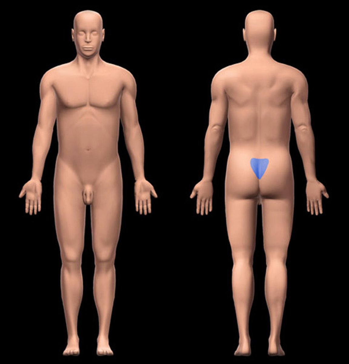
sacral region
Location:
Back (inferior)
Description:
Subdivision of back
Includes sacrum and attached muscles
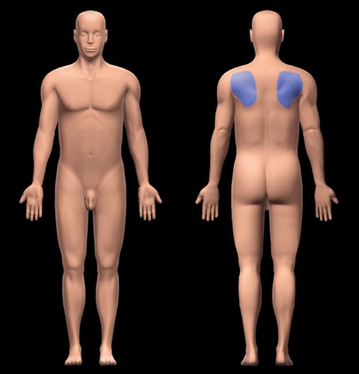
scapular region
Location:
Back (superolateral)
Description:
Subdivision of back over scapula
Comment:
Scapula also known as "shoulder blade"
sole of foot
Location:
Foot (inferior)
Description:
Plantar surface of foot (i.e., directed inferiorly in anatomical position)
Skin, muscles, nerves, and vessels on sole of foot

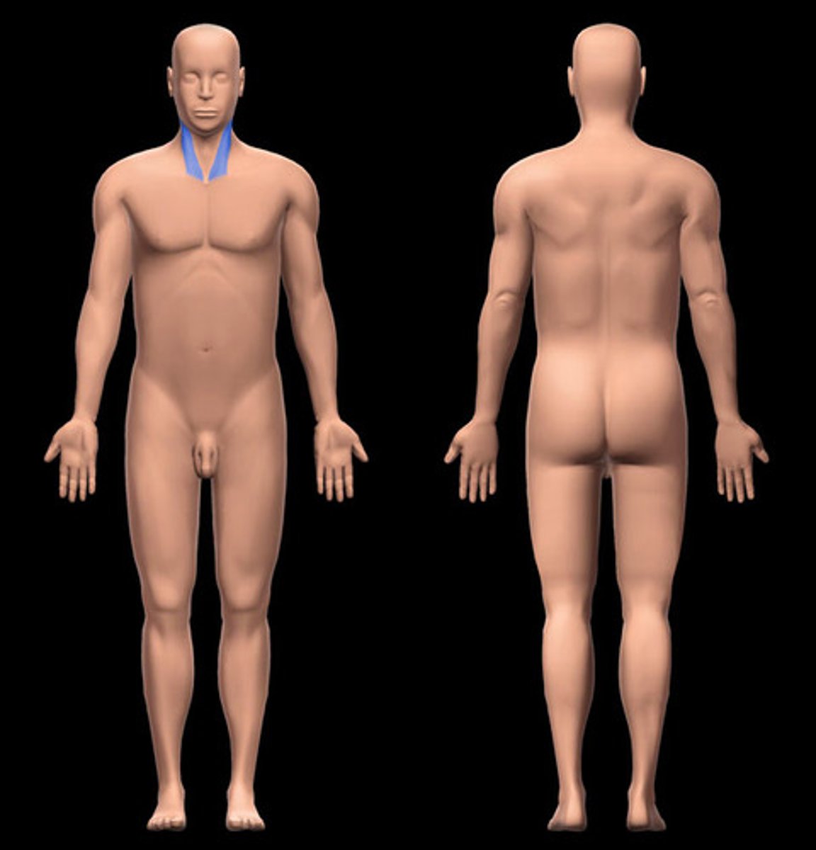
sternocleidomastoid region
Location:
Neck
Between anterior and lateral cervical regions
Description:
Subdivision of neck related to sternocleidomastoid (SCM) muscle
SCM divides anterior and lateral cervical regions
temporal region
Location:
Head (lateral)
Superior to zygomatic arch and auricular region
Description:
Part of cranial region related to temporal bone

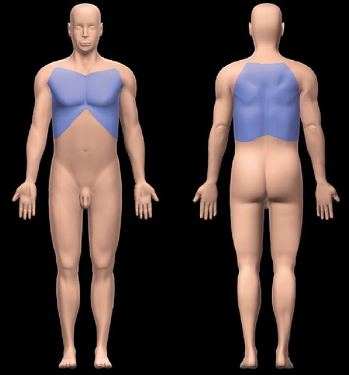
thoracic region
Location:
Superior part of trunk
Between neck and abdomen
Description:
Topographic (surface) subdivision of trunk
triangle of ausculation
Location:
Back
Description:
Small, triangular gap between trapezius and latissimus dorsi muscles and inferior part of medial scapular border
Floor of triangle formed by rhomboid major muscle and thoracolumbar fascia
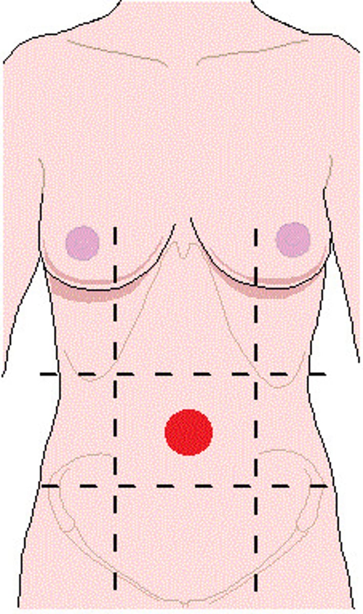
umbilical region
Location:
Abdominal wall (anterior)
Description:
One of nine regions of abdominal cavity
Median region (flanked by right and left flank regions)
Contents include parts of small and large intestines
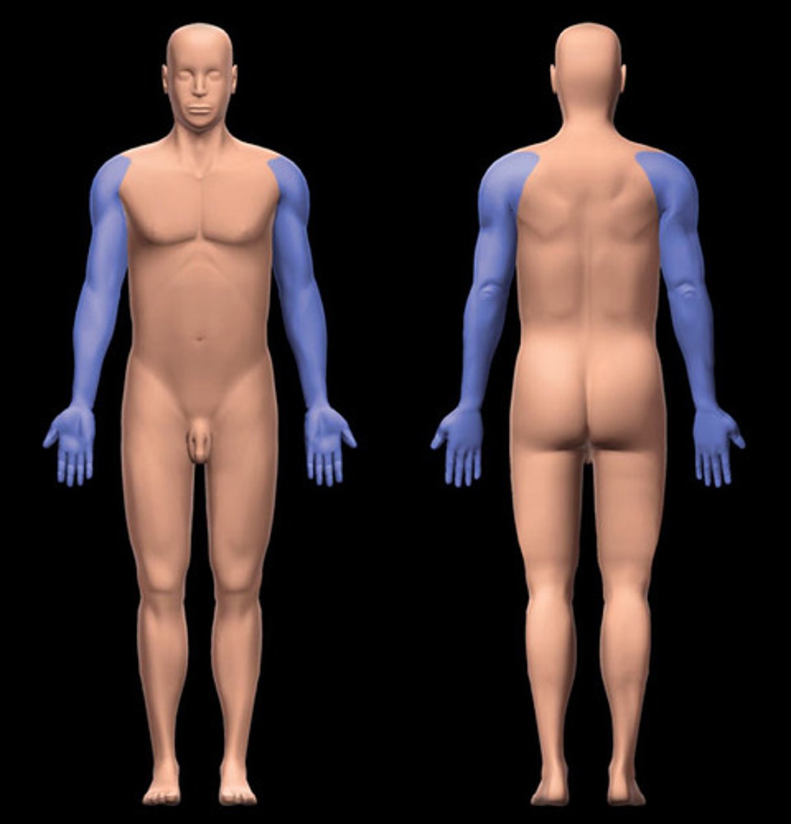
upper limb
Location:
Limb that, in anatomical position, is suspended from the shoulder and lies along lateral aspect of trunk and superior aspect of lower limb
Description:
Consists of deltoid, brachial (arm), antebrachial (forearm), and hand regions
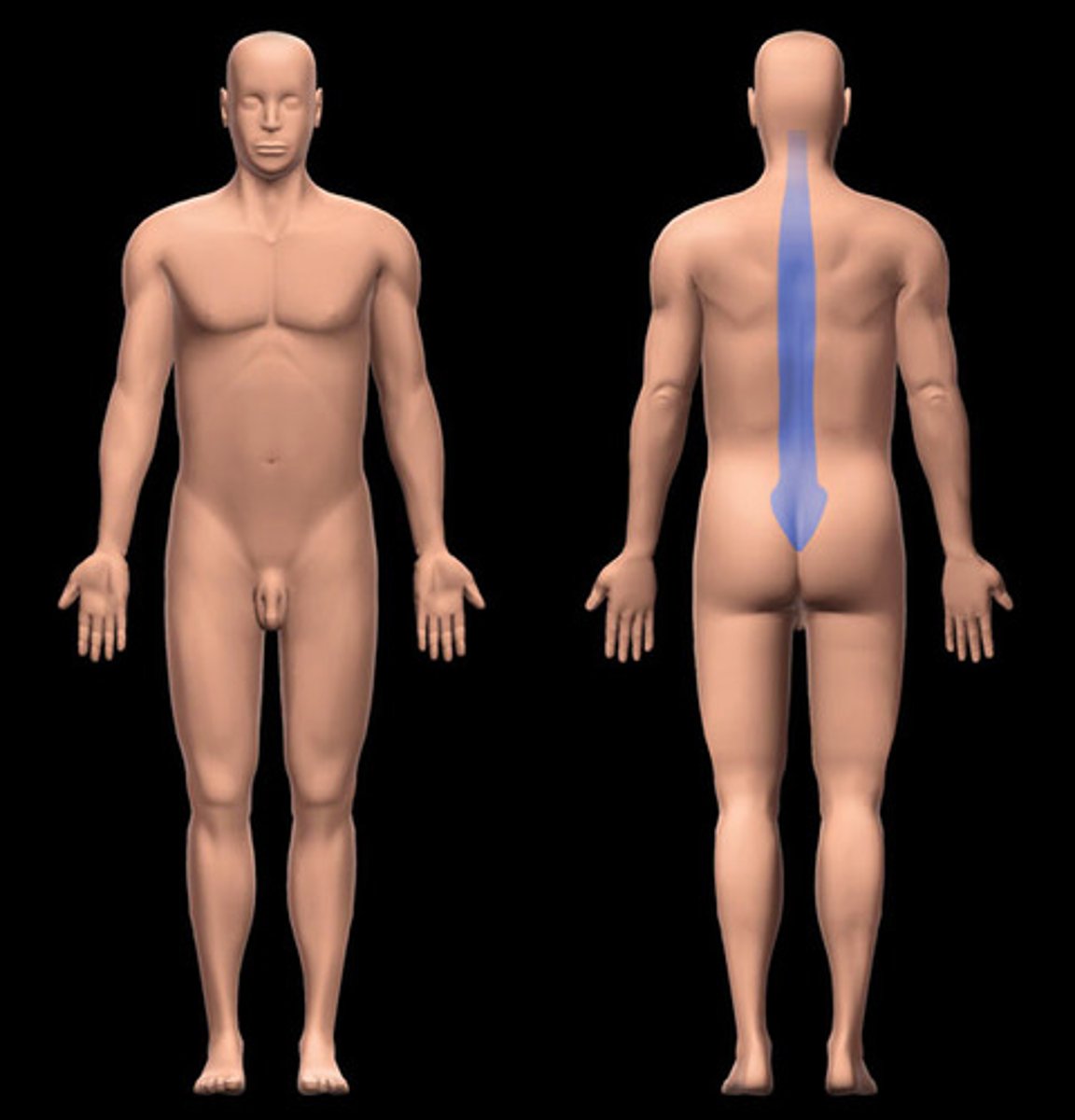
vertebral region
Location:
Posterior cervical and back regions (midline)
Description:
Subdivision of neck and back over vertebral column
Includes cervical, thoracic, lumbar, sacral and coccygeal vertebrae
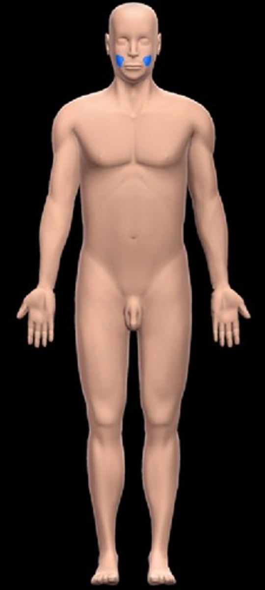
zygomatic region
Location:
Head (lateral)
Inferior to orbital region
Superior to buccal region
Description:
Part of cranial region related to zygomatic bone
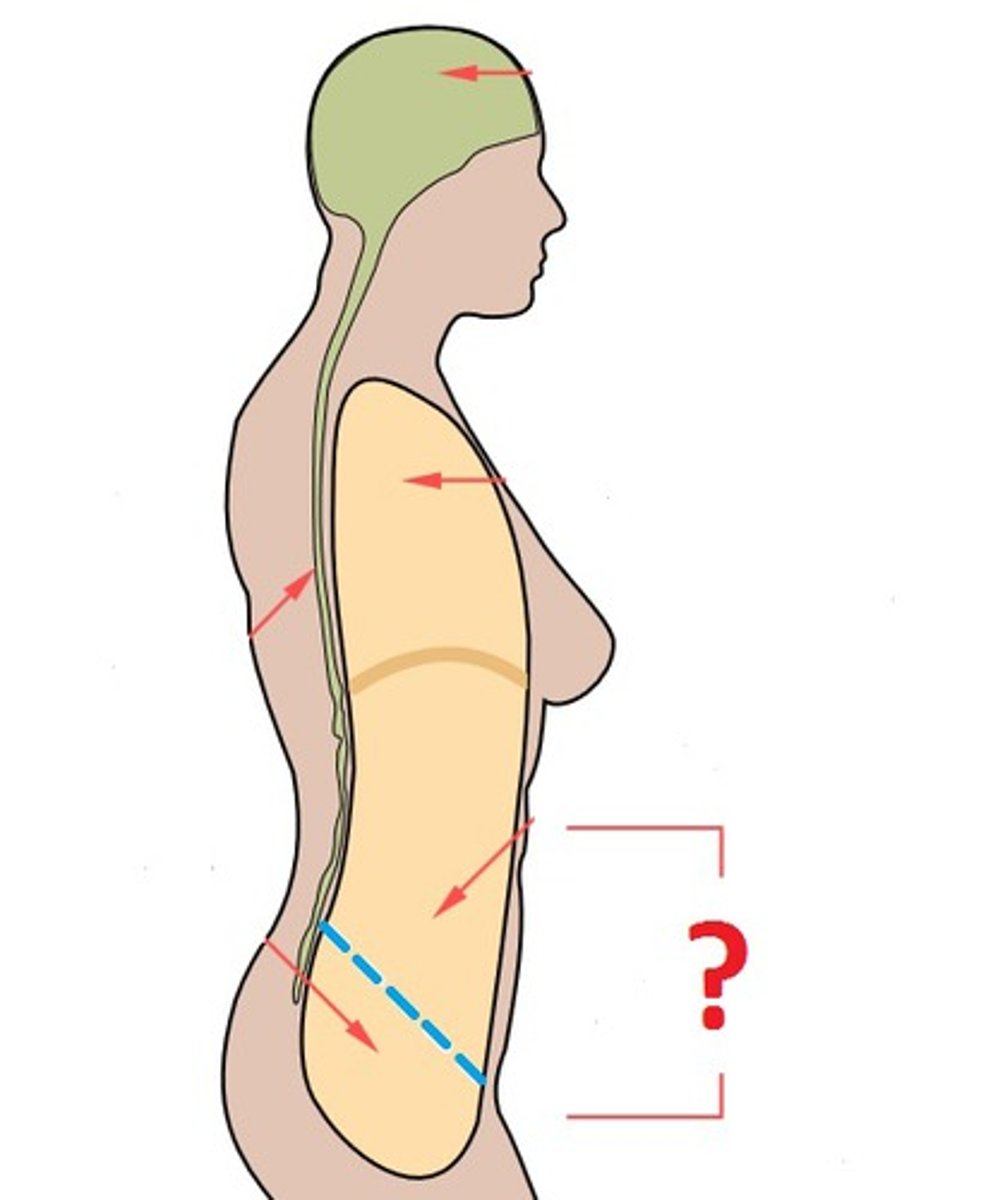
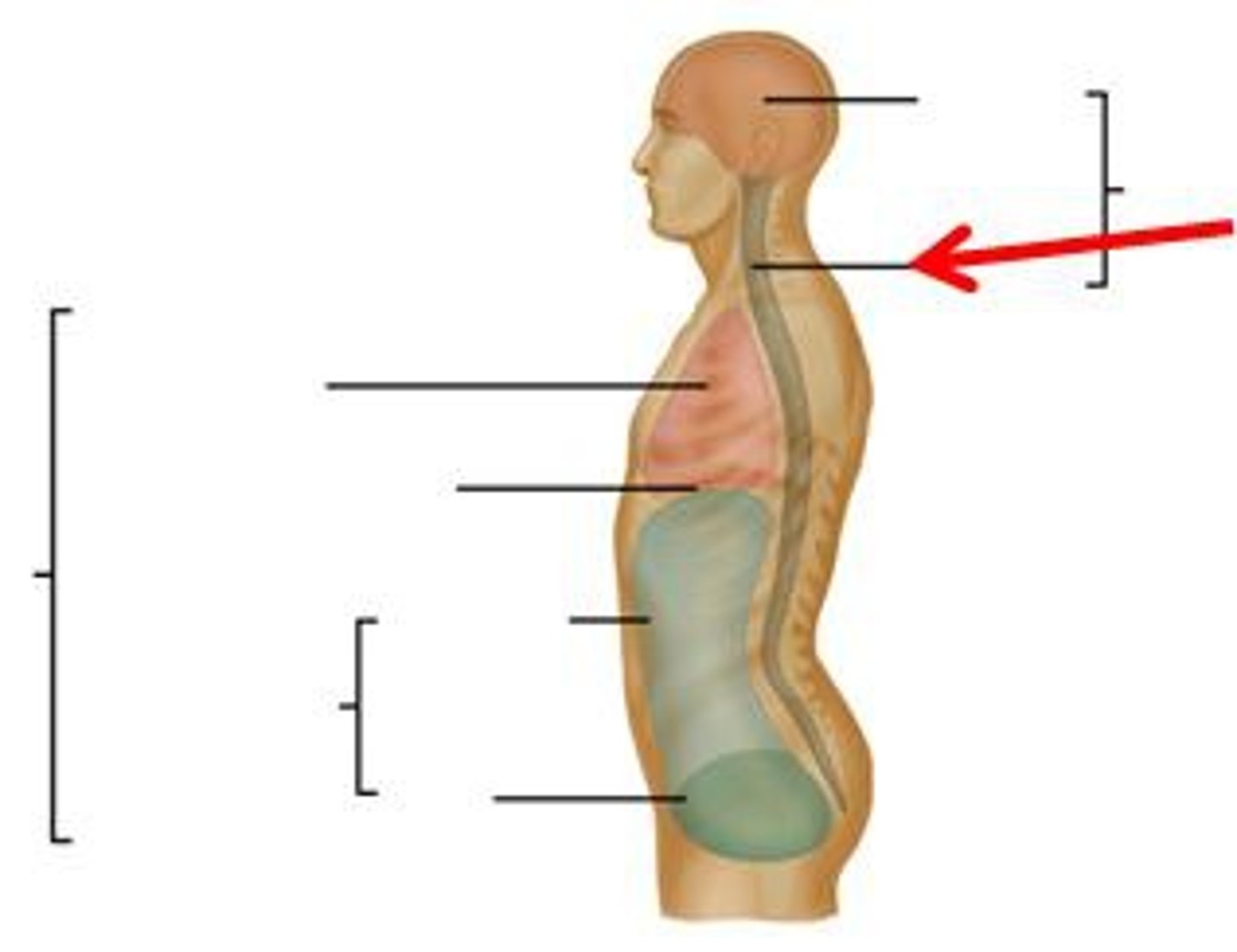
abdominopelvic cavity
Location:
Trunk, between thoracic and pelvic diaphragms
Description:
Continuous cavity formed by abdominal and pelvic cavities
Major abdominal organs include: stomach, intestines, liver, gallbladder, spleen, pancreas, kidneys and ureters, suprarenal glands, aorta, inferior vena cava, and lumbar nerve plexus
Major pelvic organs include: urinary bladder, loops of small intestine, inferior part of sigmoid colon, rectum, and reproductive organs (ovaries, uterus, vagina in female; prostate and seminal glands in male)
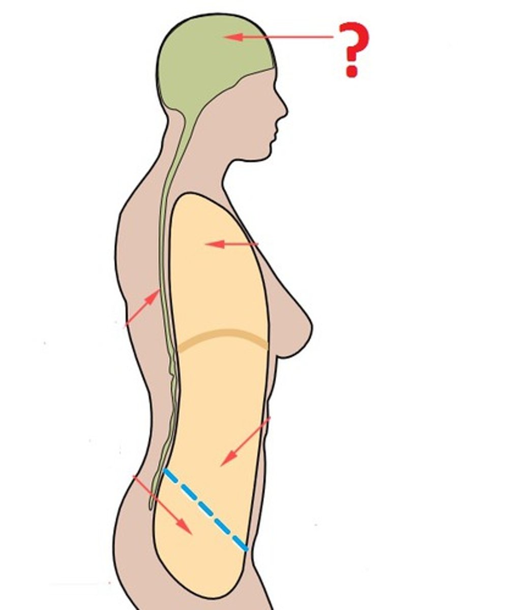
cranial cavity
Location:
Skull
Description:
Space in skull that contains brain, meninges, and cerebrospinal fluid (CSF)
Formed by frontal, occipital, sphenoid, ethmoid bones, parietal, and temporal bones
diaphragm
Action:
Dome of diaphragm flattens during inspiration
Contraction increases vertical dimension of thoracic cavity
Comment:
Primary muscle of respiration
Contraction (flattening) decreases intrathoracic pressure and increases intra-abdominal pressure
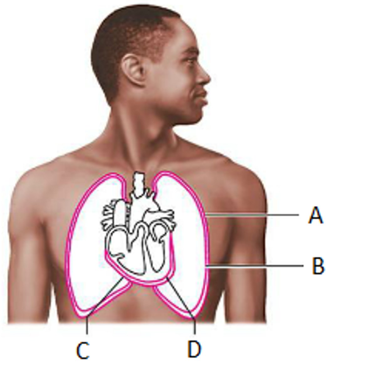
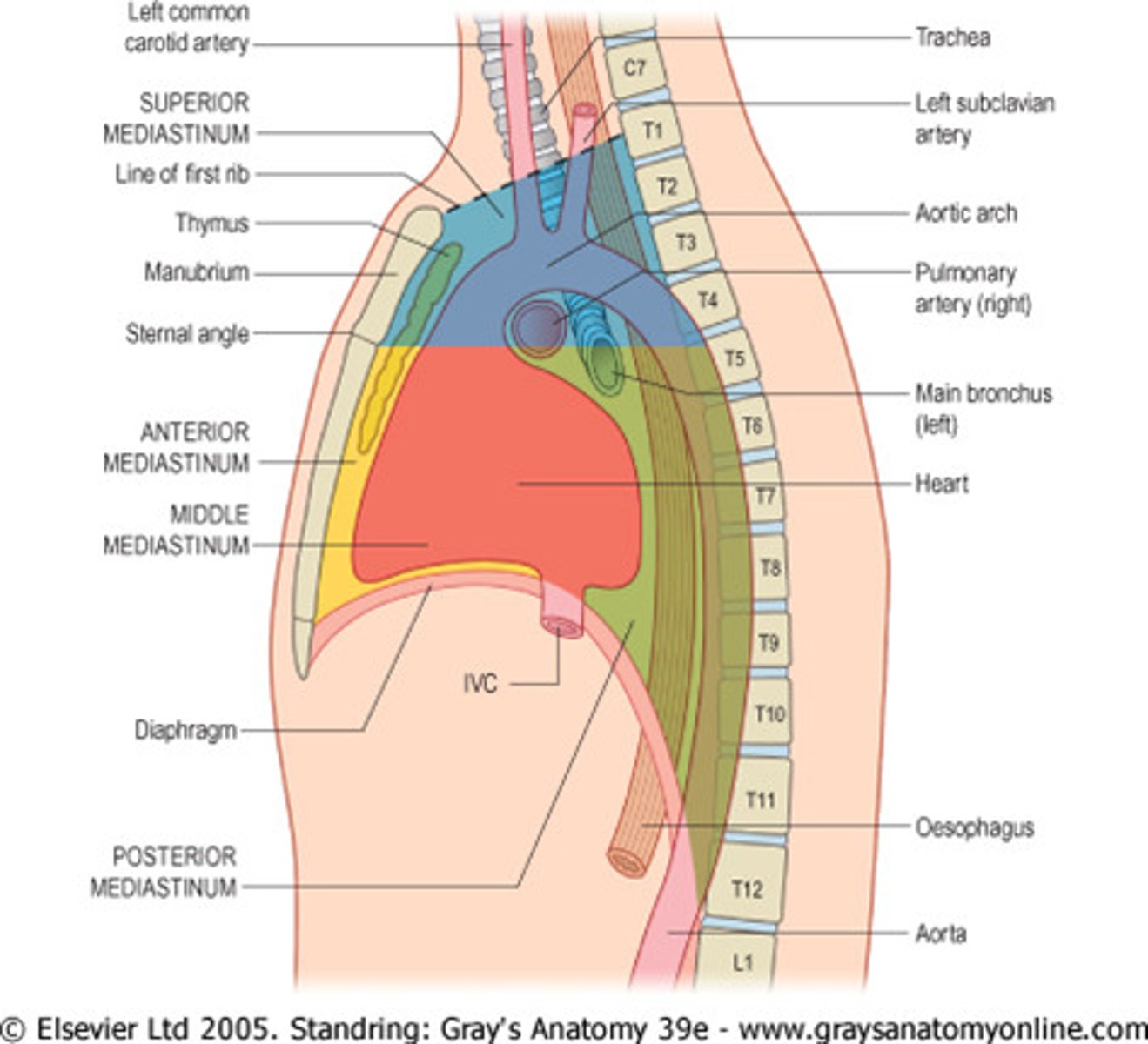
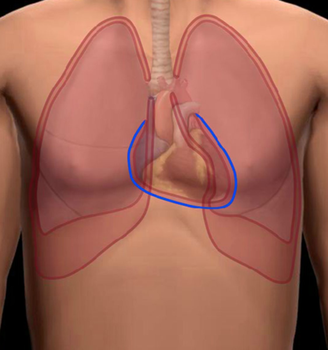
mediastinum
Mediastinum
Location:
Thoracic cavity
Description:
Middle region of thorax
Lies between sternum and thoracic vertebral bodies
Separates right and left pulmonary cavities
Divided into superior and inferior parts
Inferior mediastinum subdivided into middle, posterior, and anterior parts
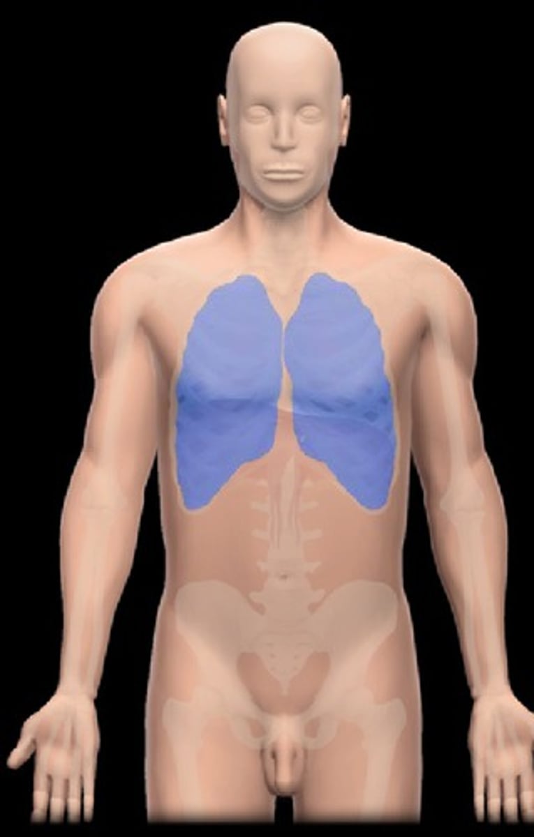
pulmonary cavity
Location:
Thorax
Description:
Bilateral subdivision of thoracic cavity (separated by mediastinum)
Contain lungs and plurae
Lined by parietal pleura
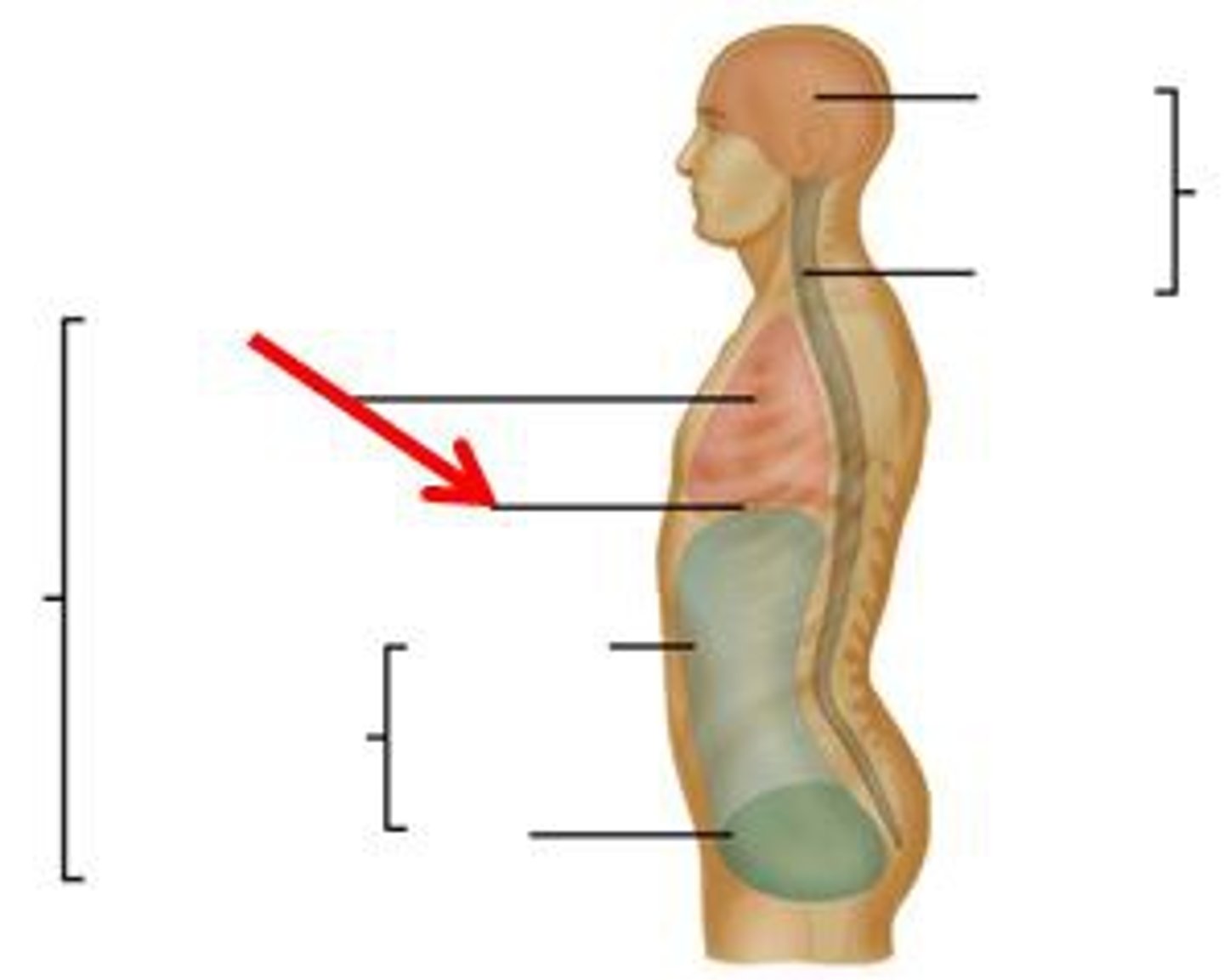
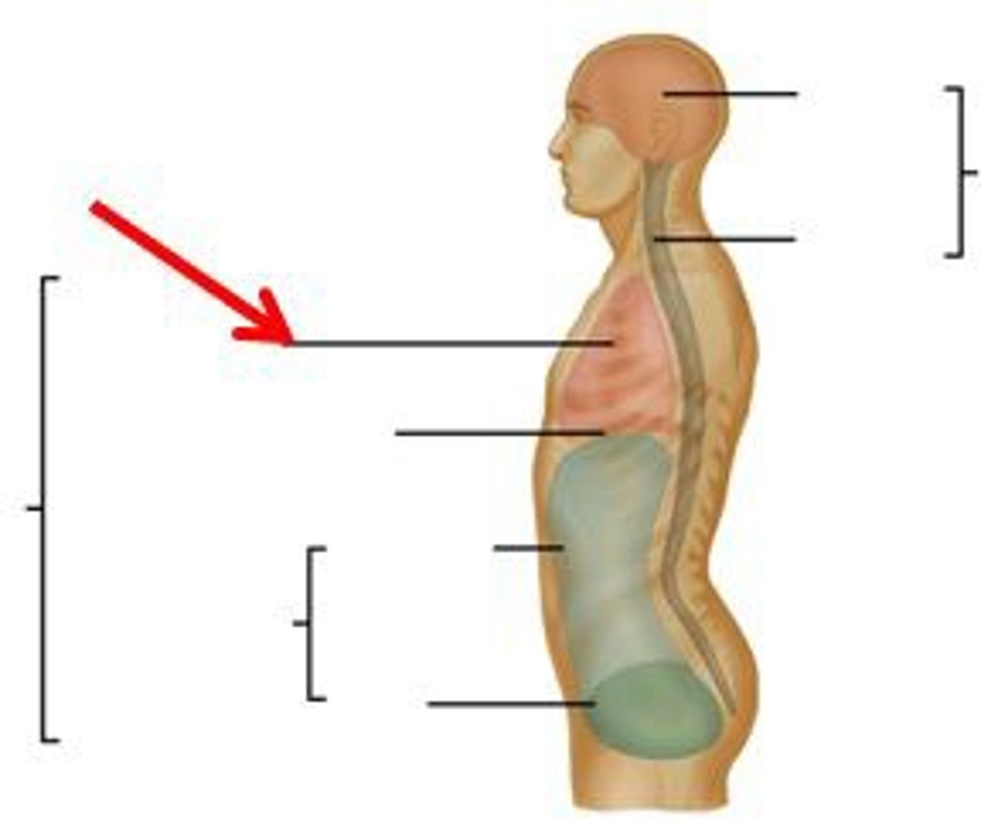
thoracic cavity
Location:
Thorax
Description:
Cavity of the chest
Bounded by sternum, ribs and costal cartilages, intercostal muscles, thoracic vertebrae, and diaphragm
Three subdivisions: a central mediastinum (contains heart and thoracic parts of great vessels, trachea, esophagus, and thymus) and bilateral pulmonary cavities (contains lungs and plurae)
vertebral canal
Location:
Vertebral column
Description:
Canal formed by combined vertebral foramina
Parietal layer of serous pericardium
Location:
Thorax (mediastinum)
Description:
Thin, serous membrane fused to inner surface of fibrous pericardium
Outer limit of pericardial cavity
Continuous with visceral layer of serous pericardium
Parietal pleura
Location:
Thorax
Description:
Thin, serous membrane
Lines pulmonary cavity
Fused to internal walls of thoracic cavity and lateral surface of mediastinum
Continuous with visceral pleura at root of lung
Regions include mediastinal, cervical, diaphragmatic, and costal
Comment:
Pleural cavity created by narrow space between parietal and visceral layers of pleura
Thorax has three subdivisions: mediastinum and right and left pulmonary cavities
Costal and peripheral diaphragmatic pleura innervated by intercostal nerves