Chapter 20 : blood
1/59
There's no tags or description
Looks like no tags are added yet.
Name | Mastery | Learn | Test | Matching | Spaced | Call with Kai |
|---|
No analytics yet
Send a link to your students to track their progress
60 Terms
Hematology
study of blood, - forming tissues, and disorders associated with them
blood
transports oxygen, carbon dioxide, and nutrients. waste, hormones
Blood
Regulates PH, body temperature, and water content ( cells ) - sem neutral 7
blood
Protection - blood loss, foreign microbes and toxins
Basic
Higher PH means
Acidic
Lower PH means
plasma
liquid matrix
Formed elements
blood cells & fragments are
hypovolemic
low blood volumes
hypervolemia
excessive blood volume
PH 7.35 - 7.45 buffered
Plasma
about 50 % of the volume of whole blood
Plasma
made of 92% of water
plasma
7% protientd ( albumins, globins, fiberogens, regulatory proteins)
Plasma
1% of other solutes, electrolytes, organic nutrients, and organic wastes
Oxygen concentration
Higher in plasma than in interstitial fluid
Oxygen → plasma → interstitial fluid
carbon dioxide concentration
whats lower in plasma than in interstitial fluid
carbon dioxide → initial fluid → plasma
Dissolved protein concentration
What is Higher in plasma? extremely low in intestinal fluid
plasma
This substance, compared to the intestinal fluid, contains:
more oxygen
less carbon dioxide
more dissolved proteins
and is also found on the inside of blood vessels
Albumins
60 %
smallest plasma protein
“ white of an egg”
multitasking protein
Albumins
contributes to the osmotic pressure of plasma
Small proteins can pass through the wall of blood vessels
But these proteins can’t, which helps maintain the pressure in blood vessels even if other proteins leave
Albumins
transports fatty acids and steroid hormones
These substances fear water, so this protein provides pockets to protect these substances from water
Blood plasma is 90% water
Globins
makes up 35% of plasma proteins
also makes hemoglobin and myoglobin in the blood
has two types
immunoglobulins
transport globlins
Immunogloblins
attack pathogens
Transport Globins
a protein that transport ions and hormones
fibrinogens
4%
Largest of the plasma proteins
involved in the blood clotting process
interacts with fibrin - makes the framework for clotting
formed elements
makes up about 45% of whole blood
erythrocytes 99.9% of whole blood.'
platelets < 0.1% of whole blood ( functions in clotting )
leukocytes < 0.1% of whole blood ( immune system )
hematocrit readings
The percentage of whole blood occupied by the formed elements
males: 45% ( 5.4 million RBCs per microliter)
females: 42% ( 4.8 million RBCs per microliter)
aka
packed cell volume ( PCV )
Volume of packed red cells ( Vpkc )
centrifuge
Red blood cells
biconcave disc with a thin central region - flexible
measure about 7.7 microns in diameter
needs to be able to fold and bend
lacks a nucleus and organelles
Anaerobic respiration (don’t use mitcrocchinda bc it counter intuitive )
Life span is 120 days
hemoglobin
Hemoglobin
a red pigment found in RBC, a gas transport
> 95% OF RBC protein
transports oxygen using a ring of iron
blood types
Determined by agglutinogens or antigens
antigens on the surface of RBC
glycoproteins or glycolipids
There are three major types of antigens
Antigen A
Antigen B
Antigen D - Rh factor the - or + ( found in resses monkeys )
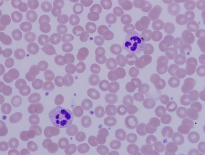
White blood cells ( WBCs)
leukocytes
nucleated, no hemoglobin ( red pigment )
short life span - hours to days
Diapedesis
chemotaxis
Diapedesis
movement of leukocytes through capillary wall - between cells
Chemotaxis
draws the leukocytes toward the invading agent using a chemical gradient
Granulocytes
class of WBCs
granular cytoplasm, lobed nuclei
Neutrophils, eosinophils, Basophiles
Agranulocytes
Classification of WBC
No granules in cytoplasm
poor staining
includes monocytes, lymphocytes
Neutrophiles
granules contain chemicals to kill bacteria
Neutrophiles
Typically, the first WBC at a bacteria site
Neutrophils
Very active phagocyte
Neutrophils
only alive for about 10 hrs

Neutrophiles
A WBC thoses nucleus is multi-lobed (which gives it the ability to fit in tight spaces )
Neutrophiles
a type of WBC that fights against bacterial infections
First on the site of a bacterial and fungi infection
activates in minutes

Eosinophils
2% - 4%
Granules release a chemical that reduces inflammation
stain acid red
Eosinophils
attack a foreign substance (parasites) that has reacted with circulating antibodies
attacks the parasite with toxin bombs
Eosinophils
Associated with allergic reactions
and contains a bilobed nucleus
( reduces allergy reactions )
Basophils
> 1 %
granules release histamine and heparin
Histamine dilates blood vessels
heparin prevents abnormal blood clotting
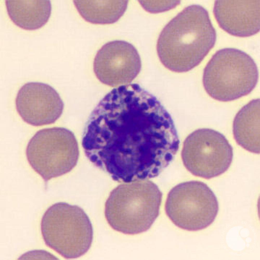
Basophils
A WBC whose nucleus is usually hidden due to all the granules ( who you’re gonna call )

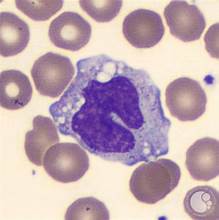
Monocytes
2 - 8 % of white blood cells
Agranular
largest WBC
the most versatile cells
contains a kidney-shaped nucleus
Monocytes
located in the kidney or a large oval-shaped nucleus

monocytes
releases chemicals to attract other phagocytic cells, fibroblasts
Fibroblasts produce collagen fibers to surround the uninfected site, scar tissue
Lymphocytes
20 -30 % of white blood cells
Agranular has little cytoplasm, and the nucleus stains purple
type of white blood cell
differentiates into :
T cells
B cells
Nk cells
a dark purple thing the same size as an RBC
specific immunity
The ability of the body to mount a counterattack against an invading pathogen or foreign protein on an individual basis
remembers a pathogen and can respond to it faster
T cells
enter peripheral tissues and attack foreign cells directly ( they do not produce antibodies )
B- cells
differentiates into plasma cells that secretes antibodies
Nk cells
responsible for immune surveillance - destruction of abnormal tissues prevents cancer
Platelets ( thrombocytes )
membrane-enclosed packets of cytoplasm
derived from megakaryocytes
large cells in bone marrow
fragments forming bits and pieces of packets
about 350,000 per microliter of blood
thrombocytopenia
lower than normal number of platelets
bleeding in digestive tract
thrombocytosis
Higher than normal number of platelets
signs for cancer
platelet
clotting response ( hemostasis )
chemical needed for the clotting release
platelet thromboplastic factor
forms a temporary patch in the wall of the damaged blood vessels
causes the contraction after a clot has formed to reduce the size of the break in the vessel wall
actin and myosin
hemopoiesis
blood formation
begins with the hematopoietic stem cells
differentiate to form two cells :
myeloid stem cells → red, white
lymphatic stem cells