Course Intro, Physics, Biology, and Safety Review
1/144
Earn XP
Description and Tags
Lecture given 3/5/2026
Name | Mastery | Learn | Test | Matching | Spaced | Call with Kai |
|---|
No analytics yet
Send a link to your students to track their progress
145 Terms
What did Wilhelm Rontgen do?
Discovered X-rays in november, 1895
won nobel prize in physics in 1901
When were X-rays discovered ?
1895
Who is Otto Walkhoff?
Obtained first intraoral radiograph with exposure time of 25 mins
Wilhelm Koenig
obtained 14 radiographs of his own teeth using exposure time of 9 mins/tooth
Edmund C. Kells
took the first radiographs in the United states, 1896
Frank Harrison
first dentist to use radiology in england
published first adverse effects of radiation in 1896
William H. Rollins
among first to warn about adverse effects of radiation
1st commercial dental radiographic apparatus introduced in ____
Germany, 1905
Howard R. Raper
Introduced radiology into dental training in 1909, wrote first textbook abt dentistry in 1913
radiation
emission of energy as electromagnetic waves or as moving subatomic particles, especially high-energy particles which cause ionization
particulates
small fast-moving particles that have both energy and mass
particulate radiation is primarily produced by …
disintegration of an unstable atom and includes alpha and beta particles
t/f small, fast-moving particles that has only mass is called particulate radiation
false- has both energy and mass
electromagnetic
energy with no mass, vibrating or pulsating waves of electrical and magnetic energy
t/f radiation can be ionizing or non-ionizing
true
ionizing radiation changes the number of electrons of an atom
what are the types of ionizing radiation?
x-rays, ultraviolet radiation, alpha/beta/neutron particles, gamma rays
non-ionizing radiation / electromagnetic radiation
does not carry enough energy
visible light, microwaves, radio waves
t/f radio waves have the highest energy and shortest wavelength, while gamma rays have lowest energy and longest wavelength
false- opposite
R, M, IR, V, UV, X, G
what are the units of current?
measured in milliAmperes (mA)
what are the units of voltage?
measured in kiloVoltage (kV)
voltage as peak value/max voltage supplied (kVp)
what are the units of time?
measured in milliSeconds (s)
integrated exposure
product of mA and s: (mAs)
what are the components of an x ray machine?
cathode, anode, oil, power supply
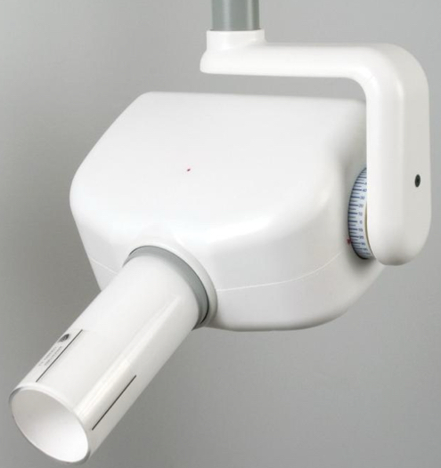
what is this?
tube head
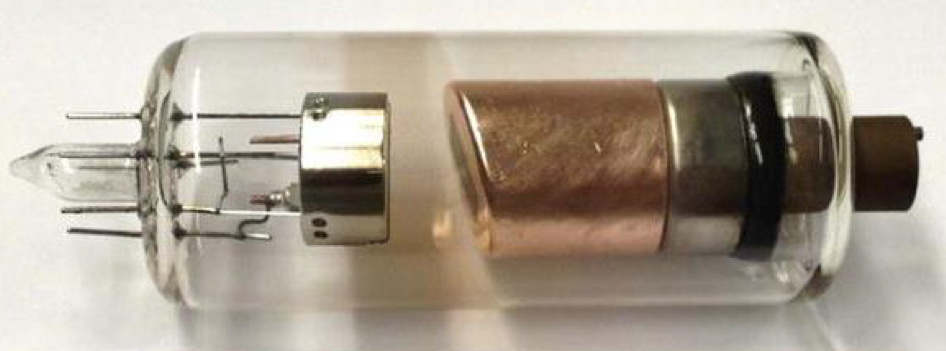
what is this?
xray tube
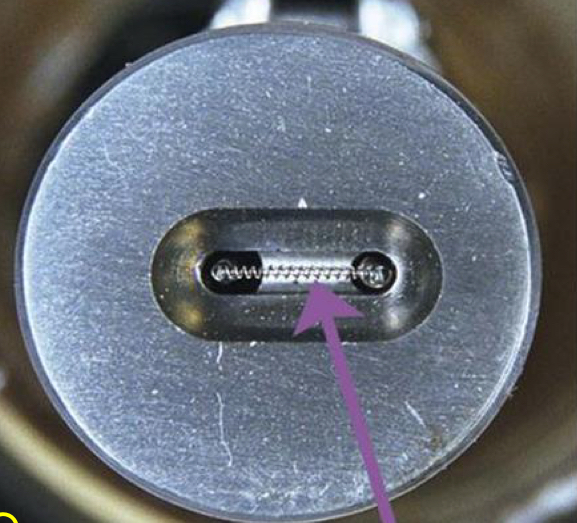
what is this?
cathode
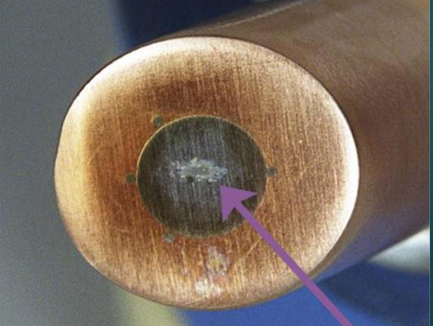
what is this?
anode
focusing cup
focuses the electrons on to anode, cup is negatively charged, helps converge the electrons to the andode (target)
What charge is the focusing cup ?
negatively charge, helps to converge the electrons to the anode
what direction do electrons move- cathode to anode or anode to cathode?
cathode to anode
cathode
negative electrode, electrode source (aka filament of tungsten)
*what are the advantages of tungsten?
high atomic number (74), high melting point (3370C), good absorber and dissipater of heat, easily available, economical
what is the tungsten target (anode) bonded to?
large copper block- for better thermal distribution
which is an advantages of tungsten?
A.Low atomic number 55
B. Low Melting point 2270
C. Not easily available
D. Good absorber and distributer of heat
E. tungsten target (anode) bonded to large silver block for better thermal distribution
D. Good absorber and distributer of heat
electron production
filament is heated by allowing a current to flow through it
filament offers resistance, gets heated, then e- emission occurs (boils off)
filament current
current used to heat filament, controls # of electrons boiled off
tube current
electron flows from cathode to anode, uses mA units
filament transformer → voltage → heat → electrons emitted by cathode
electron flow from cathode to anode
quantity of electrons (mA)
increase in time increses the quantity of electrons and increases the number of x rays
beam quantity or beam intensity
what is the electron cloud/space charge?
cloud of electrons around filament from thermionic emission
fewer electrions being focused on the target
need to potential difference to pull these away from the cathode to the anode
anode
positive electrode at high potential difference, small tungsten plate (target) embedded in a large block of copper
major source of heat production
*stationary or rotating
what is major source of heat production ?
anode
*what are the characteristics of a stationary anode?
2-3 mm thick tungsten embedded in Cu, 1×1 cm dimensions
high atomic number/melting pt (3370 C), good heat absorber/dissipater, easily available, not very expensive
The wear and tear of a ____________ is less than half of the ____________
rotating anode / stationary anode
rotating anode
more heat tolerant/efficient, lesser cooling time/damage to anode, 3,600-10,000 rpm makes it more efficient at dissipating heat
*what are characteristics of an ideal material?
high atomic number, high melting point, high thermal conductivity, low vapor pressure
how are x-rays formed through electrons ?
e- accelerated from cathode to anode by high voltage (potential difference); e- strike anode, suddenly decelerated, energy lost is converted to x rays
what does the glass enclosure do?
provides a vacuum
if e- collide with gas molecules, lose energy, produce secondary e-, change #/energy of e-
is the goal to have a large or small focal spot?
small
*what is the line focus principle?
anode surface must be angled away from cathode, increased surface area presented to e- beam
apparent/effective focal spot size remains small, heat produced still distributed over same large area
effective focal spot
aka the x ray beam, want small
actual focal spot
electrode from cathode to anode, want big
the line focus principle is an area it the ___ of the anode
tilted
why is the tilt of the anode important in the line focus principle?
there is a better heat spread without compromising on resolution and maintaining smaller penumbra (shadow)
is the production of x-rays an energy effective proccess?
no
99% of energy converted to heat, only 1% to xray
why is heat dissipation needed?
prevent disruption of target surface: roughening, pitting, cracking etc.
what are the heat dissipation mechanisms?
radiation (though vacuum), convection (though surrounding oil and tube housing), conduction (through solid tube parts like copper)
what does the power supply do?
provides low voltage current to the filament, has high potential difference to accelerate the e-
tube voltage
increasing → kVp → electron energy → autotransformer
timer controls x ray exposure
what is the typical voltage of intra oral/ceph/pano x rays?
60-90 kVp
what is the typical voltage of CBCT?
90-120 kVp
bremsstrahlung radiation
electron passes by the nucleus, giving off photons of differing energies
electron collides with the nucleus, oblitering the electron
electron hitting target atom may be completely stopped (max energy x ray) OR deflected (lower energy photon)
continuous spectrum of photon energy ranging from near zero to a maximum
*energy is measured in keV
maximum energy will be equal to kVp
characteristic radiation
e- collides with an inner shell e-
photons with the same amount of energy
what are x-rays made of?
photons
what is the percentage of photon production and heat production?
1% photon, 99% heat- not an effective process
what factors affect controlling beam quality?
mA (tube current), kVp (energy), filtration (energy), exposure time, collimation (shape), distance (intensity)
what is the effect of current on x-ray production?
more current = more quantity of xrays, but energy stays the same
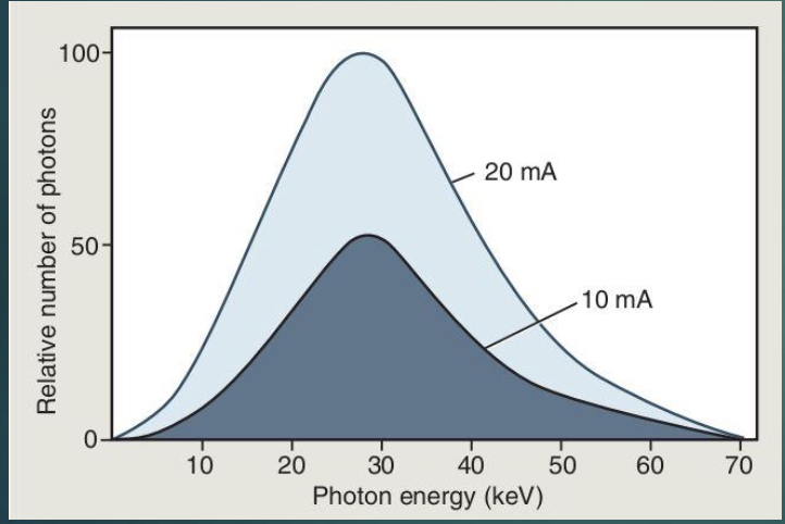
Define tube voltage peak (kVp)
increasing kVp- potential difference between cathode and anode
photon energy, number of photons generated
beam quality
how does energy of x ray vary with voltage?
increased voltage = high energy x rays (higher quality and density)
how does time relate to exposure?
increased time = increased exposure
filtration
the process of removing all photons that do not contribute significantly to image formation
removes most of the low energy photons that only add to the patient’s exposure, as well as some of the high energy photons
mean (average) energy of the beam goes up
*what are the benefits of filtration?
reduces patient dose, improves image contrast
inherent filtration
glass tube, oil, metal housing
equivalent of 0.5-2 mm aluminum
added filtration
aluminum disc placed over port to prevent low energy x-rays from exiting port
total filtration
inherent filtration + added filtration
*government regulations for x-ray machines 50-70 kVp are regulated to use ______ mm aluminum equiv
1.5mm
*government regulations for x-ray machines above 70 kVp are regulated to use ______ mm aluminum equiv
2.5 mm
collimation
process of shaping beam to make it no bigger than the size of receptor,
lead lined, focuses an x ray beam on the target
can be round or rectangular
protects a patient from non-diagnostic radiation
what are dental x ray beams collimated to?
a circle 2.75 inches (7cm) in diameter
what are the advantages of collimation?
less patient exposure, less operator exposure, less scatter generation and so improved contrast
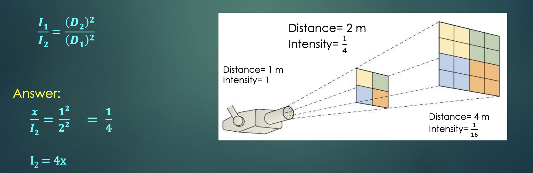
*inverse square law
intensity of beam inversely proportional to the square of the distance between the source and target
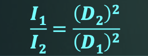
what are the variables for the inverse square law?
I1- initial beam intensity
I2- beam intensity at a new location
D1- original distance from the source when intensity was I1
D2- distance of the new location from the source
if the intensity of beam at 2m from an x-ray source is ‘x’, what will the intensity be at a distance of 1m?
I2 = 4x
what percentage of x-ray beams have no interaction?
9%
what percentage of x-ray beams scatter coherently?
7%
what percentage of x-ray beams exhibit photoelectric absorption?
27%
what percentage of x-ray beams compton scatter?
57%
what type of beam attenuation is desired?
photoelectric absorption (27%)
what type of beam attenuation is unwanted?
compton scattering (57%)
what is coherent scattering?
incoming x ray photon is deflected by the electrostatic field of an outer orbital electron
resultant photon changes direction without loss of energy
only interaction where no energy loss occurs with change in direction
8% of all interactions
classical, rayleigh, or elastic scattering
compton scattering?
caused when an x ray photon scatters off an electrion, ejecting it from orbit
recoil electron, scattered photon (may exit tissue or interact with more material)
decreases contrast (62% of all interactions)
probability directly proportional to electron density
probability of occurance increases with energy of incident photon
attenuation of the beam is greater in bone than soft tissue
creates fog on the images
where do most interactions of photoelectric absorption take place?
k shell because the electron density is highest here
photoelectric absorption
gives the characteristic x-ray formation, most interactions take place in the k shell
approx 30% of all interactions
how does photoelectric absorption work?
x-ray photon strikes an electron near the nucleus with enough energy to knock it off its orbit, overcoming the binding energy
the photon is totally absorbed and its energy is transferred to the ejected / recoil electron
tldr how can a photon with e- interact with matter?
no interaction
coherent- causes vibration, photon with same energy but different direction, no information gained
compton- outer shell e-, creates fog
photoelectric- inner shell e-, characteristic radiation, photons with the same amount of energy, what we want!
beam attenuation
the reduction in the intensity of x-ray beams as it traverses matter by either absorption or deflection of photons from the beam
measured using the half value layer (HVL)
HVL
the thickness of any material required to reduce the intensity of the beam in half
beam hardening
mean energy of the beam is increase
what factors affect attenuation?
energy of radiation, atomic number (z) of material through which the beam passes, tissue density, electron density
differential attenuation
image contrast
different body parts attenuate the x ray beam to different extents depending on their density, thickness, energy of the beam, beam size, ect
exposure
ability of x rays or gamma rays to ionize air, measured as a charge per mass of air
traditional unit was roentgen (R) which was described as 1R = 2.58 × 10C/kg
measures the intensity of the radiation field
R replaced by the SI equivalent of air kerma
kerma
kinetic energy released in matter
measure of the energy transferred from photons to electrons
measured in gray (Gy)
1 gray = 1 J/kg