Introduction to Oral Pathology
1/107
There's no tags or description
Looks like no tags are added yet.
Name | Mastery | Learn | Test | Matching | Spaced | Call with Kai |
|---|
No analytics yet
Send a link to your students to track their progress
108 Terms
Base of Lesion : flat base
sessile
Base of Lesion : between sessile and pedunculated
polypoid
Base of Lesion : stalk like
pedunculated
Surface Texture : wrinkled
corrugated
Surface Texture : a cleft or groove, normal or otherwise, show prominent depth
fissure
Surface Texture : resembling small projection or elevation found in clusters
papillary
Surface Texture : a term used to describe the surface texture of a lesion
folded

a color different from that of the surrounding tissue; it is flat and does not protrude above the surface of the normal tissue (i.e. freckle)
macule
a small, circumscribed lesion usually <1cm in diameter that is elevated or protrudes above the surface of normal surrounding tissue
papule
a palpable solid lesion up to 1 cm in diameter found in soft tissue; it can occur above, level with, or beneath the skin surface
nodule
a segment or lobe that is a part of the whole lesion; these lobes sometimes appear fused together
lobule
a small, elevate lesion <1cm in diameter that contains serous fluid
vesicle
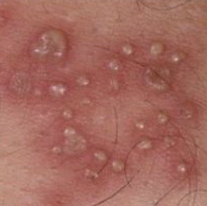
variously sized circumscribed elevations containing pus
pustule
a circumscribed, elevated lesion >5mm in diameter, usually contains serous fluid, looks like a blister
bulla
Radiographic Borders : borders that are not well demarcated, making it difficult to detect exact parameters
ill defined
Radiographic Borders: borders that are defined and in which one can clearly see the exact margins and extent of pathology
well circumscribed

Radiographic Density : less dense tissue (e.g. pulp, periapical lesions)
radiolucent
Radiographic Density : a mix of dense tissues (e.g. odontogenic lesions)
mixed radiopacity
Radiographic Density : highly dense structure (calcifications, bone, tooth)
radiopaque
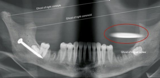
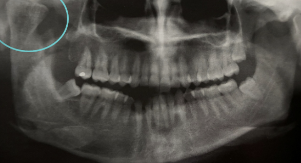
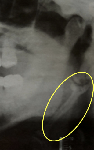
what is shown in the red circle
an artifact
Radiographic Shape : multiple single-chambered lesions that are somewhat fused together, making up the entire lesion; sometimes described as resembling soap bubbles
multilocular
Radiographic Shape: having on compartment or unit that is well defined or outlined
unilocular
Radiographic Adjacent Structures : the apex of the tooth appears shortened or blunted and irregularly shaped; occurs as a response to stimuli, which can include a cyst, tumor, trauma, malignancy
resorption
Radiographic Adjacent Structures : radiolucent lesion that extends between the roots, as seen in a traumatic bone cyst (TBC)
scalloping
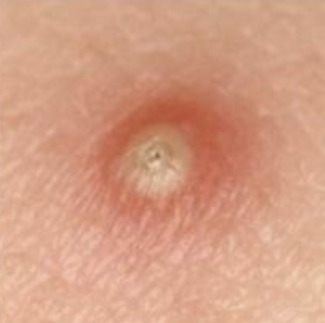
What is the term used to describe a pus filled lesion?
pustule
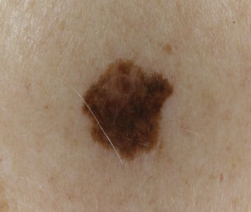
how would you describe this lesion?
a flat brown macule

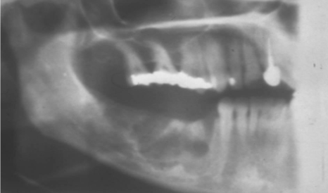
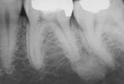
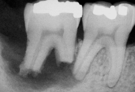
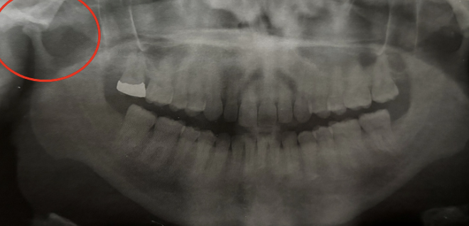
how would you describe this radiographic lesion?
an apical well-defined radiopaque lesion

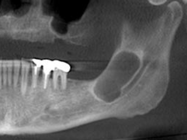
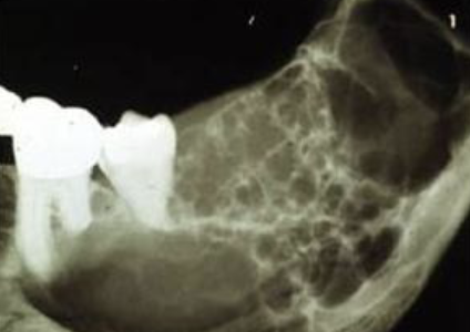
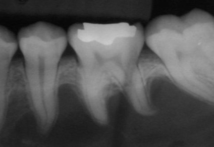
how would you describe this radiographic lesion?
a multilocular radiolucent lesion
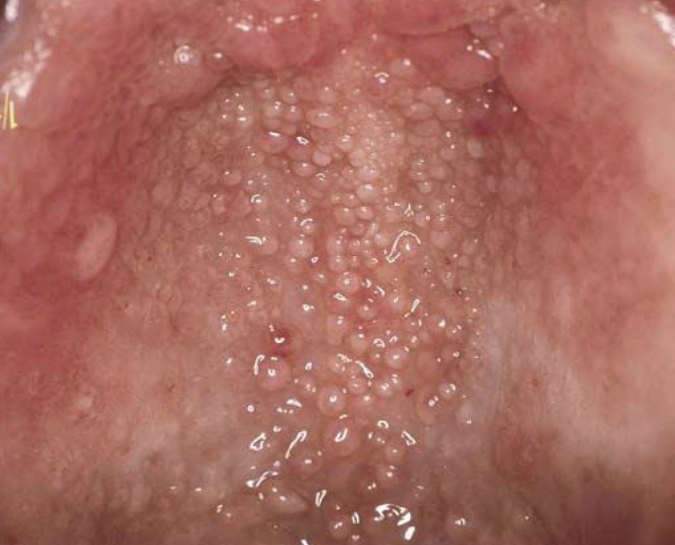
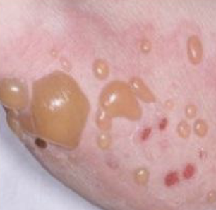
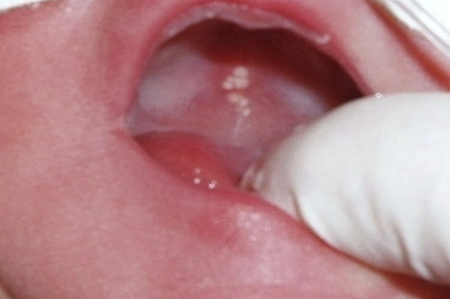
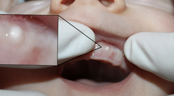
clusters of “ectopic” sebaceous glands; 80% of the population
fordyce granules
fordyce granules appearance
yellow or yellow-white papular lesions
fordyce granules most common locations
buccal mucosa and lateral portion of vermillion of upper lip
Do fordyce granules require tx?
no

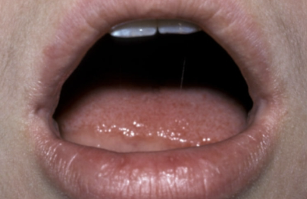
variation of normal; up to 90% of Black adults and 50% children (does not rub off)
leukoedema
leukoedema appearance
diffuse, gray-white, milky, opalescent
leukoedema most common locations
bilaterally on buccal mucosa
Does leukoedema require tx?
no
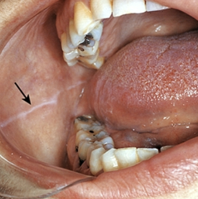
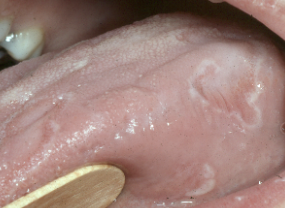
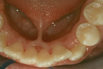
may be bilateral, can be more prominent in patients who have a clenching/ bruxing habit
linea alba
Linea Alba most common locations
anteroposterior on the buccal mucosa along the occlusal plane
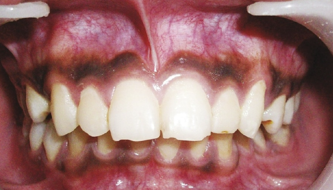
most commonly observed in patients of color, variant of normal
melanin pigmentation
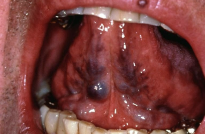
not associated with other systemic diseases, most commonly observed in individuals +60 years
lingual varicosities
lingual varicosities most common locations
ventral and lateral surfaces of the tongue
used to determine if a lesion is caused by blood within vessels (erythema) or hemorrhage (petechiae/purpura); plastic slide is pressed against lesion to produce temporary blanching by forcing blood out of superficial tissues
diascopy (positive test = blanching; redness fades, indicating inflammatory or vascular lesions)
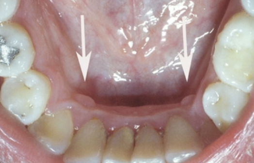
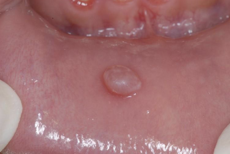
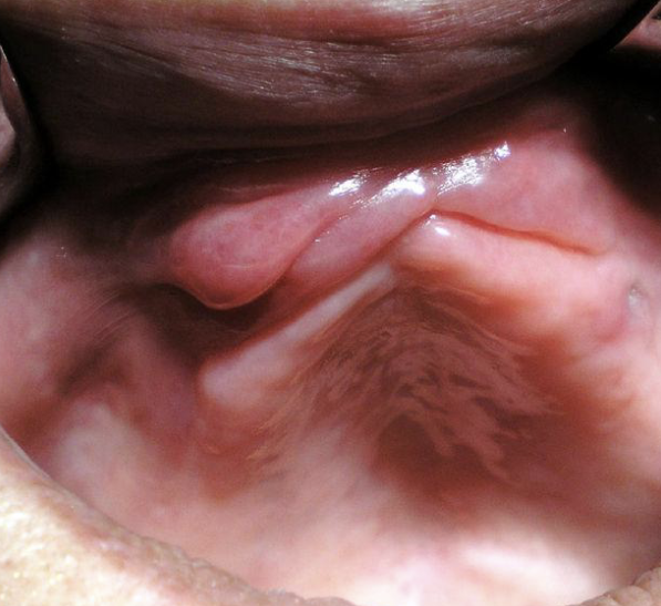
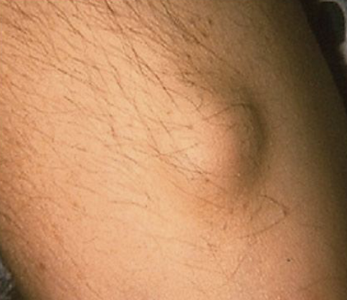
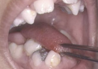
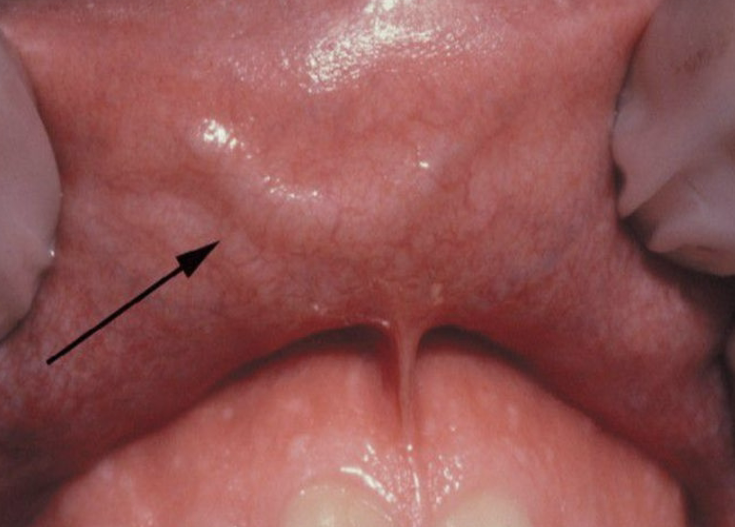
a sessile nodule on the gingival margin of the lingual aspect of the mandibular cuspids
retrocuspid papilla
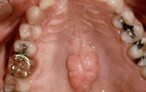
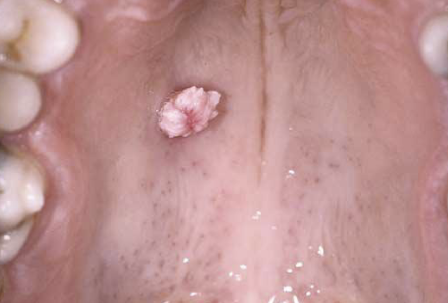
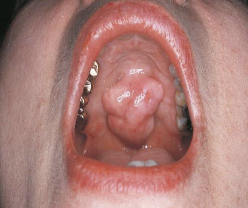
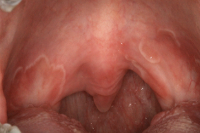
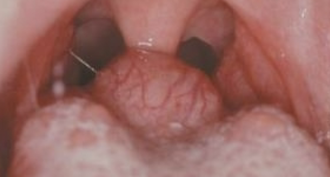
exophytic growth of normal compact bone on the hard palate
torus palatinus
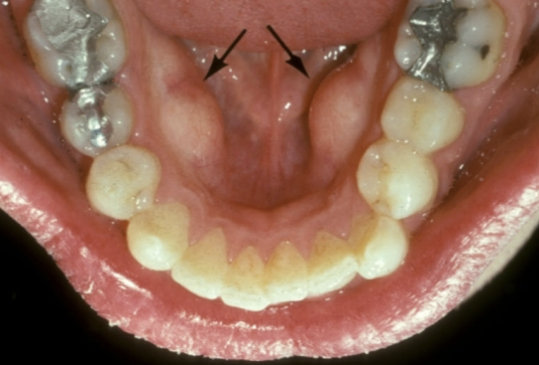
outgrowths of normal dense bone found on the lingual aspect of the mandible premolars are
mandibular tori
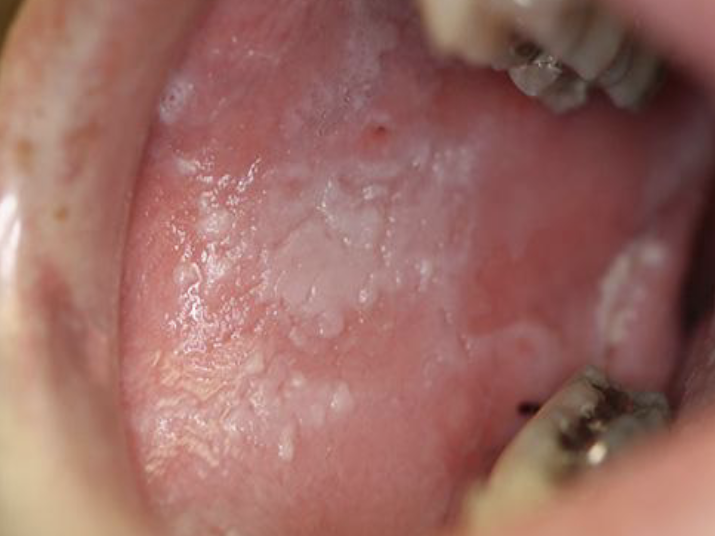
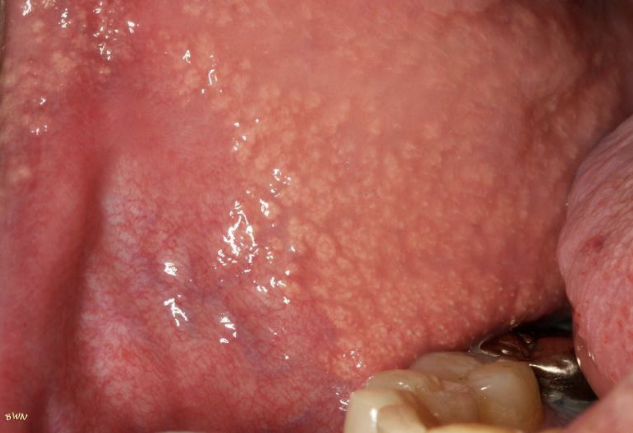
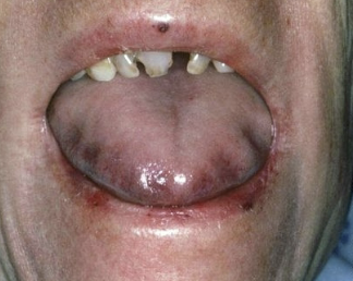
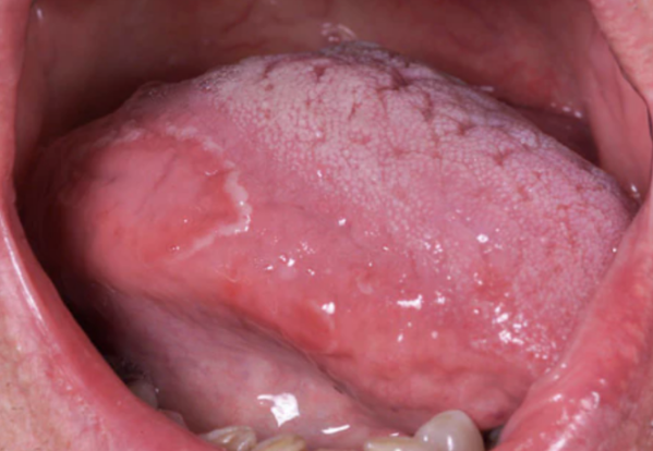
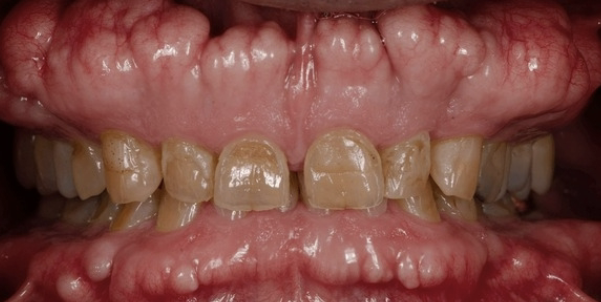
how would you describe this lesion?
generalized grey/ white lesion

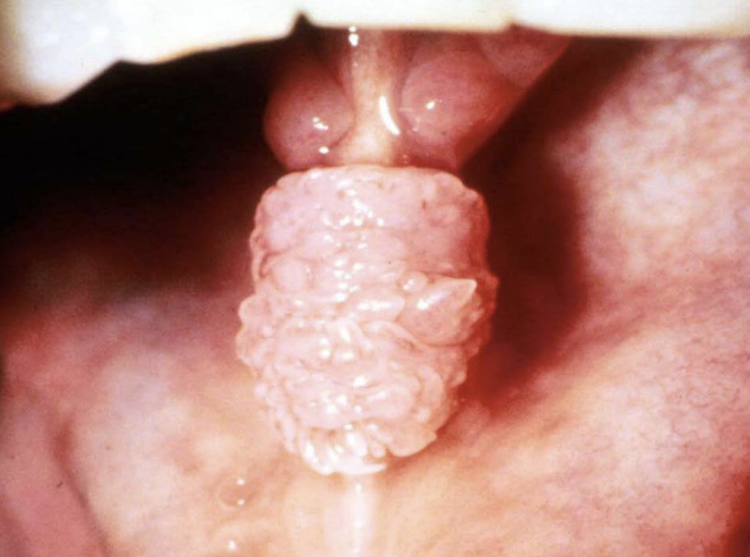
lingual thyroid
thyroid gland develops __ and descends into the neck by __
3-4 wks of life; 7th wk

__ % of ectopic thyroids are in this position
90
lingual thyroid is more common in
females (7:1)
In cases of lingual thyroid, this is the only thyroid tissue __% of the time
70
Is tx needed for lingual thyroid?
no tx needed unless a problem develops (adenoma or adenocarcinoma)
etiology unknown; if symptomatic, burning sensation may be due to candida; 10% of psoriasis patients have it, more common in females (2:1)
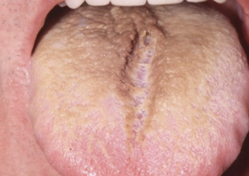
geographic tongue (benign migratory glossitis)
erythematous patches surrounded by white/ yellow serpentine borders
erythema migrans
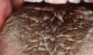
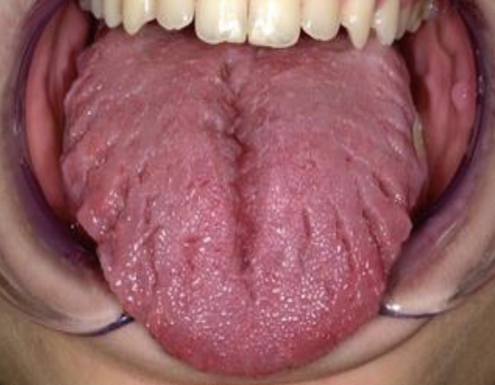
excess keratin on surface of filiform papillae; starts white, may become black, brown, orange, green, yellow
hairy tongue
cause of hairy tongue
uncertain but can be due to smoking, drugs, xerostomia, hx of radiotherapy to H&N, poor oral hygiene
tx of hairy tongue
remove offending agent, brush/ scrape tongue
stain is from food/ drink stains, chromogenic bacteria, may cause halitosis
hairy tongue
abnormally small tongue
microglossia
commonly associate w/ hypoplasia of the mandible; oromandibular-limb hypogenesis syndromes
microglossia
abnormally large tongue
macroglossia
most commonly caused by vascular malformations, muscular hypertrophy; other etiologies are down syndrome, amyloidosis, angioedema, tumors
macroglossia
abnormally short, thick lingual frenum resulting in limitation of tongue movement
ankyloglossia
tx for ankyloglossia
frenectomy
mucosal invagination that occur at the corners of the mouth on the vermillion border, not associated with facial or palatal clefts, no tx required
commissural lip pits
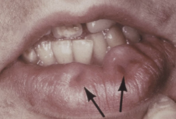
common vascular anomaly seen in the upper or lower lip of adults
caliber persistent artery
unique feature of caliber persistent artery
pulsation
is tx recommended for caliber persistent artery
no, tx may cause brisk bleeding
Which of the following is FALSE regarding lingual thyroid?
biopsy is recommended
what is the management?
none (geographic tongue)
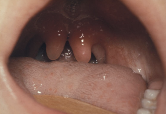
Cyst of Newborn: occurs along the median palatal raphe; due to epithelial entrapment by palatal fusion
epstein’s pearls
Cyst of Newborn: occur along the buccal and lingual aspects of the alveolar ridge
bohn’s nodule
localized bony protuberance that arise from the cortical plate; may be due to stress placed on the bone
exostosis
excessive growth of condyle; due to neoplasms and endocrine disturbances
condylar hyperplasia
underdeveloped condyle; due to congenital or acquired
condylar hypoplasia
elongation of styloid process or mineralization of the stylohyoid ligament; pain during opening, swallowing, turning head sideways; usually bilateral
eagle syndrome
Which of the following is TRUE for eagle syndrome?
pain when turning head sideways
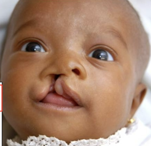
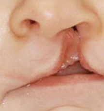
defective fusion of the medial nasal process with the maxillary process
cleft lip
failure of the palatal shelves to fuse
cleft palate
disturbances in the growth of tissue processes or their fusion may result in __
orofacial cleft
Cleft Lip + Cleft Palate
45%
Cleft Palate only
30%
Cleft Lip only
25%
most common major congenital defects
CL + CP is more common in
males
CP is more common in
females
__% of CL is unilateral
80
__% of unilateral CL are associated with CP
70
__% of bilateral CL are associated with CP
extends to the nostril
complete cleft lip
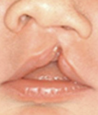
does not involve the nose
incomplete CL
complete clefts involving the alveolus usually occur between __ and __
lateral incisor and canine
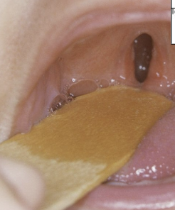
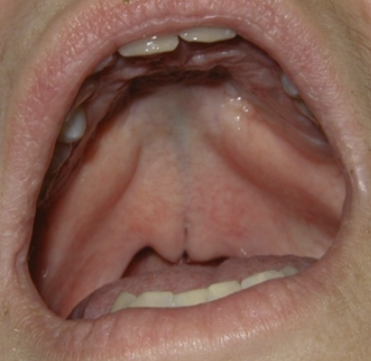
minimal manifestation of CP
bifid uvula
surface is intact but defect exists in the underlying musculature of the soft palate
submucous palatal cleft
cleft palate, mandibular micrognathia, glossoptosis (post. motion of tongue)
pierre robin syndrome (sequence)
congenital invagination of the lower lip, usually bilateral, no tx except to evaluate for van der woude syndrome
paramedian lip pits
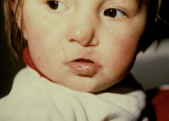
AD, CL+CP or CP only, paramedian lip pits
van der wounde syndrome
syndromes with orofacial cleft lip
pierre robin and van der woude
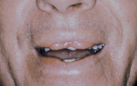
double lip