Week 5 - Psychobiology & Neuroscience
1/39
There's no tags or description
Looks like no tags are added yet.
Name | Mastery | Learn | Test | Matching | Spaced | Call with Kai |
|---|
No analytics yet
Send a link to your students to track their progress
40 Terms
The Nervous System
the pathway along which the brain sends and receives information about the body and environment.
The Nervous System - Central Nervous System (CNS)
Brain
Spinal cord
Responsible for processing and integrating information.
The Nervous System - Peripheral Nervous System (PNS)
Acts as a relay between CNS and:
Organs
Limbs
Skin
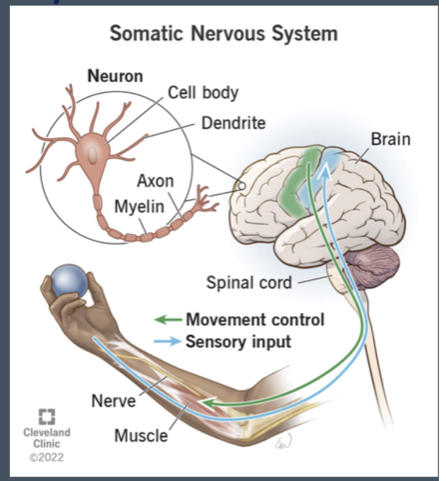
Divisions of the PNS - Somatic Nervous System
Controls voluntary movement.
Sensory (afferent) neurons → carry information TO CNS
Motor (efferent) neurons → carry information FROM CNS
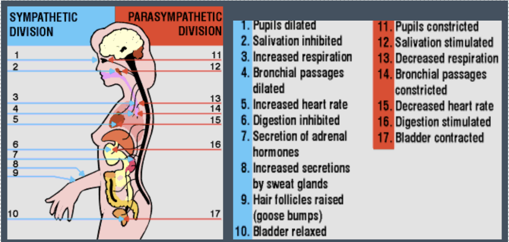
Divisions of the PNS - Autonomic Nervous System
Controls involuntary functions.
Sympathetic system → prepares body for fight or flight
Parasympathetic system → calms body after danger
Neurons
there are ~85 billion neurons in the brain
Receive, integrate, and transmit information
Many types but similar structure
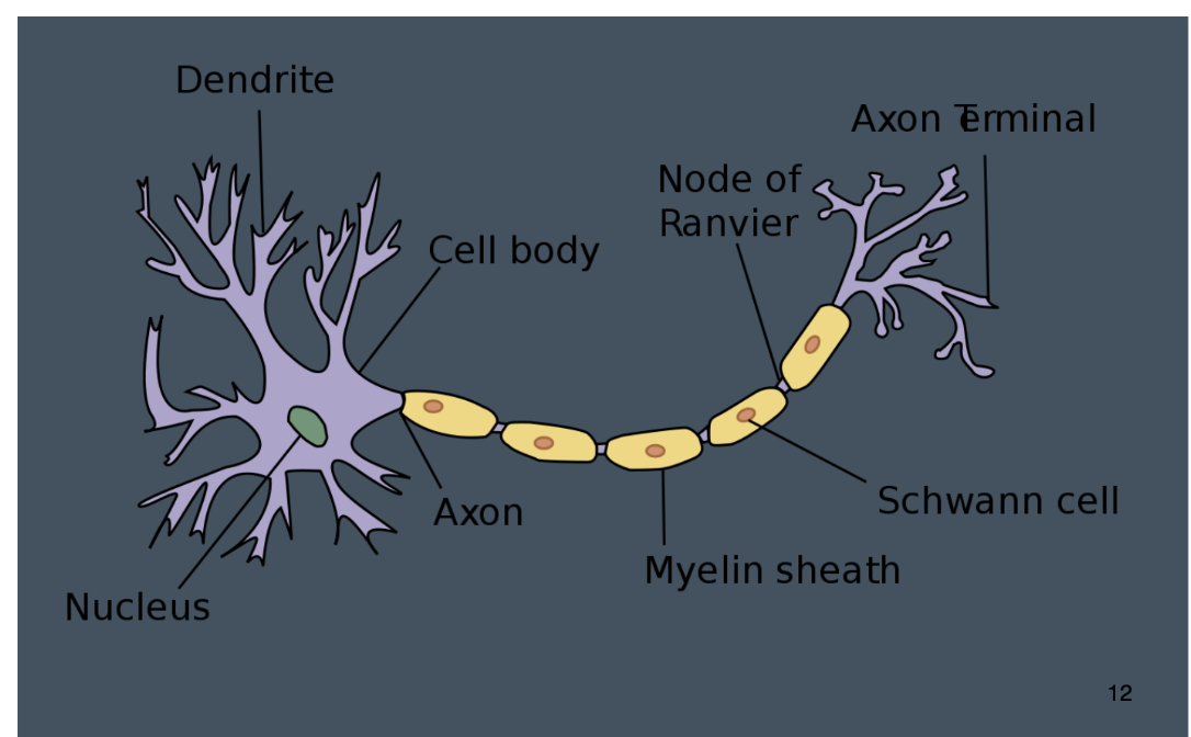
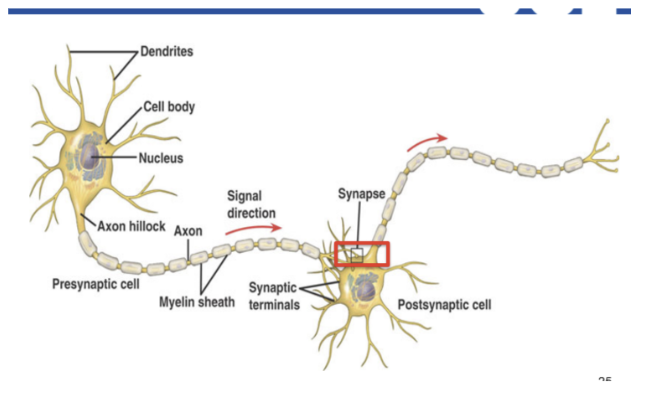
Structure of a Neuron - Dendrites
Receive information from other neurons
Structure of a Neuron - Cell Body (Soma)
Contains nucleus and genetic code
Structure of a Neuron - Axon
Carries electrical signal away from soma
Structure of a Neuron - Myelin Sheath
Fatty covering axon
Increases speed of transmission
Structure of a Neuron - Axon Terminal
Where neurotransmitters are released
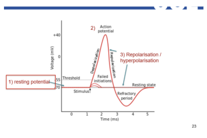
The Neuronal Impulse/Action Potential
An electrical signal sent down the axon to the axon terminal
All-or-nothing event
Either fires fully or not at all
The Neuronal Impulse/Action Potential - Stages
Resting Potential: The neuron is "charged" and ready, but quiet (at -70mV).
Depolarisation: The "fire" button is pressed. Sodium rushes in, and the charge spikes up to +40mV.
Repolarisation: The reset begins. Potassium rushes out, and the charge drops back down.
Hyperpolarisation: The reset "overshoots," making the neuron briefly more negative than usual so it can't fire again too quickly.
Return to Rest: The cell uses its internal "pumps" to get back to the original -70mV level.
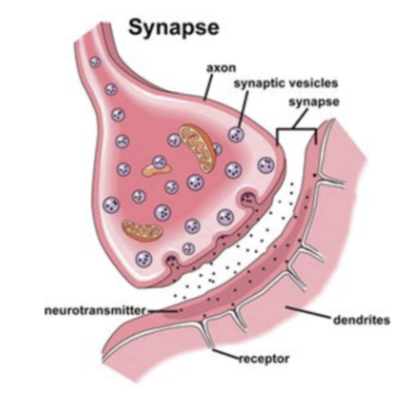
The Synapse & Synaptic Transmission
Vesicles & Transport: Synaptic vesicles (tiny sacs) carry chemical messengers called neurotransmitters to the very end of the neuron (the axon terminal).
The Release: The electrical impulse pushes these vesicles to the edge, where they release the neurotransmitters into the synapse (the tiny gap between neurons).
The Binding (Lock & Key): The neurotransmitters float across the gap and bind to receptors on the next neuron. Like a lock and key, each chemical only fits its specific receptor to pass the signal along.
Drugs and Neurotransmission - Amphetamines
Stimulate dopamine release (entire supply of dopamine is released into synaptic gap)
Block reuptake
Hence ‘high’ is even more intense than with cocaine → leads to stronger mood crash
Drugs and Neurotransmission - Cocaine
Blocks reuptake of dopamine & noradrenaline
Drugs and Neurotransmission - SSRIs
Block serotonin reuptake
Increase serotonin in synaptic cleft
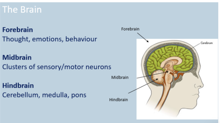
The Brain - Major divisions - Forebrain
Thought
Emotion
Behaviour
The Brain - Major divisions - Midbrain
Sensory and motor clusters
The Brain - Major divisions - Hindbrain
Cerebellum
Medulla
Pons
Hindbrain Structures - Medulla & Pons
Essential functions
Cardiac function (pump for the circulatory system)
Breathing
Sleep-wake cycle
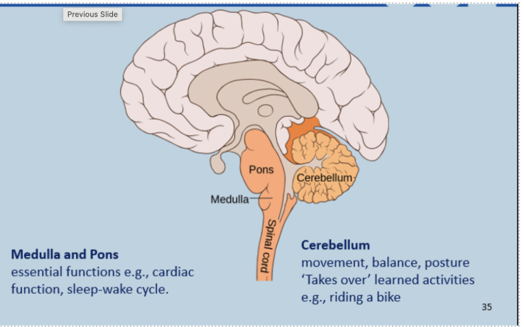
Hindbrain Structures - Cerebellum
Movement
Balance
Posture
Learned motor skills (e.g., riding a bike)
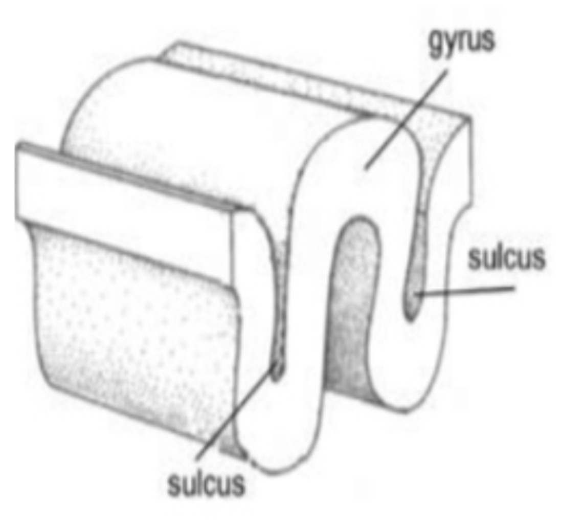
Cerebral Cortex
Spread out = ~2500 cm²
Convoluted surface
Gyrus = ridge
Sulcus = groove/valley
Cerebral Cortex - Grey vs White Matter
Grey matter → cell bodies, dendrites, synapses
White matter → axons connecting grey matter
Lobes of the Cerebrum - Frontal Lobe
Largest lobe
Executive functions:
Planning
Inhibition
Working memory
Attention shifting
Goal-directed behaviour
Lobes of the Cerebrum - Temporal Lobe
Auditory processing
Primary auditory cortex
Speech comprehension (left hemisphere)
Lobes of the Cerebrum - Parietal Lobe
Integrates sensory information
Spatial processing
Navigation
Number processing (intraparietal sulcus)
Lobes of the Cerebrum - Occipital Lobe
Visual cortex
Vision:
Form
Motion
Colour
Limbic System - Amygdala
Emotion processing
“Danger detector”
Triggers fight or flight
Fear conditioning
Social cue processing
Emotionally arousing memories
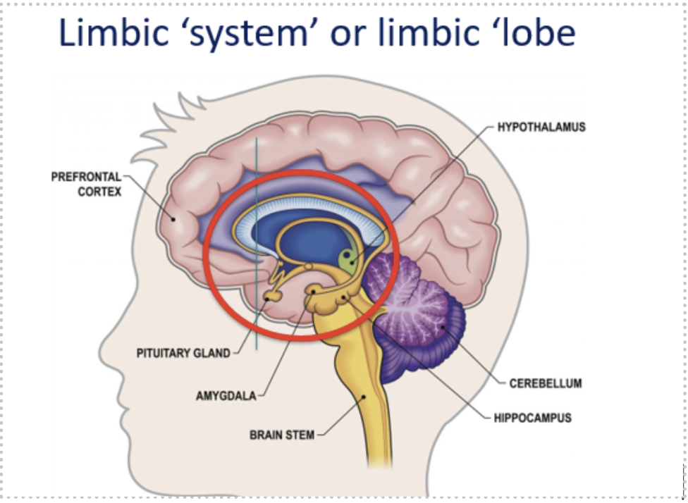
Limbic System - Hippocampus
Memory processing
Learning
Spatial recognition
Imagining the future
Damage → anterograde amnesia
Case Study: H.M.
Hippocampus removed for epilepsy
Personality unchanged
Could not form new memories
Brain Organisation - Localisation
Specific cognitive abilities located in specific brain areas.
Brain Organisation - Lateralisation
Some functions dominant in one hemisphere.
Brain Organisation - Lateralisation - Left Hemisphere
Controls right side of body
Language dominant
Brain Organisation - Lateralisation - Right Hemisphere
Controls left side of body
Spatial orientation
Face recognition
Creativity
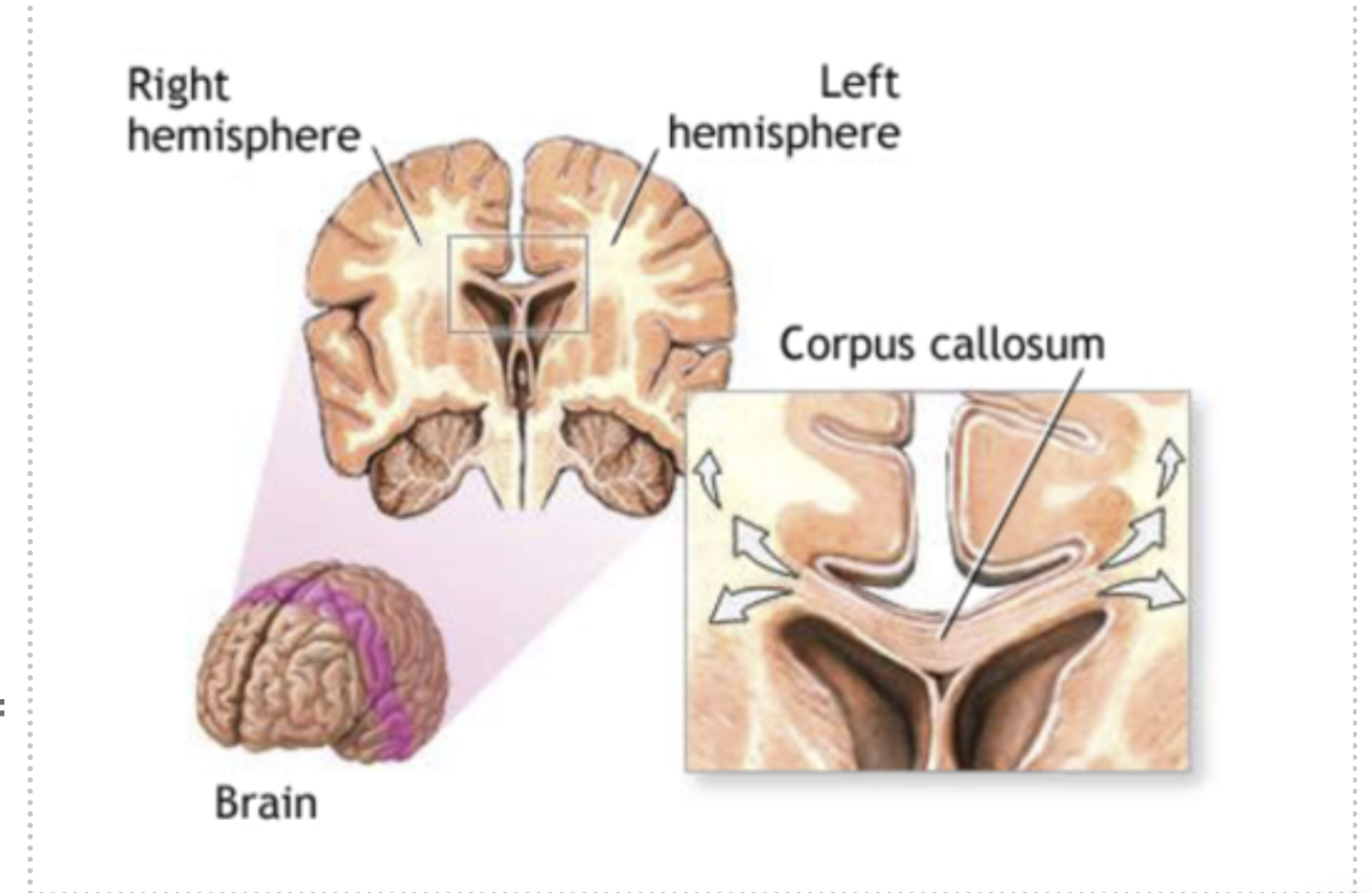
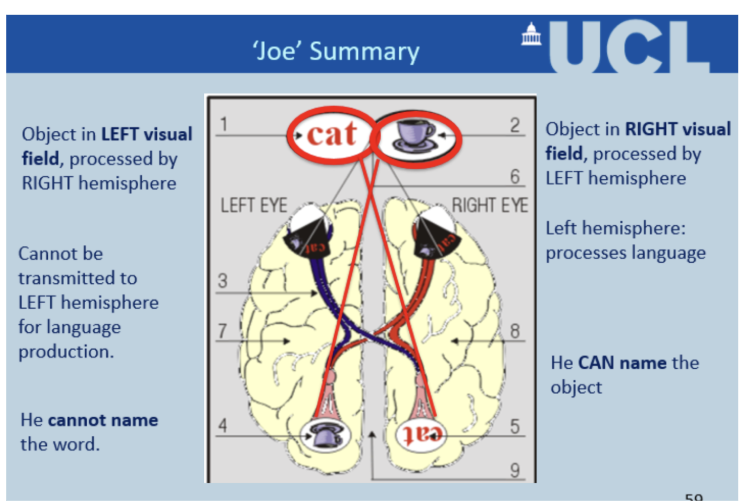
Split Brain Research (Sperry)
Corpus callosum (bundle of nerve fibers that allow your brain's left and right hemispheres to communicate) cut to treat epilepsy. Joe stares at center dot and objects are flashed either to the right side of the dot really or left
If object in:
Right visual field → Left hemisphere → Can name it
Left visual field → Right hemisphere → Cannot name it
Demonstrates hemispheric specialisation.
Neural Plasticity
Brain’s ability to change structure and function in response to experience or damage.
Higher in childhood but continues in adulthood.
e.g. London taxi drivers (Maguire et al. 2001): Taxi drivers who had completed "The Knowledge" (memorising thousands of London routes) had significantly larger posterior hippocampi than non-taxi drivers. Size correlated with years of experience. Demonstrates that sustained cognitive activity produces structural brain changes.
e.g. Musicians (Bengtsson et al., 2005)
Larger temporal cortex
Evidence for Plasticity
London Taxi Drivers (Maguire et al., 2001)
Larger posterior hippocampus
Musicians (Bengtsson et al., 2005)
Larger auditory cortex
Brain Damage Recovery (Thiel et al., 2006)
Right hemisphere compensates for left hemisphere language damage
Research Methods in Neuroscience - Lesion Studies
Study individuals with brain damage.
Advantages:
Stronger causal inference
Identify structure-function relationships
Disadvantages:
Often post-event
Lack experimental control
Ethical limitations
Research Methods in Neuroscience - EEG (Electroencephalogram)
Measures electrical activity via scalp electrodes.
Excited state:
Low amplitude
High frequency
Relaxed state:
High amplitude
Low frequency
Advantages:
Excellent temporal resolution
Inexpensive
Child-friendly
Disadvantages:
Poor spatial resolution
Research Methods in Neuroscience - fMRI
measures brain activity by detecting changes associated with blood flow
Active brain areas use more oxygen → detectable via magnetic changes.
fMRI Advantages:
Good spatial resolution
Examines brain networks
Disadvantages:
Poor temporal resolution
Expensive
Motion artefacts
Hard with children
Can cause anxiety