microscope quiz
1/39
There's no tags or description
Looks like no tags are added yet.
Name | Mastery | Learn | Test | Matching | Spaced | Call with Kai |
|---|
No analytics yet
Send a link to your students to track their progress
40 Terms
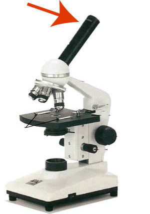
Eyepiece/Ocular Lens
What you look through, magnifies 10x
Revolving Nosepiece
Switches between objective lenses
Objective Lenses
Magnifies specimen (low, 4x) (medium, 10x) (high, 40x)
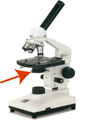
Stage
Where you put the specimen
Stage clips
Holds the specimen in place
Lamp
Supples light to view the specimen
Diaphragm
Opens/closes to control how much light passes through the specimen
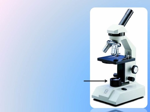
Coarse Adjustment Knob
Moves stage up/down to bring specimen roughly into focus
Fine adjustment knob
Brings specimen into sharper focus
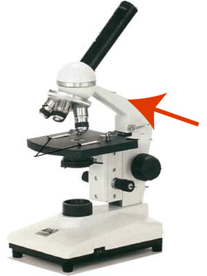
Arm
What you hold when carrying a microscope
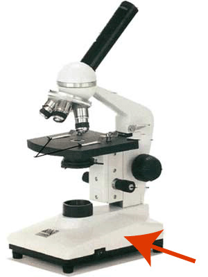
Base
Bottom of the microscope where you hold when carrying.
Eyepiece
Arm
Coarse Adjustment Knob
Fine Adjustment Knob
Revolving Nosepiece
Objective Lenses
Stage
Stage Clips
Diaphragm
Base
Lamp
Aristole (384 - 322 BC)
The Father of Biology; one of the first to use the scientific method.
Janssn Bothers (1590)
Dutch eyeglass makers. They made the first microsscope.
Galileo (1609)
Improved the microscope with better magnification
Schleidan and Schwann (1830)
Observed cells and came up with the idea that all living things are composed of cells.
Van Leeuwenhoek (1674)
First person to observe the movement of living cells
Hooke (1665)
Added lensess and light to Janssens design and came up with the idea of cells.
Virchow (1885)
Came up with the idea that new cellss arise fom pre-existing cells
Ernst and Knoll (1931)
Invented electron microscope
Zernik (1953)
Won nobel prize for phase contast method
The Cell Theory
All living thing are made up of cells
The cell is the smallest unit of life
All cells come from pre-existing cells
Magnification
How much larger the picture is than the real size of the specimen
HPFD/LPFD = LPM/HPM (how to find fov on high power)
formula you need to know
Scale = size of object in diagram/actual size of object (must be in the same units)
formula you need to know
Contrast
The difference in colour/light between the specimen and its background; it allows us to see depth.
Light Microscopes
Light passes through the specimen. It is cheaper compared to most microscops, small, and easy to use. Although, it has low magnitude, and it is hard to view live specimen.
Confocal Microscope
Uses lasers and computers to focus on a small area. It has highe resoultion, 3D imagery, and can view live specimen. Although, it is quite expensive, large and has low magnification.
Scanning Electron Microscope (SEM)
Beam of electrons is reflected off the specimen. It produces info about the surface of the specimen, can create 3D imagery, and has a high magnification (1 millionX). Although, its very large and expensive, does not view live specimen and is black and white.
Transmission Electron Microscope (TEM)
Beam of electrons is passes through the specimen. It produces info about the inner structure of the specimen, can create 3D imagery, and has a high magnification (1 millionX). Although, its very large and expensive, does not view live specimen and is black and white.