mass transport
1/47
There's no tags or description
Looks like no tags are added yet.
Name | Mastery | Learn | Test | Matching | Spaced | Call with Kai |
|---|
No analytics yet
Send a link to your students to track their progress
48 Terms
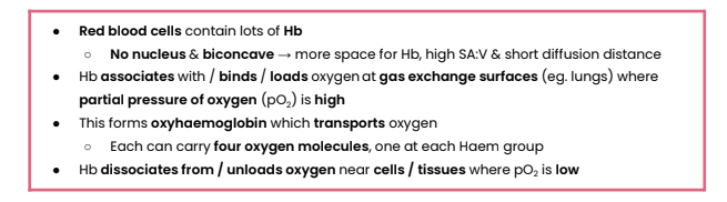
Describe the role of red blood cells & haemoglobin (Hb) in oxygen transport
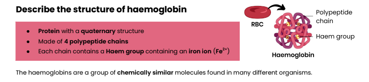
Describe the structure of haemoglobin
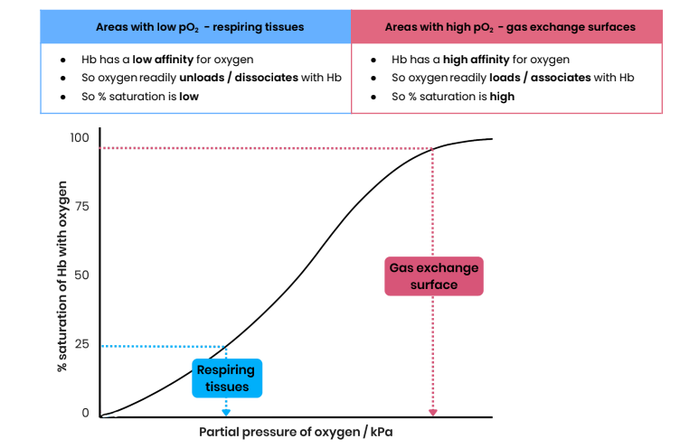
Describe the loading, transport and unloading of oxygen in relation to the
oxyhaemoglobin dissociation curve
Explain how the cooperative nature of oxygen binding results in an
S-shaped (sigmoid) oxyhaemoglobin dissociation curve
1. Binding of first oxygen changes tertiary / quaternary structure of haemoglobin
2. This uncovers Haem group binding sites, making further binding of oxygens easier
Describe evidence for the cooperative nature of oxygen binding
● At low pO2, as oxygen increases there is little / slow increase in % saturation of Hb with oxygen
○ When first oxygen is binding
● At higher pO2, as oxygen increases there is a big / rapid increase in % saturation of Hb with oxygen
○ Showing it has got easier for oxygen to bind
What is the Bohr effect?
● Effect of CO2 concentration on dissociation of oxyhaemoglobin
● Oxyhaemoglobin dissociation curve shifts right
Explain the effect of CO2 concentration on the dissociation of
oxyhaemoglobin
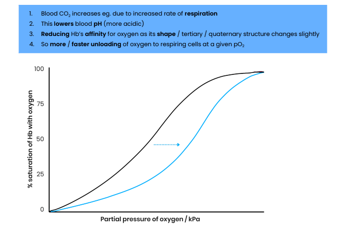
Describe evidence for the Bohr effect
At a given pO2 %, the saturation of Hb with oxygen is lower
Explain the advantage of the Bohr effect (eg. during exercise)
More dissociation of oxygen → faster aerobic respiration / less anaerobic respiration → more ATP produced
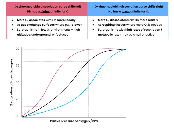
Explain why different types of haemoglobin can have different oxygen
transport properties
● Different types of Hb are made of polypeptide chains with slightly different amino acid sequences
● Resulting in different tertiary / quaternary structures / shapes
● So they have different affinities for oxygen
Explain how organisms can be adapted to their environments by having
different types of haemoglobin with different oxygen transport properties
shifting right= e.g. birds and rodents, which have higher metabolic rates
shifting left= e.g. llamas/ fetal Hb
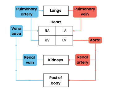
Describe the general pattern of blood circulation in a mammal
Closed double circulatory system - blood passes through heart twice for every circuit around body:
1. Deoxygenated blood in right side of heart pumped to lungs; oxygenated returns to left side
2. Oxygenated blood in left side of heart pumped to rest of body; deoxygenated returns to right
Suggest the importance of a double circulatory system
● Prevents mixing of oxygenated / deoxygenated blood
○ So blood pumped to body is fully saturated with oxygen for aerobic respiration
● Blood can be pumped to body at a higher pressure (after being lower from lungs)
○ Substances taken to / removed from body cells quicker / more efficiently
Draw a diagram to show the general pattern of blood circulation in a
mammal, including the names of key blood vessels
Name the blood vessels that carry oxygenated blood to the heart muscle
Coronary arteries - located on surface of the heart, branching from aorta
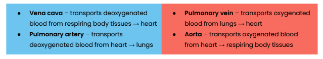
Name the blood vessels entering and leaving the heart and lungs
Name the blood vessels entering and leaving the kidneys
● Renal arteries – oxygenated blood → kidneys
● Renal veins – deoxygenated blood to vena cava from kidneys
Label a diagram to show the gross structure of the human heart (inside)
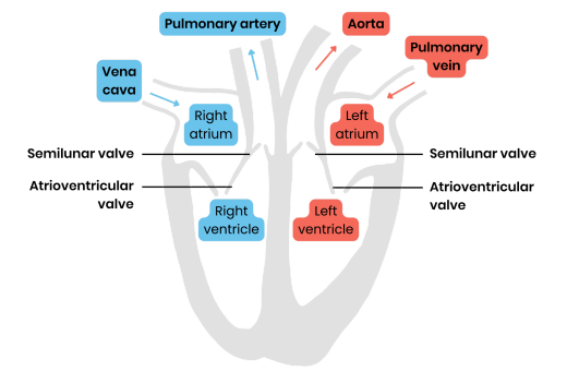
Suggest why the wall of the left ventricle is thicker than that of the right
● Thicker muscle to contract with greater force
● To generate higher pressure to pump blood around entire body
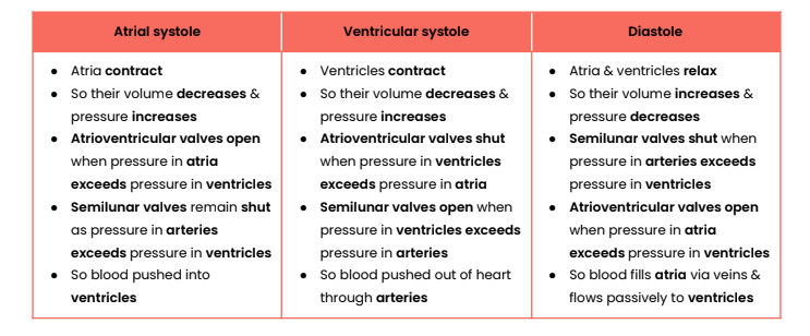
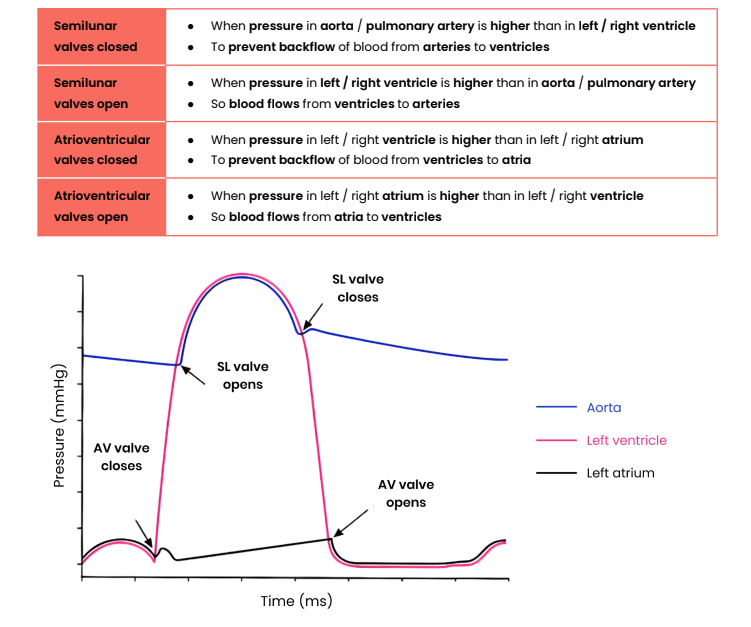
Explain the pressure & volume changes and associated valve movements
during the cardiac cycle that maintain a unidirectional flow of blood
Explain how graphs showing pressure or volume changes during the cardiac
cycle can be interpreted, eg. to identify when valves are open / closed
How can heart rate be calculated from cardiac cycle data?
Heart rate (beats per minute) = 60 (seconds) / length of one cardiac cycle (seconds)
Describe the equation for cardiac output
Cardiac output (volume of blood pumped out of heart per min)
= stroke volume (volume of blood pumped in each heart beat) x heart rate (number of beats per min)
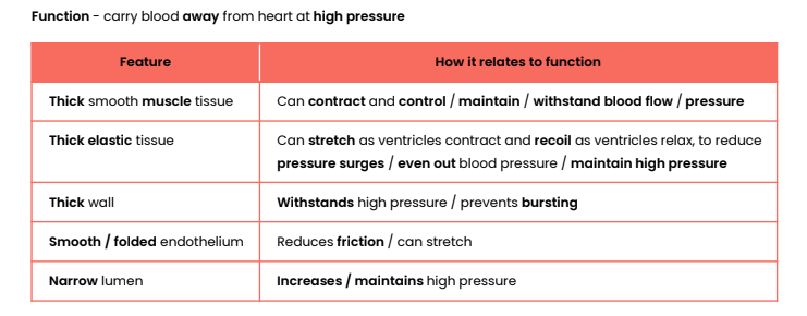
Explain how the structure of arteries relates to their function
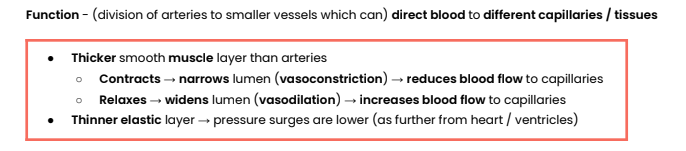
Explain how the structure of arterioles relates to their function
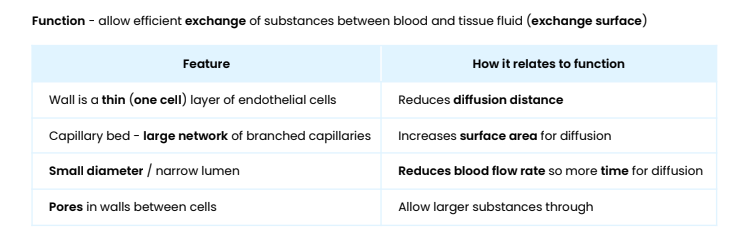
Explain how the structure of capillaries relates to their function
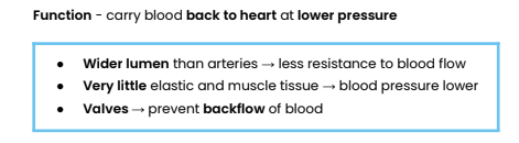
Explain how the structure of veins relates to their function
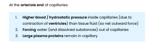
Explain the formation of tissue fluid
Explain the return of tissue fluid to the circulatory system
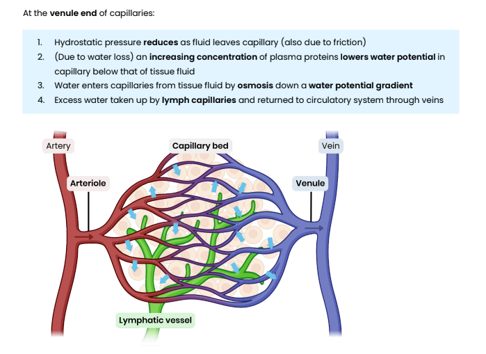
Suggest and explain causes of excess tissue fluid accumulation
● Low concentration of protein in blood plasma
○ Water potential in capillary not as low → water potential gradient is reduced
○ So more tissue fluid formed at arteriole end / less water absorbed at venule end by osmosis
○ Lymph system may not be able to drain excess fast enough
● High blood pressure (eg. caused by high salt concentration) → high hydrostatic pressure
○ Increases outward pressure from arteriole end AND reduces inward pressure at venule end
○ So more tissue fluid formed at arteriole end / less water absorbed at venule end by osmosis
○ Lymph system may not be able to drain excess fast enough
● Blockage in lymph system → excess fluid builds up and cannot be reabsorbed
What is a risk factor? Give examples for cardiovascular disease
● An aspect of a person’s lifestyle or a substance in a person’s body / environment
● That has been shown to be linked to an increased rate of disease
● Examples - age, diet high in salt or saturated fat, smoking, lack of exercise, genes
The principles of analysis, interpretation and evaluation of data covered in ‘3.2 gas exchange’ also apply here.
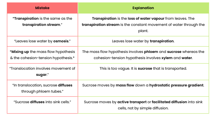
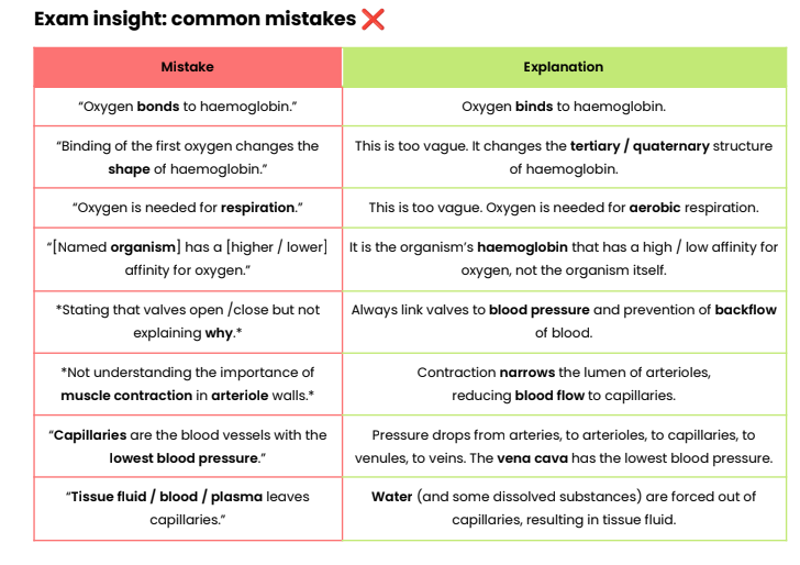
Exam insight: common mistakes ❌
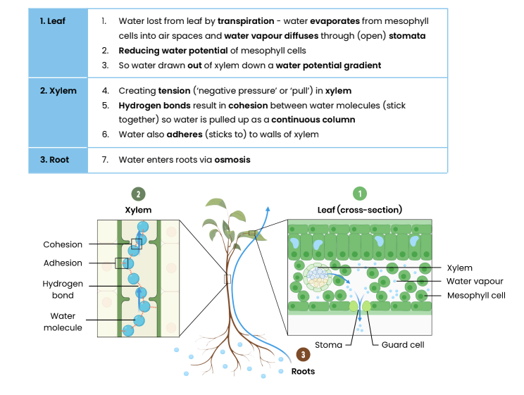
Describe the function of xylem tissue
Transports water (and mineral ions) through the stem, up the plant to leaves of plants
Suggest how xylem tissue is adapted for its function
● Cells joined with no end walls forming a long continuous tube → water flows as a continuous column
● Cells contain no cytoplasm / nucleus → easier water flow / no obstructions
● Thick cell walls with lignin → provides support / withstand tension / prevents water loss
● Pits in side walls → allow lateral water movements
Explain the cohesion-tension theory of water transport in the xylem
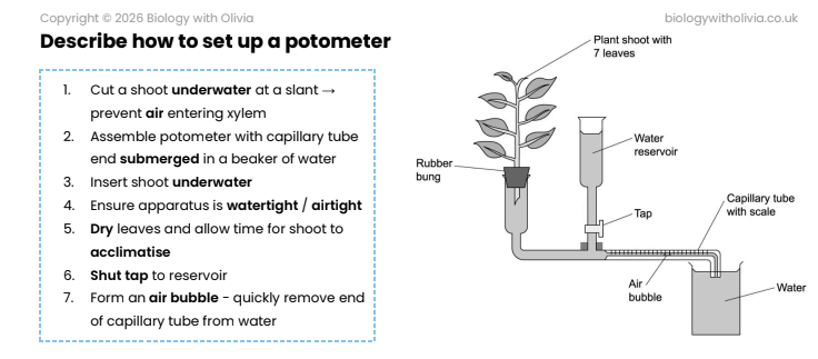
Describe how to set up a potometer
Describe how a potometer can be used to measure the rate of transpiration
Potometer estimates transpiration rate by measuring water uptake:
1. Record position of air bubble
2. Record distance moved in a certain amount of time (eg. 1 minute)
3. Calculate volume of water uptake in a given time:
○ Use radius of capillary tube to calculate cross-sectional area of water (πr2)
○ Multiply this by distance moved by bubble
4. Calculate rate of water uptake: divide volume by time taken
Describe how a potometer can be used to investigate the effect of a named
environmental variable on the rate of transpiration
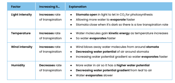
● Carry out the above, change one variable at a time (wind, humidity, light or temperature)
○ Eg. set up a fan OR spray water in a plastic bag and wrap around the plant OR change
distance of a light source OR change temperature of room
● Keep all other variables constant
Suggest limitations in using a potometer to measure rate of transpiration
● Rate of water uptake might not be same as rate of transpiration
○ Water used for support / turgidity
○ Water used in photosynthesis and produced during respiration
● Rate of movement through shoot in potometer may not be same as
rate of movement through shoot of whole plant
○ Shoot in potometer has no roots whereas a plant does
○ Xylem cells very narrow
Suggest how different environmental variables affect transpiration rate
Describe the function of phloem tissue
Transports organic substances eg. sucrose in plants
Suggest how phloem tissue is adapted for its function
1. Sieve tube elements
○ No nucleus / few organelles → maximise space for / easier flow of organic substances
○ End walls between cells perforated (sieve plate)
2. Companion cells
○ Many mitochondria → high rate of respiration to make ATP for active transport of solutes
What is translocation?
● Movement of assimilates / solutes such as sucrose
● From source cells (where made, eg. leaves) to sink cells (where used / stored, eg. roots) by mass flow
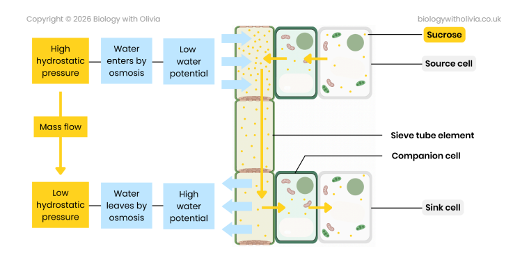
Explain the mass flow hypothesis for translocation in plants
1. At source, sucrose is actively transported into phloem sieve tubes / cells
2. By companion cells
3. This lowers water potential in sieve tubes so water enters (from xylem) by osmosis
4. This increases hydrostatic pressure in sieve tubes (at source) / creates a hydrostatic pressure gradient
5. So mass flow occurs - movement from source to sink
6. At sink, sucrose is removed by active transport to be used by respiring cells or stored in storage organs
Describe the use of tracer experiments to investigate transport in plants
1. Leaf supplied with a radioactive tracer eg. CO2 containing radioactive isotope
14C
2. Radioactive carbon incorporated into organic substances during photosynthesis
3. These move around plant by translocation
4. Movement tracked using autoradiography or a Geiger counter
Describe the use of ringing experiments to investigate transport in plants
1. Remove / kill phloem eg. remove a ring of bark
2. Bulge forms on source side of ring
3. Fluid from bulge has higher conc. of sugars than below, showing sugar is transported in phloem
4. Tissues below ring die as cannot get organic substances
Suggest some points to consider when interpreting evidence from tracer &
ringing experiments and evaluating evidence for / against the mass flow
hypothesis
● Is there evidence to suggest the phloem (as opposed to the xylem) is involved ?
● Is there evidence to suggest respiration / active transport is involved?
● Is there evidence to show movement is from source to sink? What are these in the experiment?
● Is there evidence to suggest movement is from high to low hydrostatic pressure?
● Could movement be due to another factor eg. gravity?
Exam insight: common mistakes ❌