Biology cell membranes
1/40
There's no tags or description
Looks like no tags are added yet.
Name | Mastery | Learn | Test | Matching | Spaced | Call with Kai |
|---|
No analytics yet
Send a link to your students to track their progress
41 Terms
Define what a eukaryotic organism is
Eukaryotic organisms are made of eukaryotic cells and have a nucleus
Define what a prokaryotic organism is
Prokaryotic organisms are made of prokaryotic cells and do not have a nucleus
What are the cell walls in plant cells made of?
It is a cellulose cell wall with plasmodesmata
What are the cell walls of algal cells made of? Do they have a chloroplast?
Their cell walls are made of chitin. Nah. Loser.
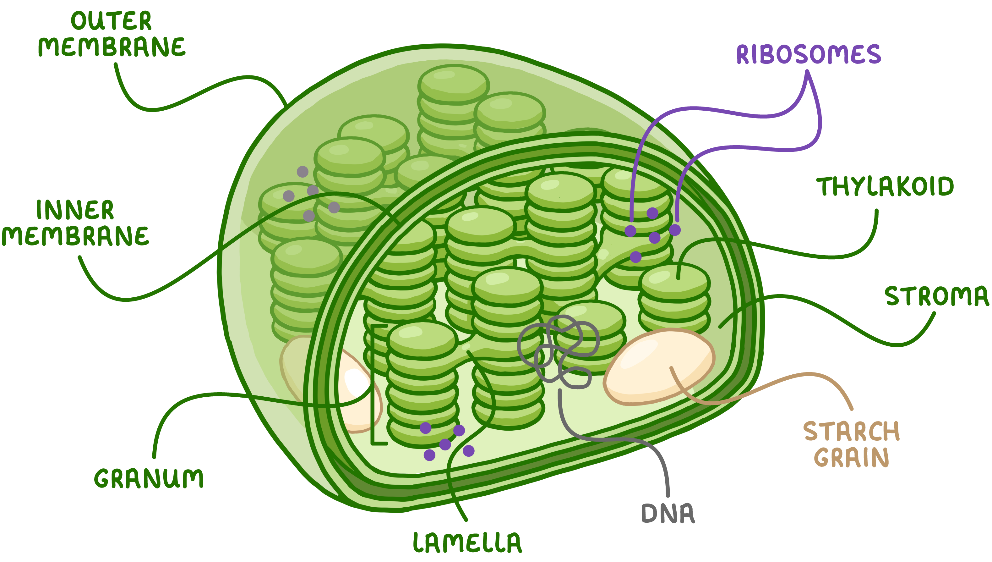
List the adaptations of chloroplast
Contains granal membrane which has a large surface area for photosynthesis as well as electron carriers and enzymes which are essential for the 1st stage of photosynthesis
Stroma contain fluid for the second stage of photosynthesis
thylakoid membrane Contains DNA and ribosomes to enable quick and easy manufacturing of proteins
The DNA in the chloroplast is circular
The function of chloroplast and a brief description of internal structure
Chloroplast- The site of which photosynthesis takes places
It is full of fluid which contains starch grain
The chloroplast envelope has a double plasma membrane which contains what gas goes in and out
Contains grana which has stacked thylakoids which contain chlorophyll
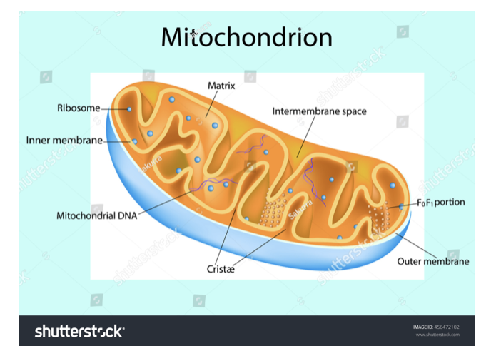
The function of mitochondria and its internal structure
Mitochondria are the sites of aerobic respiration
They provide all of the energy a cell requires - so more active cells (eg. muscles) will have greater numbers of mitochondria
They have a double membrane where the inner is folded to form cristae where ATP is produced.
Inside is the matrix which contains enzymes involved in respiration
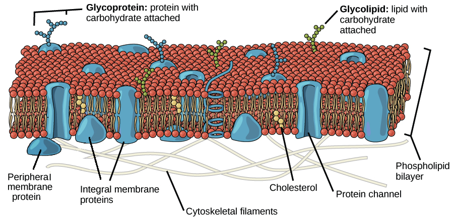
The function of plasma membrane and its internal structure
The plasma membrane controls the entry and exit of substances in and out of the cell
The plasma membrane is a phospholipid bilayer and has proteins embedded within it which are able to move (hence why it is called the phospholipid bilayer)
It has receptor molecules on it which allow it to respond to chemicals like hormones
Has cholesterol molecule which stabilises phospholipid membrane
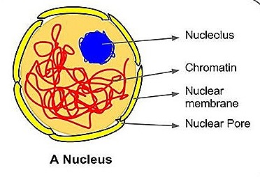
The function of nucleus’
The nucleus is the largest organelle and is surrounded by its own membrane called the nuclear envelope
The nucleus contains the DNA of the cell and is the site of DNA replication
The nucleus contains a small dark structure called the nucleolus which synthesises ribosomes
The DNA in a nucleus exists as chromatin due to its association with the histone protein allowing more DNA to fit into the nucleus
The nucleus controls all the activities of the cell
The pores of the nuclear envelope allows substances to move between the nucleus and the cytoplasm
Production of mRNA/tRNA
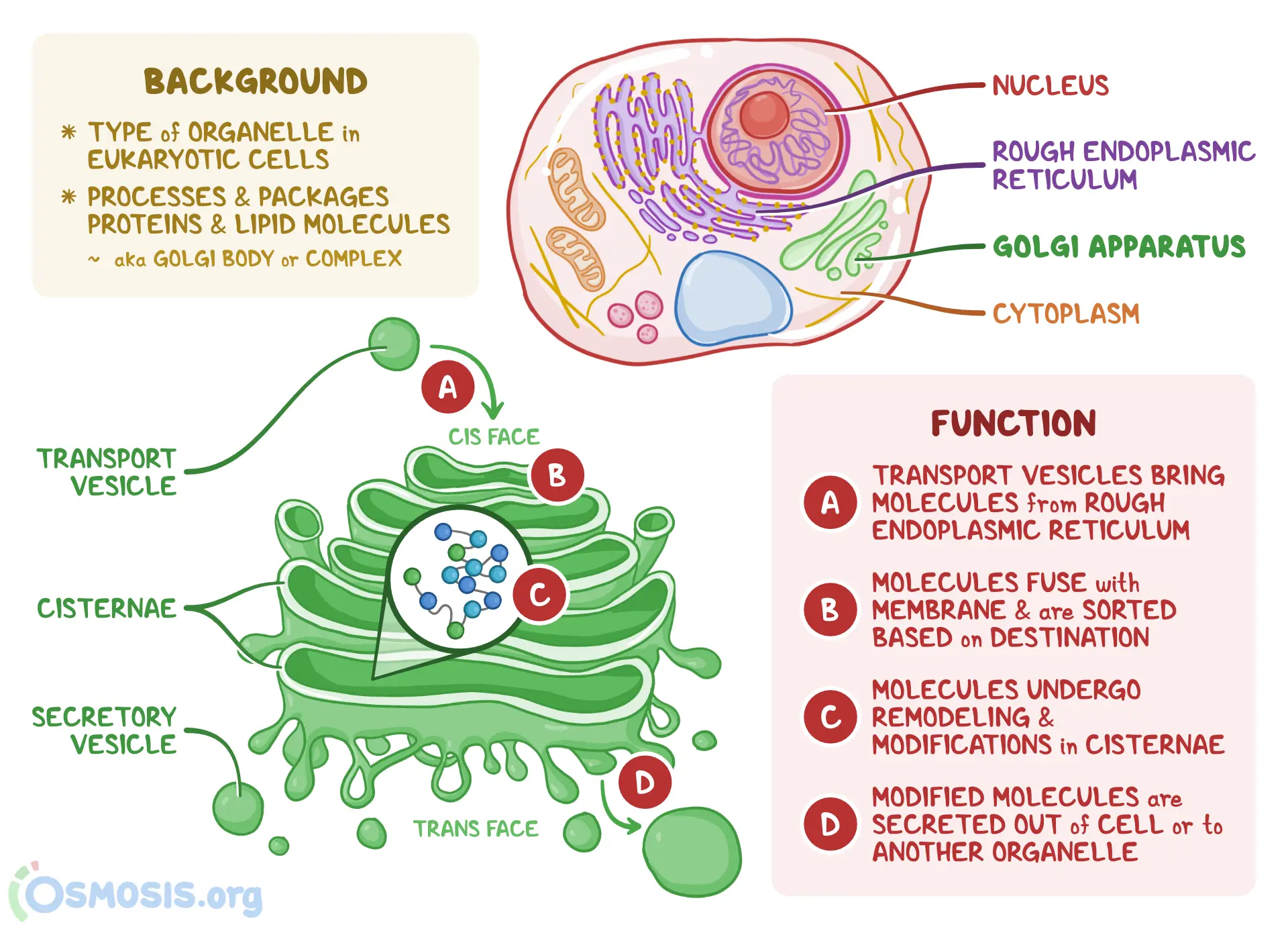
Name the function of the Golgi Apparatus
1. Golgi (apparatus)
Modifies / processes triglycerides
Combines triglycerides with proteins
Packaged for release / exocytosis
Name the function and internal structure of the ribosome
A ribosome is a tiny organelle that either float free in the cytoplasm or attatch to the rough endoplasmic reticulum
Ribosomes are the site of protein synthesis
Ribosomes are made up of proteins and ribosomal RNA
it consists of a light sub unit on the bottom and heavy subunit at the top

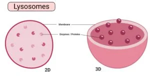
Lysosome
Lysosome are small round organelles which contain hydrolytic enzymes which are kept seperate from the rest of the cell by a membrane
The digestive enzymes of the lysosome are used to digest invading cells or even destroy the cell itself when needed
Name the function of endoplasmic reticulum
The endoplasmic reticulum is a series of membrane folds that connect to the nucleus envelope
the space between these folds is filled with fluid which transports substances around within the substance
Describe the function of smooth endoplasmic reticulum and rough endoplasmic reticulum
the smooth endoplasmic reticulum processes and packages lipids
This type of RER is studded with ribosomes and its function is to process proteins produced by the ribosome
Describe the function and composition of cell walls
Plant cells are formed of microfibrils of cellulose
Within a cell wall a thick layer called the middle lamella which is a boundary between plant cells that cements them all together
The cell wall in an algae cell is made of cellulose/glycoprotein
The cell wall of fungi is made of chitin
The vacuole keeps the cell rigid
The role of a cell wall is to provide strength to the plant and prevent the plant cells from bursting and to allow water to move through the plant
Explain structure prokaryotic cells
The cytoplasm in a prokaryotic cell has no membrane bound organelles, unlike a eukaryotic cell
it may have a flagellum which is a long hair like structure that rotates to make the prokaryotic cell move
Prokaryotic cells do not have a nucleus and instead the DNA floats free in the cytoplasm. Its circular DNA present are long coiled up strands not attatched to any histone protein.
The cell wall of prokaryotes are made up of murein. murein is a glycoprotein
Whats the deal with plasmids?
Plasmids are small loops of DNA that arent a part of the main circular DNA molecules
Plasmids contain genes for things like antibiotic resistance and can be passed between prokaryotes
not all prokaryotes have plasmids
What do some prokaryotes also have capsule ?
They have a capsule made up of secreting slime which helps to protect bacteria from attack by cells of the immune system.
What is a virus? What does it internal structure consist of?
Viruses are just nucleic acids surrounded by protein and are smaller than bacteria
Viruses have no plasma membrane no cytoplasm and no ribosome
it has a core of genetic material of either DNA or RNA
it has an attatchment protein which sticks out from the edge of the capsid
the protein coat around the core is called the capsid
What is the function of viruses
Viruses invade and hijack the internal structure of cells and reproduce inside of them
What is the difference between prokaryotic and eukaryotic dna
Eukaryotic DNA is long and linear whereas prokaryotic DNA is circular
Eukaryotic DNA is associated with the histone protein which tightly pack DNA allowing more to be stored in the nucleus, prokaryotic DNA is not associated with any protein
Eukaryotic DNA is found in the nucleus prokaryotic DNA is found in the cytoplasm
Eukaryotic DNA can be found in the mitochondria whereas Prokaryotic DNA cannot
What is binary fission? Name the steps of binary fission aswell
Binary fission is how prokaryotic cells replicate to form two daughter cells
-The circular DNA and plasmids replicate. The main DNA loop can only replicate once however the plasmid can replicate many times
The cell gets bigger and the DNA loops move to opposite pairs of the cell
The cytoplasm begins to divide and new cell walls begin to form
The cytoplasm divides and two daughter cells are produced. Each daughter cell has one copy of the circular DNA but can have a variable number of plasmids
Whats the deal with viral replication?
Viruses have attatchment proteins which are complementary to the receptors on the host cells.
The viruses use these attatchment proteins to bind to these receptors
This enables the virus to inject its genetic material into the host cell
Replicates the genetic material and creates more proteins
Then, it bursts out of the host cell and repeats the process
What is the equation for magnification?
magnification = actual size/ indepth size
How do transmission electron microscopes work?
Transmission electron microscopes use electrons
Allow for a greater resolution
So small organelles can be observed
Requires thin specimen and cannot show colour
Can only use dead specimens
How do scanning electron microscopes
Scanning electron microscopes scan a beam of electrons across the specimen. This knocks off electrons from the specimen which are gathered in a cathode ray tube to form an image
The images you end up with show the surface of the specimen and they can be 3-D
SEM’s are good because they can be 3D and used on thick specimens
But they give lower resolution images
What are the two types of microscopes
There is an optical (light) microscope which use light to form an image and have a maximum resolution of about 0.2 micrometers.
This means you cant use an optical microscope to view organelles smaller than 0.2 micrometers and this includes ribosomes, the ER and lysosomes. You can see the nucleus. The maximum useful magnification of an optical microscope is 1500x
There are electron microscopes which use electrons to form an image and have a higher resolution than optical microscopes to give a more detailed image
they have a maximum resolution of about 0.00002 micrometers
the maximum useful magnification of an electron microscope is about x1500000
How do you prepare a slide?
Pipette a small drop of water onto the slide
Then use tweezers to place a thin selection of your specimen on top of the water drop
add a drop of stain which highlights the objects in a cell
eg. iodine is used to stain starch grains in plant cells
Finally add. the cover slip. To do so, stand the slip upright on the slide next to the water droplet. then tilt and lower it so it covers the specimen. Try not to get any air bubbles under there or they will obstruct your view of the specimen
Name the steps of ultracentrifugation
the solution must be kept ice cold in order to reduce enzyme activity that could digest organelles
the solution must be kept in a buffer solution to maintain the ph and make sure none of the enzymes are denatured
The solution should be kept isotonic inorder to prevent damage of the organelles due to osmosis
homohenise the cell. We do this in a number of different ways for example using a blender to break up the plasma membrane in order to release organelles
Next we do filtration where the homogenised cell solution is filtered through a guaze to seperate any large debris or tissue debris
finally we go through ultra centrifugation
the cell fragments are poured into a tube and the tube is poured into a centrifyge and spun at a low speed with the heaviest organelles like nuclei get flung to the bottom of the tube by the centrifuge
they form a thick sediment at the bottom called the pellet. The rest of the organelles stay suspened in the fluid above the sediment, the supernatant
the supernatant is drained off and poured into another tube and spun in the centrifuge at the higher speed.
repeat the process until you get the organelles you require
Heaviest to lighest organelles
Nucleus, mitochondria, lysosome, e.r, ribosomes
Name the phases of interphase
G1- Cell grows and form new organelles and proteins are made
Synthesis occurs when DNA replicates
G2 - Cell keeps growing and proteins for mitosis are made
Name the phases of mitosis
interphase- the cell carries out the normal functions but the cell starts to divide
prophase- the chromosomes condense getting shorter and fatter. the bundles of protein called centrioles start moving to opposite ends of the cell forming a a network of protein across it called the spindle
the nuclear envelope breaks down and chromosomes lie free in the cytoplasm
metaphase- the chromosome each with two chromatids line up along the middle of the cell and become attatched to the spindle by their centromere
anaphase - the centromere divide, seperating each pair of sister chromatids. The spindles contract, pulling chromatids to opposite poles of the spindle, centromere first. this makes the chromatids appear v shaped
telophase - the chromatids reach the end of the poles and become long and thin again. they are now once again called chromosomes. a nuclear envelope reforms
cytokinesis takes place dividing the cytoplasm creating two identical daughter cells
Whats the deal with mitosis and cancer?
mitosis is the controlled division of cells
cancer is the uncontrolled division of cells
some cancer drugs target the cell cycle such as chemotherapy which prevent the synthesis of enzymes needed for DNA replication
List the mitosis practical
cut 1cm from the tip of a growing root. it needs to be from the tip because thats where the growth occurs and where mitosis takes place
prepare a boiling tube containing 1M hydrochloric acid and put it in a water bath at 60C
transfer the root tip into the boiling tube and incubate for about 5 minutes
use a pipette to rinse the root tip with cold water. leave the tip to dry on a paper towel
Place the root tip on a microscope and cut 2mm from the very tip of it. Get rid of the rest.
use a mounted needle to break the tip open and spread the cell out thinly
Add a few drops of stain and leave it for a few minutes. The stains will make chromosomes easier to see under a microscope. There are loads of different stains all with different mass
Place a coverslip over the cells and push down firmly to squash the tissue.This will make the tissue thinner and allow light to pass through it. Don’t smear the coverslip sideways or you’ll break the chromosomes
Look at all the stages of mitosis under the optical microscope
How do you observe cells using optical microscopes?
Clip the slide you prepared onto the stage
select the lowest powered objective lens
use the coarse adjustment knob to bring the stage up to just below the objective lens
look down the eyepiece and use the coarse adjustment knob to move the stage downwards away from the objective lens until the image is roughly in focus
adjust the focus with the fine adjustment knob until you get a clear image of whats on the slide
if you need to see the slide with greater magnification to swap to a high powered objective lens and refocus
an eyepiece graticule is filled onto the eyepiece which is like a transparent ruler with numbers but no units
what is the equation for mitotic index
mitotic index= number of cells with visible chromosomes / total number of cells observed
What is a stage piece micrometer?
the stage piece micrometer is placed on the stage - it is a microscope slide with an accurate scale and is used to work out the value of each division on the eyepiece graticule
artefacts definition
artefacts are things that you can see down the microscope that arent aoart if the cell or specimen
they are especially more common in electron micrographs
Define magnification
The magnification of an object refers ton how many times larger the image is compared to the object.
Define the resolution
The resolution of a microscope is the minimum distrance between two objects in which they can still be viewed as seperate.
What is an eyepiece graticule?
an eyepiece graticule is filled onto the eyepiece which is like a transparent ruler with numbers but no units. We need to calibrate the eyepiece to work out what each division is worth.
The scale on the stage micrometer is typical 2mm wide wiith the divisions being 10 micrometers apart