Bony spaces of the head
1/64
There's no tags or description
Looks like no tags are added yet.
Name | Mastery | Learn | Test | Matching | Spaced | Call with Kai |
|---|
No analytics yet
Send a link to your students to track their progress
65 Terms
What are the 2 broad categories of the bony spaces of the head
intra-cranial
extra-cranial
What makes up the boundaries of the intracranial space
the cranial vault/ skull cap
Contents of the cranial vault
brain
associated nerves
vasculature
cerebrospinal fluid
meninges
What is the largest bony space of the head
the main vestibule of the cranial vault
What are some of the smaller spaces within the cranial vault
tympanic cavity
epitympanic recess
facial canal
mastoid air cells
pituitary fossa
How is the cranial vault divided into compartments?
-divided into 2 continuous compartments by inward folds of the meningeal layer of dura mater
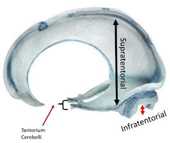
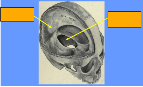
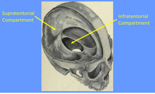
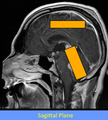
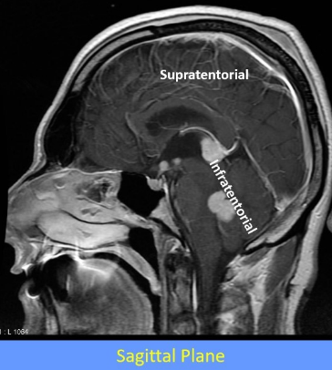
What is the tentorium cerebelli and what does it do
tent shaped fold of dura mater
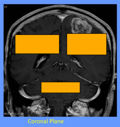
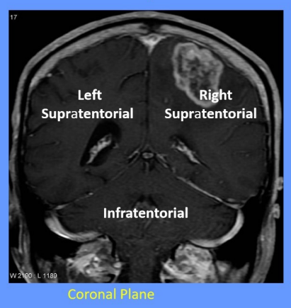
divides the intracranial cavity into supratentorial and infratentorial compartments
What are the posterior, lateral and anterior attachements of the tentorium cerebelli
posterior- internal occipital protuberance and transverse sulci
lateral- superior border of the petrous part of the temporal bone
anteriorly- anterior and posterior clinoid processes
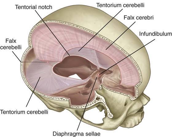
What divides the supratentorial compartment
divided by the falx cerebri into the left and right supratentorial space which can communicate with each other and with the infratentorial space
What is the falx cerebri
vertical fold of the dura mater located in the longitudinal fissure that separates the left and right cerebral hemispheres
Anterior, superior and posterior attachements of the falx cerebri
anterior- crista galli
posterior- internal occipital protuberance
superior- internal surface of calvaria along the sagittal suture
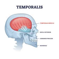
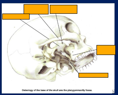
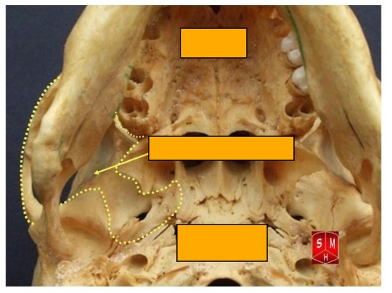
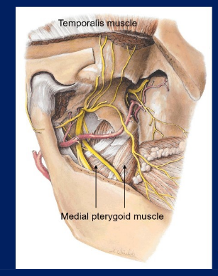
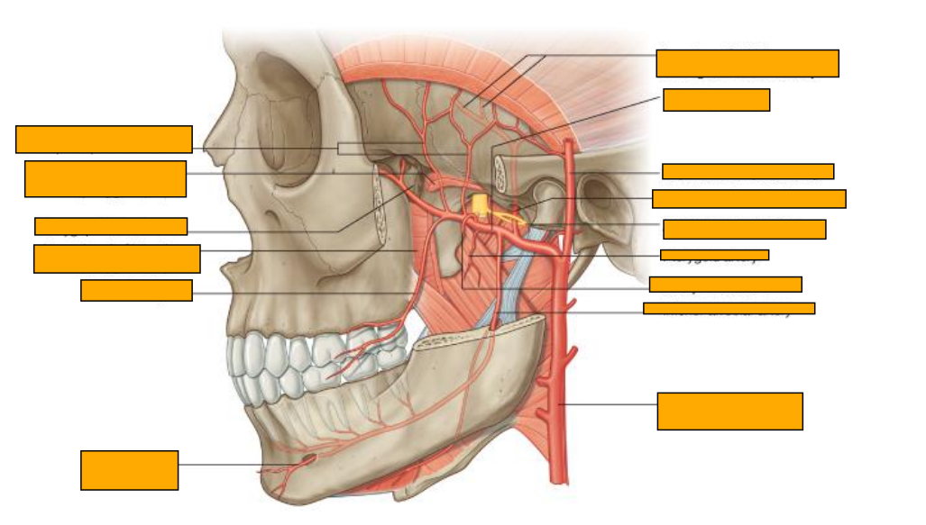
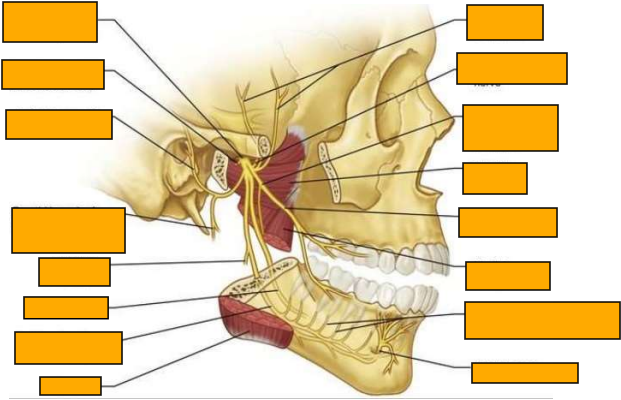
Label this image
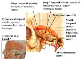
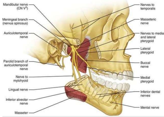
Label this image
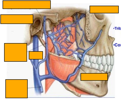
Label this image
What are the 3 main extra-cranial spaces of the head
temporal fossa
infra-temporal fossa
pterygopalatine fossa
What is the temporal fossa
shallow depression on the side of the skull bounded by temporal lines
terminates below the level of the zygomatic arch
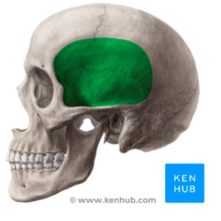
Superior and posterior borders of the temporal fossa
superior temporal line (origin of the deep temporal fascia)
Inferior border of the temporal fossa
runs along the zygomatic arch
Anterior border of the temporal fossa
posterior surface of frontal process of the zygomatic bone and the zygomatic process of the frontal bone
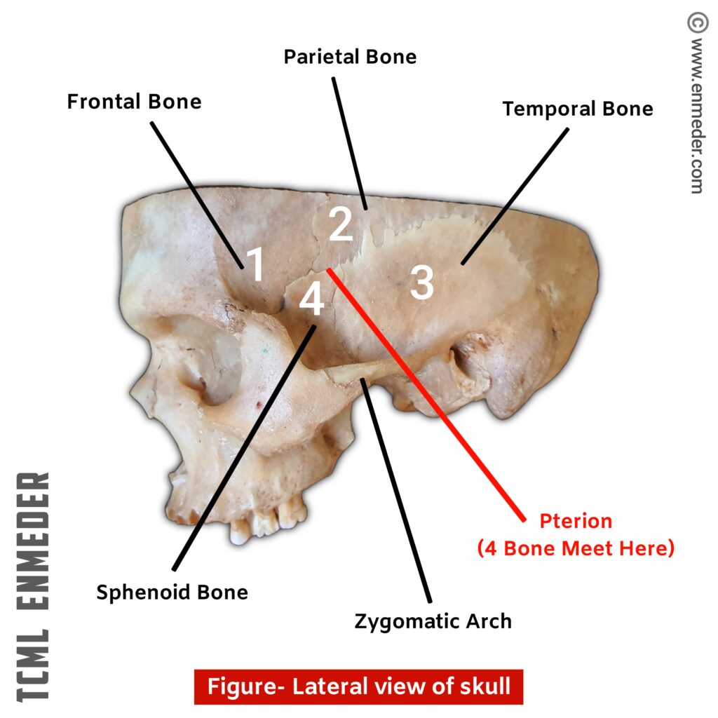
Medial borders of the temporal fossa
frontal, parietal, temporal and sphenoid bone
Lateral border of the temporal fossa
temporal fascial
Which muscle is the temporal fossa the site of origin for
temporalis muscle
What is the insertion point of the temporalis muscle
under the zygomatic arch, to the coronoid process of the mandible
Which blood vessels are found in the temporal fossa
superficial temporal artery- which is a terminal branch of the external carotid artery
Which nerves are found in the temporal fossa
-mandibular nerve
-anterior and posterior branches of the deep temporal nerve
-auricotemporal nerve
-zygomaticotemporal nerve (NCV2)
-temporal branches of the facial nerve
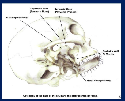
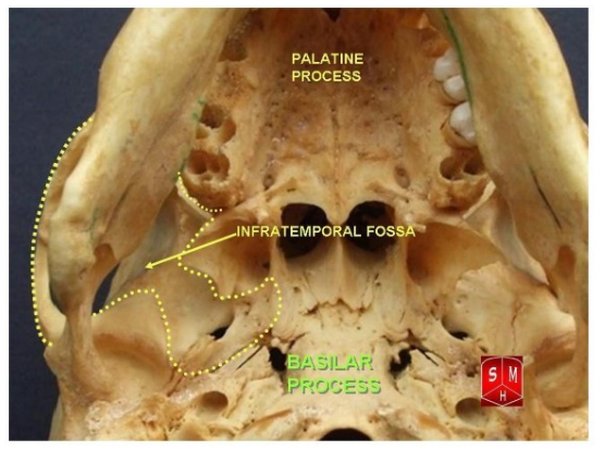
Location of the infratemporal fossa in relation to other structures
-below the middle cranial fossa
-medial and deep to the zygomatic arch
-behind the maxilla
How does the infratemporal fossa communicate with the temporal fossa
through the interval between the zygomatic arch and cranial bones
What is the basilar process of the infratemporal fossa
irregularly shaped cavity of anatomical and clinical importance
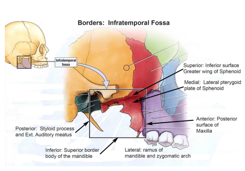
Superior border of the infratemporal fossa
infratemporal surface greater wing of the sphenoid
Anterior border of the infratemporal fossa
posterior surface of the maxilla
Medial border of the infratemporal fossa
lateral pterygoid plate
Posterior border of infratemporal fossa
styloid and mastoid processes
Lateral border of the infratemporal fossa
ramus of the mandible
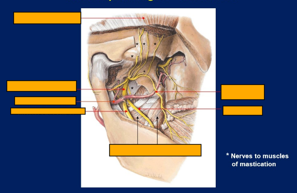
Muscular contents of the infratemporal fossa
-lower part of the medial pterygoid
-lower part of the lateral pterygoid
-lower part of the temporalis msucle
Nerve contents of the infratemporal fossa
mandibular nerve and its branches:
inferior alveolar nerve
buccal nerve
lingual nerve
(nerves to msucles of mastication)
Clinical importance of the infratemporal fossa
-it is a potential anatomical space and pathologies within this region can evolve without detection
Deep arteries of the infratemporal fossa
maxillary artery and middle meningeal artery
Superficial arteries of the infratemporal fossa
superficial temporal artery
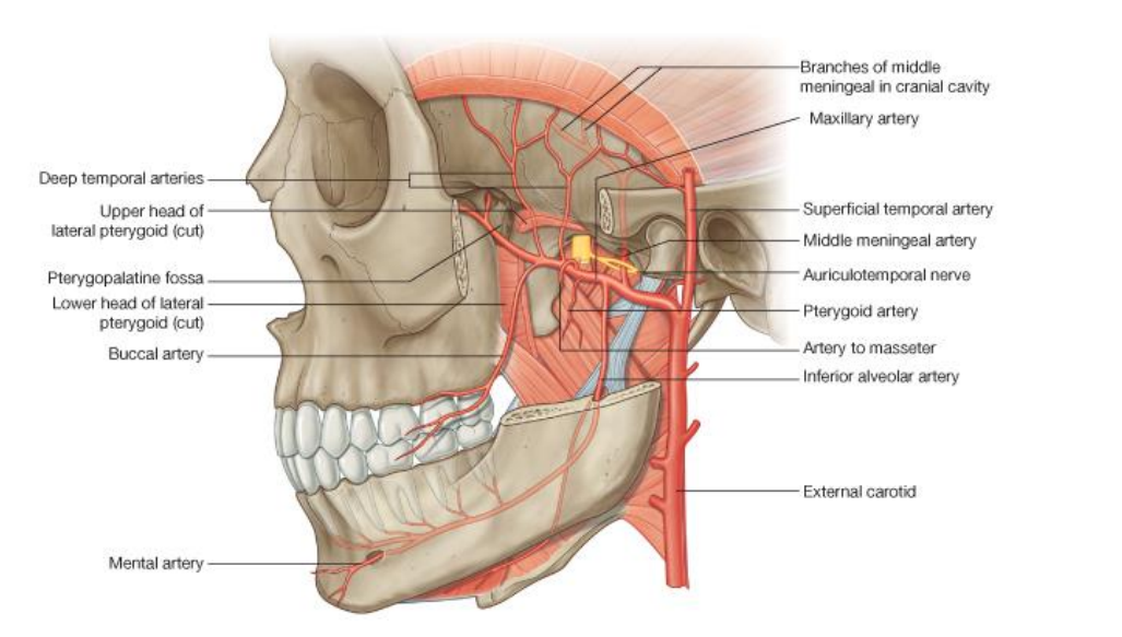
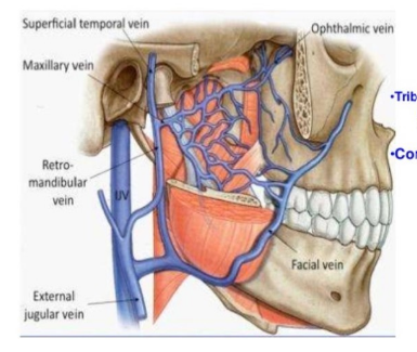
Veins of the infratemporal fossa
maxillary vein
middle meningeal vein
pterygoid venous plexus
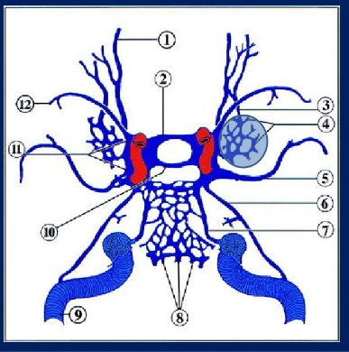
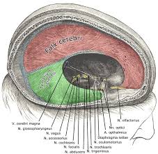
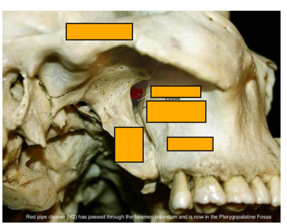
Label 1 to 5
1- superior ophthalmic vein
2- anterior intercavernous sinus
3- inferior ophthalmic vein
4- pterygoid venous plexus
5- middle meningeal vein

Openings of the infratemporal fossa
foramen ovale
foramen spinosum
alveolar canal
inferior orbital fissure
pterygomaxillary fissure
What is inferior alveolar nerve block
anaesthetic injected around the mandibular foramen which blocks the inferior alveolar nerve
What is mandibular nerve block
anaesthetic injected adjacent to the nerve as it enters the infratemporal fossa
Which nerves are affected by mandibular nerve block
inferior alveolar, lingual, buccal and auriculotemporal
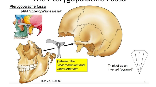
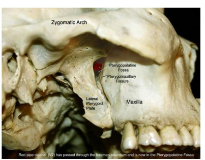
Location of the pterygopalatine fossa
small space behind and below the orbit
betweent the viscerocranium and neurocranium
Anterior boundary of the pterygopalatine fossa
posterior surface of the maxilla
Posterior boundary of the ptg fossa
pterygoid process
Medial boundary of the pterygopalatine fossa
perpendicular plate of palatine bone
Lateral boundaries of the pterygopalatine fossa
pterygomaxillary fissure
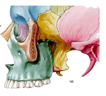
Superior and inferior boundary of the pterygopalatine fossa
superior- greater wing and body of sphenoid
inferior- pyramidal process of palatine bone
Why is the pterygopalatine fossa of clinical importance
-communicates with many other sites
-can be a major route of spread of infections and metastases (spread of cancer)
How does the pterygopalatine fossa communicate with the infratemporal fossa
via the pterygomaxillary fissure
How does the ptg fossa communicate with the middle cranial fossa
via the foramen rotundum
How does the ptg fossa communicate witht he nasal cavity
via the sphenopalatine foramen
How does the ptg fossa communicate with the orbital cavity
via inferiororbital fissure
How does the ptg fossa communicate with the palate
via the palatine canal
How does the ptg fossa communicate with the nasopharynx
via the pharyngeal and pterygoid canals