Phys M3 - Tissue Level & Integumentary System
1/46
There's no tags or description
Looks like no tags are added yet.
Name | Mastery | Learn | Test | Matching | Spaced | Call with Kai |
|---|
No analytics yet
Send a link to your students to track their progress
47 Terms
Epithelial Tissues
Simple Squamous Epithelium
Simple Cuboidal Epithelium
Simple Columnar Epithelium
Stratified Squamous Epithelium
Stratified Cuboidal Epithelium
Stratified Columnar Epithelium
Pseudo-stratified Columnar Epithelium
Transitional Epithelium
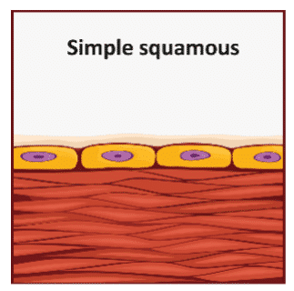
Simple Squamous Epithelium
Function: Allows simple diffusion of gases, nutrients, and waste across a thin membrane.
Structure: A delicate single layer of flat, thin, and smooth cells closely fitted together (tile floor).
Location: Air sacs of lungs, capillary walls, interior of heart (endothelium), lining ventral body cavities (mesothelium).
Simple Cuboidal Epithelium
Function: Secretion, absorption, and protection (glandular cells).
Structure: Single layer of cubed/hexagonal-shaped cells.
Location: Kidney tubules, secretory portion of the thyroid, salivary glands.
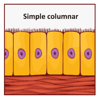
Simple Columnar Epithelium
Function: Secretion of enzymes, mucous or other substances; absorption--microvilli increases surface area.
Structure: Single layer of taller-than-wide cells; may contain mucous-secreting glands, microvilli, or cilia.
Location: Digestive tract (stomach for secretion, intestines for absorption).
Goblet cells: A type of columnar cell that is a unicellular, mucous-secreting gland found in digestive and respiratory tracts.
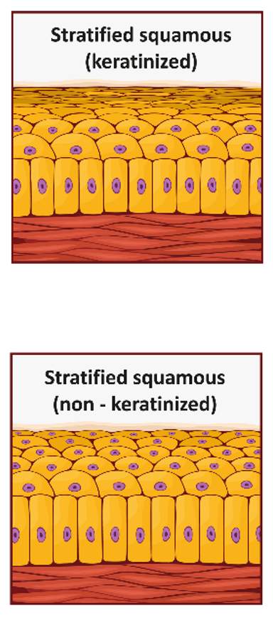
Stratified Squamous Epithelium
Function: Found where mechanical stresses are harsh; protection against wear and tear of constant friction.
Structure: Thick membrane with several layers of cells; top layers squamous, bottom layers typically cuboidal or columnar.
Keratinized: Bottom layers of cells are living, completing mitosis, and producing keratin. Top layers are flat, dead, and full of keratin.
Non-keratinized: Top layers of cells are living and not keratinized.
Location: Keratinized: skin; Non-keratinized: mouth, tongue, vaginal canal, pharynx, esophagus, anus.
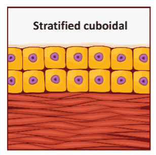
Stratified Cuboidal Epithelium
Function: Duct lining; secretion, absorption, and protection.
Structure: Rare tissue, multiple layers of cubed/hexagonal-shaped cells.
Location: Sweat glands, mammary glands.
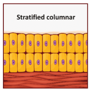
Stratified Columnar Epithelium
Function: Protection.
Structure: Rare; either two layers or multiple layers.
Location: Conjunctiva covering the eye, salivary ducts, mammary glands, urethra.
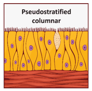
Pseudo-stratified Columnar Epithelium
Function: Protection, secretion, movement of mucous.
Structure: Several cell types with varying shapes and functions; appears to be layered because nuclei are seen at different heights but not truly stratified, since every cell comes into contact with basement membrane; typically possess cilia.
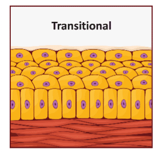
Transitional Epithelium
Function: Allows for distension without tearing the membrane.
Structure: Modified-stratified epithelium (cells change shape from round to flat).
Location: Urinary bladder.
Glands
Glands within epithelial tissue are specialized structures that produce and secrete substances essential for various bodily functions.
two main types:
exocrine glands: release their products onto epithelial surfaces
endocrine glands: secrete hormones directly into the bloodstream.
Play a crucial role in maintaining homeostasis by regulating processes such as digestion, temperature control, and metabolism
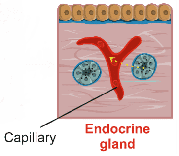
Endocrine Gland
Endocrine glands: Ductless glands that secrete hormones into the extracellular space; secretions absorbed into the blood to be circulated throughout the body.
Location: Thyroid gland, adrenal gland, pituitary gland, pancreas.
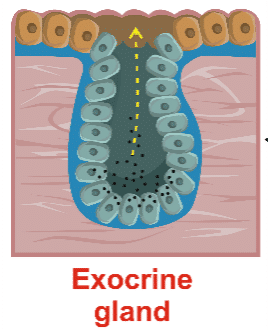
Exocrine Gland
Exocrine glands: Contain ducts or tubes to take the secretion away from the gland to an epithelial surface.
Location: Salivary glands (saliva to the oral cavity), sweat glands (sweat to skin surface), gastric glands (gastric juice to the cavity of the stomach), pancreas (enzymes to the intestines).
Types of Secretions:
Serous Glands: Watery solution with enzymes.
Mucous Glands: Mucins that hydrate to form mucous.
Mixed Exocrine Glands: Different types of gland cells secrete different exocrine secretions.
3 main modes of secretion used by exocrine glands:
holocrine, merocrine, and apocrine
Holocrine secretion:
Gland cell bursts when a cell is packed with secretion; stem cells go through cell division to replace lost cells.

Mecrocrine Secretion
Most common; released from secretory vesicles by exocytosis.
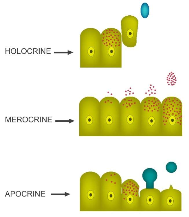
Apocrine secretion
Loss of cytoplasm along with the secretion through packed vesicles in the apical portion of the cytoplasm; must rebuild before the next secretion.

Roles of connective tissues in bodily functions
Provide structural support, store energy, insulate the body, and protect organs.
They are composed of specialized cells, fibers, ground substance, and extracellular matrix …
which work together to perform various functions essential for maintaining the body's integrity and homeostasis.
Loose connective tissue
Areolar tissue
Adipose tissue
Reticular tissue
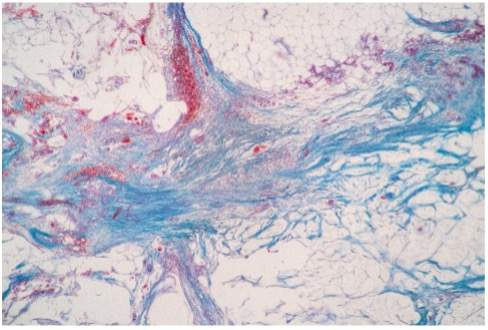
Loose Connective Tissue
Loose connective tissue provides support and flexibility and is found in the skin and around organs.
Areolar Tissue
Function
Holds tissue fluid.
Absorbs shock.
Provides space for immune cells.
Protects underlying tissue (muscle).
Allows muscle to contract without pulling on the skin.
Good blood flow (point of injections for this reason).
Allows gas exchange from tissue capillaries.
Structure: Gel-like matrix; many or all fibers and cells present, making it unspecialized; macrophages and white blood cells present for immune function.
Location: Beneath the surface of the dermis and epithelial tissues of the body that have openings to the environment.
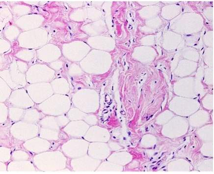
Adipose Tissue
Function
Fat storage for reserve fuel.
Insulates against heat loss.
Cushions and protects organs.
Adipocytes: Fat cells containing a single, large, lipid droplet; all other organelles squeezed into one side.
Location: Under the skin (especially in flanks and buttocks), around kidneys and eyeballs, in bones, abdomen, and breasts.
White fat: Pale yellow color (adults).
Brown fat: Increased vascularization gives adipose tissue a deeper color (children use this to increase metabolism for heat production; this is why babies don’t shiver).
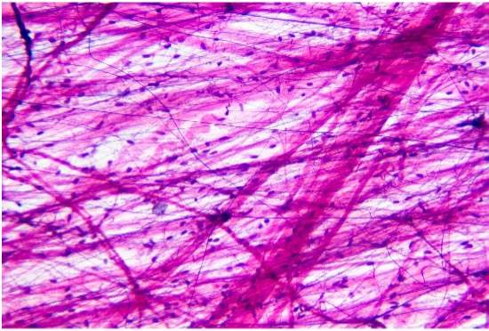
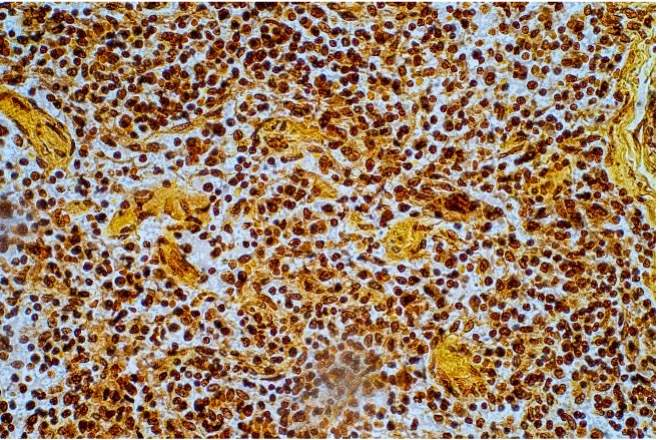
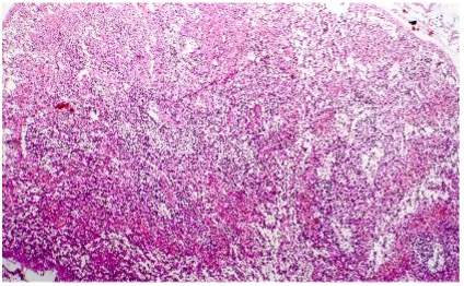
Reticular Tissue
Function: Supporting framework.
Structure: Create a stroma (support structure) to the parenchyma (functional cells).
Location: Spleen and liver.
Dense Connective Tissue
offers strong support and is found in tendons and ligaments.
Dense regular
Dense irregular
Elastic connective tissue
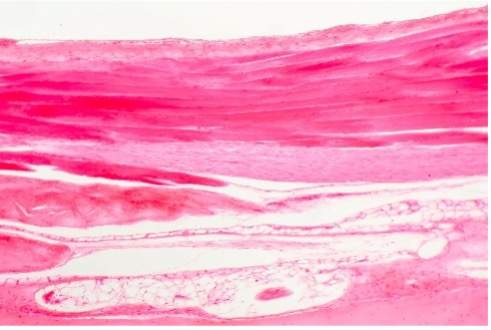
Dense Regular
Function
Provides strong attachment. Contributes to the formation of tendons.
Resists strong mechanical forces from two directions.
Structure: Collagen fibers parallel and lined up with the direction of applied force.
Fibroblasts: Produces the gel-like ground substance of matrix and secretes the precursor protein for collagen synthesis.
Dense Irregular
Function: Strengthens/supports areas subject to stress from many directions.
Structure: Interwoven network with an irregular pattern of collagen.
Fibroblasts: Produces the gel-like ground substance of matrix and secretes the precursor protein for collagen synthesis.
Location: Dermis layer of skin, capsule around liver and kidneys, surrounding cartilage of joints (perichondrium), surrounding bone (periosteum).
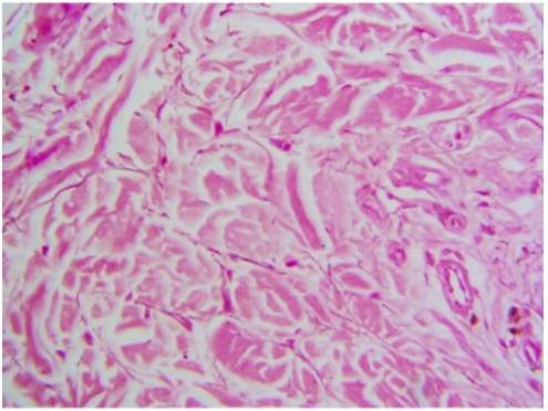
Elastic Connective Tissue
Function: Stabilizes positions of the vertebrae, permits expansion and recoiling of organs and structures.
Structure: Dense connective tissue dominated by elastin fibers.
Location: Trachea, artery walls, elastic cartilage, vocal cords.
Supportive Connective Tissue
provides structural or flexible support and is found in the bones, joints, ears, and nose.
Cartilage
Bone
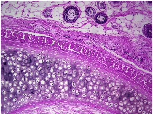
Cartilage
Function
Supports and reinforces (trachea).
Prevents friction and cushions (joints).
Supports skin (ear and nose).
Absorbs shock (vertebrae).
Structure
Cells: Chondrocytes are the only cells present found within chambers called lacunae.
Avascular: No blood flow, so nutrients and wastes exchange by diffusion; the chemical anti-angiogenesis produced in cartilage depresses vessel growth.
Matrix: Firm but flexible proteoglycan matrix.
Outer layer: Surrounded by perichondrium (dense irregular outer layer for support and protection, cellular inner layer for growth and maintenance).
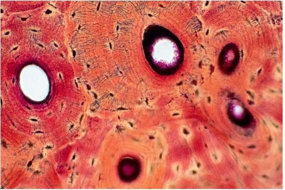
Bone
Function
Supports and protects.
Provides levers for muscles to act on.
Stores calcium, fat, and other minerals.
Site for blood cell formation.
Structure
Cells: Osteocytes (bone cells) found in lacunae and communicate with other cells via canaliculi.
Vascular: Vascularized and capable of repair.
Outer layer: Covered by periosteum with outer dense layer and inner cellular layer.
Matrix: Hard matrix composed of calcium salts and flexible collagen, making bones strong and resilient; resistant to shattering.
Location: Skeletal system.
Fluid Connective Tissue
transports nutrients, gases, wastes, and immune cells as it circulates throughout the body.
Blood
Lymph
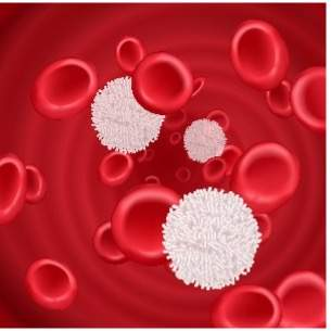
Blood
Function: Transport of gases, nutrients, wastes, and other substances.
Structure: Fluid matrix called plasma; parenchyma consists of cells and fragments of cells (formed elements).
Red blood cells: Carry oxygen.
White blood cells: Destroy pathogens.
Platelets: Prevent blood loss and clotting.
Location: Contained within blood vessels; the heart moves it through 1. arteries, 2. capillaries, and 3. veins and then returns to the heart.
Lymph
Structure: Forms from interstitial fluid.
Location: Within lymph vessels.
Function: Immune system monitor; maintains blood volume and homeostasis of blood solutes.
Significance of Bodily Membranes
Body membranes are thin layers of tissue that cover surfaces, line body cavities, and divide spaces or organs. They play crucial roles in protecting internal structures, facilitating movement, and reducing friction between organs.
Mucous Membranes: Line body cavities open to the exterior; involved in absorption and secretion.
Serous Membranes: Line closed body cavities and cover organs; secrete serous fluid to reduce friction.
Cutaneous Membrane: Also known as the skin; protects underlying tissues and regulates body temperature. Protects against external environment.
Synovial Membranes: Line joint cavities; acts as shock absorbers and produce synovial fluid for joint lubrication.
Functions of the integumentary system:
Protection: From external environment (bacteria, pathogens, extreme temperatures, friction).
Excretion: Salts, water, and wastes.
Maintains internal temperature: Sweating
Warm: The blood vessels of the skin dilate to release heat.
Cold: The blood vessels constrict to keep heat within the body.
Melanin production: Protects from ultraviolet rays.
Water resistant: Keratin production protects against abrasion and water loss.
Vitamin D synthesis: Helps calcium metabolism.
Sensation: Includes touch, pressure, pain, and temperature sensation.
Epidermis
Protects against pathogens, UV radiation, and water loss; involved in the production of keratin and melanin.
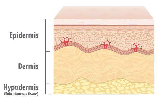
Dermis
Provides strength and elasticity to the skin; contains collagen and elastin fibers, blood vessels, nerve endings, and glands.

Layers of the Epidermis:
Stratum corneum Old
Stratum lucidum
Stratum granulosum
Stratum spinosum ⬆
Stratum basale
Dermis Young
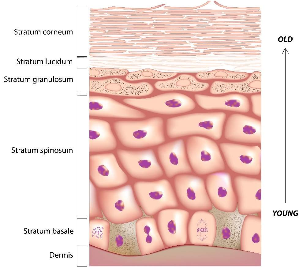
Stratum Basale
create new keratinocytes through cell division that move upward toward the surface of the skin.
Stratum Spinosum
Living keratinocytes from Stratum Basale continue moving through Stratum Spinosum, no cell division occurs here.
Stratum Granulosum
Keratinocytes enter Stratum Granulosum start to die, and go through a chemical process where they are dehydrated by keratohyaline and structurally reinforced by keratin (imagine the cells being dried out and filled with re-bar, to make them thin but strong).
Stratum Corneum
Thin and strong dead keratinocytes enter the Stratum Corneum and help form the protective outer layer of skin.
Layers of the Dermis
Papillary Layer: Areolar tissue with capillaries, lymphatics, and sensory neurons; named because of the dermal papillae lock in with the epidermal ridges.
Reticular Layer: Deep to the papillary layer and composed of dense irregular connective tissue with collagen and elastin fibers.
Cells of the Dermis
Collagen: Gives skin strength and flexibility.
Elastin: Gives skin resilience; age, overstretching, and UV radiation can damage or destroy elastin leaving wrinkles or sagging skin.
Wrinkles: Due to excessive sun exposure, fibroblasts are damaged, and maintenance of the dermal layer is compromised.
Cleavage (tension) lines: Direction collagen and elastin fibers are bundled within the dermis; surgeons cut parallel to cleavage lines to minimize scarring.
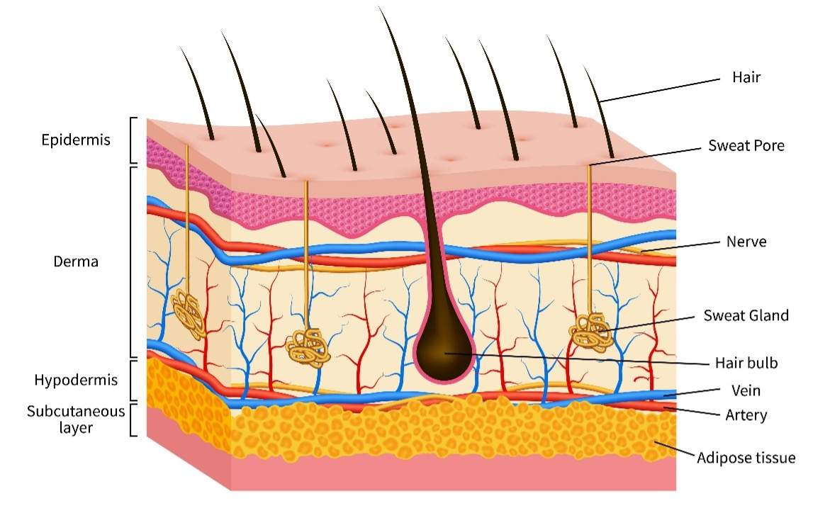
Dermal Blood Flow
Cutaneous Plexus: Arteries bordering the hypodermis and reticular layer of dermis; supplies subcutaneous tissues (adipose tissue), hair follicles, sweat glands, and other structures within the dermis.
Papillary Plexus: Feed the contours of the epidermis.
Arterioles: Small arteries with smooth muscle in the walls, which permits constriction or dilation. Remember that blood carries heat.
Vasodilation: Arterioles dilate (increase in diameter), which increases blood flow in a warm environment; this allows extra heat to be closer to the surface of the skin where it can be emitted.
Vasoconstriction: Arterioles constrict (decrease in diameter), which decreases blood flow in cooler environments; this keeps the heat centralized in the core of the body (to maintain homeostasis in organs of consequence).
“Fight or Flight” Response: Stress induces this response; the body shunts blood from the dermis to the muscles, heart, and brain for combat in a stressful situation; cold skin and stress-induced sweating may occur causing a “cold sweat.”
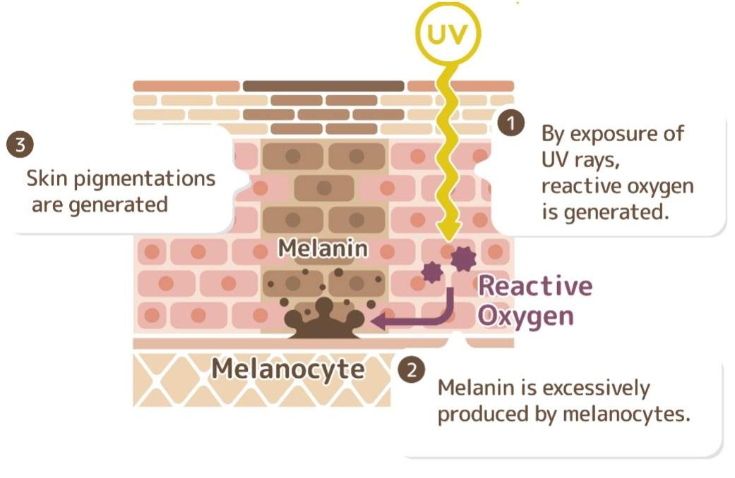
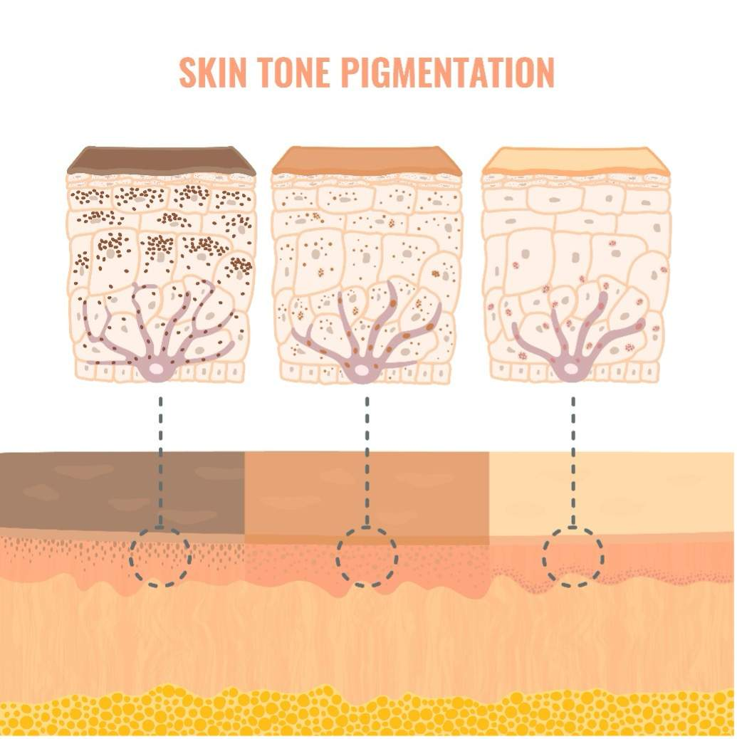
Melanin Production
occurs in melanocytes, which are found in the basal layer of the epidermis. It is responsible for skin pigmentation and provides protection against UV radiation.
Skin tone is determined by the amount of melanin that is produced by melanocytes. More melanin results in darker skin tones.
Skin Pigments
Carotene: Orange-yellow pigment that accumulates in the epidermis; derived from orange vegetables; converts to vitamin A, which aids in eye function.
Melanin: Brown, yellow-brown, or black pigment produced by melanocytes within the stratum basale; packaged in melanosomes and transferred into keratinocytes.
Vitamin D Conversion
UVB radiation (from sunlight) converts 7-dehydrocholesterol in the skin to vitamin D3 (cholecalciferol). (Hint: cholecalciFERol is FIRst)
Vitamin D3 is then transported to the liver, where it is converted to calcidiol. (Hint: calciDIol where DI means 2, or step 2)
It then goes to the kidneys, where it is converted to its active form, calcitriol. (HINT: calciTRIol, where TRI is 3, or step 3)
Finally, calcitriol then influences blood calcium levels by increasing calcium absorption in the small intestine, decreasing the excretion of calcium by the kidney, and causing the bones to release stored calcium.
How does melanin production protect the body?
By absorbing UV radiation to prevent DNA damage