Microbiology Lab Exam II
1/74
There's no tags or description
Looks like no tags are added yet.
Name | Mastery | Learn | Test | Matching | Spaced | Call with Kai |
|---|
No analytics yet
Send a link to your students to track their progress
75 Terms
What is the most effective wavelength of UV light for germicidal lamps?
At a wavelength of 264 nm, where the wavelength maximizes the formation of pyrimidine dimers. Highly effective in inactivating microorganisms by damaging their DNA and RNA.
What mechanisms of UV light lead to DNA damage?
Primarily by inducing covalent bonds between adjacent pyrimidine bases, creating lesions that distort the DNA helix and interfere with replication and transcription.
Why would bacteria and viruses be more prone to quick death following exposure to UV light compared to us?
They are small, simple, and poorly protected, while humans have multiple layers of shielding and powerful DNA‑repair systems.
Some bacteria have mechanisms to help repair the damage introduced by UV light. Be able to explain one such method, mediated by photolyase enzymes.
Photoreactivation: Photolyase binds to UV‑induced pyrimidine dimers (usually thymine dimers) and uses energy from visible light to break the abnormal covalent bond, restoring the original bases without cutting the DNA backbone.
While UV light does not affect humans as immediately as microbes, it can still cause things like sunburn, increased signs of aging, and increased cancer risks. What are the mechanisms by which UV causes sunburn, skin aging, and increased cancer risks?
Sunburn (acute inflammation) Direct DNA damage → inflammation — driven mainly by UV‑B
Skin aging (photoaging, chronic structural damage) ROS → collagen breakdown — driven mainly by UV‑A
Increased cancer risk, mutations + immune suppression — UV‑B and UV‑A both contribute
Psychrophiles
Cold‑loving microbes that grow best at very low temperatures (≈ –10 to 15°C).
They require cold environments like glaciers, deep oceans, and Arctic soils.
Psychrotrophs
Cold‑tolerant microbes that grow slowly in the cold (0–30°C) but prefer moderate temperatures.
These are the classic food‑spoiling organisms in refrigerators.
Mesophiles
Moderate‑temperature microbes that grow best at 20–45°C. Includes most human pathogens because 37°C is ideal.
Thermophiles
Heat‑loving microbes that grow best at 50–60°C. Common in hot springs, compost piles, and geothermal soils.
Hyperthermophiles
Extreme heat‑loving microbes that grow optimally at 80°C or higher. Often found in volcanic vents and deep‑sea hydrothermal systems.
Why are higher temperatures more detrimental to bacteria than lower temperatures?
Heat kills because it destroys structure (denatures proteins). Cold preserves structure but slows function.
Which of these growth categories can be involved in human disease as human pathogens?
Mesophiles since it is around the perfect human body temperature. Includes most of all clinically important bacteria, S. aureus, S. pyogenes, E. coli, Salmonella, Mycobacterium tuberculosis, and Neisseria spp.
Definitions of acidic and basic on the pH scale.
Acidic (pH < 7)
A solution is acidic when it has more hydrogen ions (H⁺) than hydroxide ions.
pH below 7
Higher H⁺ concentration
Tastes sour, can be corrosive
Examples: stomach acid, lemon juice
Basic / Alkaline (pH > 7)
A solution is basic when it has more hydroxide ions (OH⁻) than hydrogen ions.
pH above 7
Lower H⁺ concentration
Feels slippery, tastes bitter
Examples: bleach, baking soda solution
Acidophile
Grow best in acidic environments (optimal pH ~1–5).
Thrive in low‑pH habitats like acid mine drainage or the stomach.
Example: Helicobacter pylori (acid‑tolerant, not a strict acidophile).
Neutrophile
Grow best at neutral pH (optimal pH ~6.5–7.5).
Most human pathogens fall here because human tissues are near neutral pH.
Example: E. coli, Staphylococcus aureus, Streptococcus spp.
Alkalinophile/Alkaliphile
Grow best in basic/alkaline environments (optimal pH ~8–11).
Found in soda lakes, alkaline soils, and industrial settings.
Example: Bacillus alcalophilus.
Identify Antiseptic Alcohols: Recognize the two alcohols mentioned as effective antiseptics and specify their optimal concentrations for use.
Ethanol (ethyl alcohol) and Isopropanol (isopropyl alcohol) both work best at 70%. Water is still required for protein denaturation.
Mechanism of Action: Understand how alcohols destroy bacterial cells.
Alcohols kill bacteria by denaturing proteins, dissolving membrane lipids, and dehydrating the cell — with protein denaturation being the most important mechanism.
Pros and Cons of Alcohols: Discuss one advantage and one disadvantage of using alcohols as antiseptics.
Alcohols kill bacteria quickly by denaturing proteins and disrupting membranes. They provide rapid antisepsis on skin before injections or procedures.
Alcohols evaporate quickly, so they leave no lasting antimicrobial effect. They also work poorly when organic material (blood, dirt, feces) is present.
Antibiotic History: Name the first antibiotic used clinically to treat bacterial infections.
Penicillin (discovered by Alexander Fleming, 1928; widely used by the early 1940s)
Using the study materials, understand the classification structure used to indicate the level of risk of certain microbes due to their antibiotic resistance.
The WHO Bacterial Priority Pathogens List (BPPL) 2024 identifies 15 families of antibiotic-resistant bacteria, categorized into critical, high, and medium priority groups. This classification aims to guide research and development (R&D) for new antibiotics and promote international cooperation to combat antimicrobial resistance (AMR).
Acinetobacter baumannii
CRITICAL PRIORITY, carbapenem-resistant
Enterobacterales
CRITICAL PRIORITY, third-generation cephalosporin-resistant | carbapenem-resistant
Enterococcus faecium
HIGH PRIORITY, vancomycin-resistant
Pseudomonas aeruginosa
HIGH PRIORITY, carbapenem-resistant
Staphylococcus aureus
HIGH PRIORITY, methicillin-resistant (MRSA)
Antibiotic Spectrum: Differentiate between narrow-spectrum and broad-spectrum antibiotics.
Narrow- Antibiotics that target a small, specific group of bacteria. Effective against either Gram‑positive OR Gram‑negative, but not both. Cause less disruption to normal microbiota. Useful when the pathogen is known.
Broad- Antibiotics that target a wide range of bacteria. Effective against both Gram‑positive AND Gram‑negative organisms. Useful when the pathogen is unknown (empiric therapy). Higher risk of disrupting normal flora → can lead to superinfections (e.g., C. difficile).
Kirby-Bauer Method: Explain the procedure and interpretation of results in the Kirby-Bauer method.
To determine whether a bacterium is susceptible, intermediate, or resistant to specific antibiotics.
Inoculate a Mueller-Hinton agar, apply antibiotic disks, incubate, antibiotics diffuse outward. After, measure diameter in mm and compare to chart.
Susceptible (S, large zone) - Antibiotic works; bacteria inhibited at achievable drug levels
Resistant (R, small or no zone) - Antibiotic does not inhibit the bacterium
aminoglycosides
inhibit protein synthesis (30s)
cephalosporins
inhibit cell wall synthesis
penicillins
inhibit call wall synthesis
tetracyclines
inhibit protein synthesis (30s)
quinolones
inhibit DNA replication (topoisomerase 2 and 4)
macrolides
inhibit protein synthesis (50s)
sulfonamides
inhibit folate synthesis
glycopeptides
inhibit call wall synthesis
Mannitol salt agar (MSA)
Tests for salt tolerance and mannitol fermentation, mainly staphylococcus species. Contains high salt concentration, mannitol, and phenol red. It was inoculated by a line streak with loop. Color change and growth are observed. Yellow = acidic pH (mannitol fermentation), Red = no fermentation. Growth means salt tolerance.
Staphylococcus medium 110 (SM110)
This test is very similar to MSA, but without the color change due to the present of a pH indicator. These test can tell you if a bacteria is salt-tolerant and if they produce a pigment (golden). However, pigment result can only be known IF the bacteria are salt-tolerant. Otherwise, they won't grow. Inoculated as line streak with loop.
Sheep's blood agar (SBA)
Tests for hemolysis pattern and swarming. Inoculated with line streak and optional stabs. Bacteria produce hemolysins, which can: Completely lyse RBCs, partially damage them, or leave them intact. Beta leaves halos around inoculation sites, alpha is in-between, and gamma is none.
DNase agar (without indicator)
Tests for DNase hydrolysis. Inoculated by line streak with loop. If the organism produces DNase, it breaks DNA into smaller fragments. Hydrochloric acid 1M is added, and a positive exhibits a halo, while a negative does not.
Novobiocin Susceptibility
Tests for antibiotic resistance. Inoculated in class with lawn streak on MHA. A paper disc is placed on and diffuses outward to create a concentration gradient. A resistant result shows a zone size of 16mm or less. A susceptible result looks like a zone size bigger than 16mm.
Staphylococcus epidermidis bio
Staphylococcus epidermidis is a Gram‑positive, coagulase‑negative coccus that is one of the most abundant members of the normal skin flora. Although typically harmless, it becomes clinically important in hospital settings due to its ability to form robust biofilms on implanted medical devices such as catheters, prosthetic joints, and heart valves. These biofilms protect the organism from antibiotics and immune clearance, making infections persistent and difficult to treat. It is novobiocin‑susceptible, non‑hemolytic, and does not ferment mannitol on MSA. S. epidermidis is a leading cause of nosocomial bloodstream infections, especially in immunocompromised patients or those with indwelling devices.
Staphylococcus saprophyticus bio
Staphylococcus saprophyticus is a Gram‑positive, coagulase‑negative coccus best known for causing uncomplicated urinary tract infections, particularly in sexually active young women. It colonizes the genitourinary tract and uses specialized adhesins to bind tightly to uroepithelial cells, allowing it to persist in the urinary environment. A key diagnostic hallmark is its novobiocin resistance, which distinguishes it from other coagulase‑negative Staphylococci. It is typically non‑hemolytic and does not ferment mannitol. While less virulent than S. aureus, its predictable association with UTIs makes it clinically significant.
Staphylococcus aureus bio
Staphylococcus aureus is a Gram‑positive, coagulase‑positive coccus and one of the most clinically important human pathogens. It commonly colonizes the anterior nares and skin, often asymptomatically, but can cause a wide spectrum of disease—from skin and soft‑tissue infections to pneumonia, osteomyelitis, endocarditis, sepsis, and toxin‑mediated illnesses such as toxic shock syndrome.
Its virulence stems from numerous factors, including protein A, coagulase, hemolysins, leukocidins, and various exotoxins. On MSA, it ferments mannitol (yellow colonies) and shows beta‑hemolysis on blood agar. Methicillin‑resistant strains (MRSA) pose major public‑health challenges due to their antibiotic resistance and ability to spread in both community and healthcare settings.
Viridans Streptococci
Examples: S. mutans, S. sanguinis
Major Diseases:
Dental caries (tooth decay)
Subacute bacterial endocarditis (especially after dental work)
Virulence Factors:
Biofilm formation → stick to teeth (plaque)
Dextran production → helps adhere to damaged heart valves
Low virulence overall, but opportunistic
Streptococcus pneumoniae
Major Diseases:
Pneumonia (classic “rust-colored sputum”)
Otitis media (middle ear infection)
Sinusitis
Meningitis
Virulence Factors:
Polysaccharide capsule → MOST IMPORTANT (prevents phagocytosis)
Pneumolysin → damages host cells
IgA protease → helps colonize respiratory tract
Streptococcus pyogenes
Major Diseases:
Pharyngitis (strep throat)
Scarlet fever
Impetigo (skin infection)
Necrotizing fasciitis (“flesh-eating disease”)
Rheumatic fever (post-infection complication)
Glomerulonephritis
Virulence Factors:
M protein → key factor (prevents phagocytosis)
Streptolysins (O & S) → destroy RBCs/WBCs
Exotoxins (Spe toxins) → cause scarlet fever & toxic shock
Hyaluronidase → spreads through tissues
Streptococcus agalactiae
Major Diseases:
Neonatal sepsis
Neonatal meningitis
Pneumonia in newborns
Virulence Factors:
Polysaccharide capsule → prevents phagocytosis
Beta-hemolysis
Ability to colonize vaginal tract → transmission during birth
Enterococcus spp.
Major Diseases:
Urinary tract infections (UTIs)
Endocarditis
Intra-abdominal infections
Nosocomial (hospital-acquired) infections
Virulence Factors:
Intrinsic antibiotic resistance (VERY important clinically)
Biofilm formation
Aggregation substance → helps adherence
Can survive harsh conditions (bile, salt, etc.)
Bile Esculin (BE) Hydrolysis
Tests for esculin hydrolysis. Can the bacterium survive a bile salt environment. Inoculated with S streak as slant. A positive shows blackening of media, a negative has no change.
6.5% Salt Tolerance Test
Tests for salt tolerance. Can the bacterium survive a hypertonic environment. Inoculated by loop. A positive exhibits turbidity, a negative does not.
CAMP Test
The CAMP test looks for synergistic hemolysis between: CAMP factor (produced by S. agalactiae), β‑hemolysin (produced by S. aureus). Species are streaked on blood agar. Positive shows a sharp, arrowhead‑shaped zone of enhanced β‑hemolysis at the junction of the two organisms, negative shows no enhanced hemolysis only normal hemolysis patterns.
Bacitracin Sensitivity:
Optochin Sensitivity:
Used to differentiate Streptococcus pneumoniae from other alpha-hemolytic streptococci (such as viridans streptococci).
S. pneumoniae is sensitive to optochin, showing a zone of inhibition (≥14 mm is considered significant).
Viridans group streptococci are resistant to optochin.
SXT Sensitivity:
Used to distinguish Group A Streptococci (S. pyogenes) and Group B Streptococci (S. agalactiae) from other beta-hemolytic streptococci.
Both Group A and Group B streptococci are resistant to SXT.
Other beta-hemolytic streptococci (e.g., Groups C, G) are generally sensitive.
viridans streptococci bio
Viridans streptococci are a diverse group of α‑hemolytic or non‑hemolytic, optochin‑resistant, bile‑insoluble streptococci that inhabit the oral cavity, pharynx, and upper GI tract. They are normally harmless but become pathogenic when they enter the bloodstream, especially after dental manipulation. They are best known for causing dental caries (S. mutans) and subacute infective endocarditis on damaged heart valves (S. sanguinis, S. mitis). The S. anginosus group is notable for causing deep abscesses in the brain, liver, and abdomen. Their virulence centers on dextran production, which allows adherence to tooth enamel and exposed fibrin on damaged valves, acid production (S. mutans), and biofilm formation that supports plaque development and persistent infection.
Streptococcus pyogenes bio
Streptococcus pyogenes is a β‑hemolytic, bacitracin‑sensitive, PYR‑positive organism that colonizes the oropharynx and skin. It is responsible for a wide range of diseases, from pharyngitis and impetigo to severe invasive infections such as necrotizing fasciitis, streptococcal toxic shock syndrome, and bacteremia. Its virulence is driven by M protein (antiphagocytic and central to rheumatic fever pathogenesis), streptolysins O and S (β‑hemolysis), streptokinase, DNases, and pyrogenic exotoxins (SpeA, SpeB), which mediate toxic shock and scarlet fever. Post‑infection immune complications include acute rheumatic fever and post‑streptococcal glomerulonephritis, making GAS one of the most clinically significant streptococci.
Enterococcus spp. bio
Enterococcus species (primarily E. faecalis and E. faecium) are Gram‑positive cocci that inhabit the GI tract and are notable for their extreme environmental resilience. They are BE‑positive, 6.5% NaCl‑positive, and often highly antibiotic‑resistant, including VRE strains. Clinically, they cause urinary tract infections, bacteremia, endocarditis, and intra‑abdominal or pelvic infections, especially in hospitalized or immunocompromised patients. Their virulence arises from biofilm formation, aggregation substance (promotes adherence and plasmid exchange), cytolysin, and intrinsic resistance mechanisms that allow survival in harsh conditions and under antibiotic pressure.
Escherichia coli
Major Diseases:
Urinary tract infections (UTIs) → most common cause
Neonatal meningitis
Gastroenteritis (various strains: ETEC, EHEC, etc.)
Sepsis
Virulence Factors:
Fimbriae (P pili) → attachment in UTIs
Capsule (K antigen) → protects from immune system
Endotoxin (LPS) → septic shock
Exotoxins:
Heat-labile (LT) & heat-stable (ST) toxins → diarrhea
Shiga-like toxin (EHEC) → severe disease (HUS)
Klebsiella spp.
Major Diseases:
Pneumonia (often severe, “currant jelly” sputum)
UTIs
Liver abscesses
Virulence Factors:
Thick polysaccharide capsule → VERY important (mucoid colonies)
Endotoxin (LPS)
Biofilm formation
Proteus mirabilis
Major Diseases:
UTIs (especially catheter-associated)
Kidney stones (important association)
Virulence Factors:
Urease → raises urine pH → stone formation
Swarming motility → spreads across surfaces
Fimbriae → adhesion
Endotoxin
Pseudomonas aeruginosa
Major Diseases:
Opportunistic infections (burns, wounds)
Pneumonia (especially in cystic fibrosis patients)
UTIs
Sepsis
Hot tub folliculitis, swimmer’s ear
Virulence Factors:
Exotoxin A → inhibits protein synthesis (like diphtheria toxin)
Biofilm formation → chronic infections
Pyocyanin pigment → tissue damage
Efflux pumps & antibiotic resistance
Endotoxin (LPS)
Enterobacter/Citrobacter/Serratia
Major Diseases:
Nosocomial infections (hospital-acquired)
UTIs
Respiratory infections
Sepsis
Virulence Factors:
Antibiotic resistance (often multidrug-resistant)
Endotoxin (LPS)
Biofilm formation
Serratia: produces red pigment (prodigiosin)
Yersinia spp.
Major Diseases:
Y. pestis → plague (bubonic, pneumonic)
Y. enterocolitica → gastroenteritis (can mimic appendicitis)
Virulence Factors:
Antiphagocytic proteins (Yops) → block immune response
Capsule (in Y. pestis)
Endotoxin
Ability to survive in macrophages
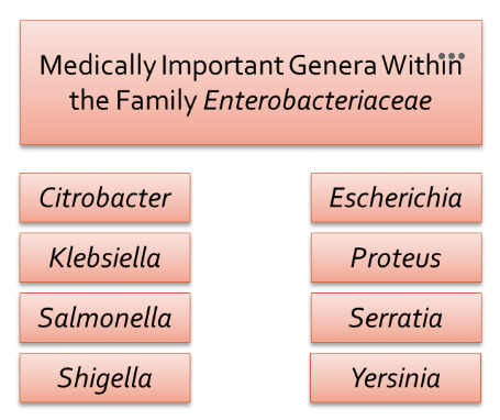
There are several major genera within the family Enterobacteriaceae that cause human disease.
MacConkey agar
Tests for lactose fermentation. Bile salts + crystal violet → inhibit Gram‑positive organisms. Lactose → carbohydrate source. Neutral red → pH indicator. Inoculated by isolation streak. A positive shows pink/red colonies, a negative is colorless.
Eosin Methylene Blue (EMB) agar
Tests for lactose fermentation. Eosin Y + methylene blue dyes: inhibit gram‑positive organisms, act as pH indicators. Inoculated by streaking for isolation on EMB plate. Positive shows dark purple or green metallic colonies. Negative is colorless.
Hektoen Enteric Agar
Tests for H2S and sugar fermentation. Bile salts inhibit Gram‑positive organisms and many non‑pathogenic Gram‑negatives. Carbohydrate fermentation → acid production. Inoculated with isolation streaks. Positive shows pink, black, or green colors. Negative shows no colors.
Tryptone/Indole Test
Tests for indole production. Presence of the enzyme tryptophanase, which breaks down tryptophan → indole + pyruvate + ammonia. Inoculated with loop. Kovac’s reagent is added, positive shows red ring, negative shows no change.
Urea medium
Tests for urease production. Inoculated with loop. Presence of the enzyme urease. Ability to hydrolyze urea → ammonia + CO₂. Positive turns barbie pink, negative has no color change.
Kligler’s iron agar
Tests for glucose/lactose fermentation, H2S, and gas. Inoculated with S slant and stab. Phenol red = pH indicator, Acid → yellow, Alkaline → red. If only glucose is fermented → butt stays yellow, slant reverts to red (K/A). If lactose is fermented → slant and butt stay yellow (A/A). Gas production → cracks, bubbles, or lifted agar. H2S shows black precipitate.
SIM medium
Tests for H2S, indole, and motility. Inoculated with stab needle. Ability to reduce sulfur compounds (thiosulfate) → H₂S gas. Presence of tryptophanase, which converts tryptophan → indole. Ability of bacteria to move through semisolid agar. Kovac’s is added to see if red ring is present: indole positive. Black precipitate means H2S positive, motility is observed.
Escherichia coli bio
Escherichia coli is a Gram‑negative, lactose‑fermenting, indole‑positive rod that normally inhabits the human colon as part of the healthy microbiota. While many strains are harmless, pathogenic variants cause a wide range of diseases. Extraintestinal infections include urinary tract infections, neonatal meningitis, and bacteremia, often due to strains with enhanced adhesins and capsules. Intestinal pathogenic strains include ETEC, EHEC, EPEC, EAEC, and EIEC, each defined by distinct virulence factors such as heat‑labile and heat‑stable enterotoxins (ETEC), Shiga toxin (EHEC), or bundle‑forming pili (EPEC). Key virulence factors include fimbriae (P pili) for urinary tract adherence, K1 capsule in neonatal meningitis, LPS endotoxin, and various exotoxins depending on the pathotype. E. coli is one of the most clinically important and diverse bacterial species.
Proteus mirabilis bio
Proteus mirabilis is a Gram‑negative, highly motile, urease‑positive rod known for its characteristic swarming motility on agar. It inhabits the GI tract but is best known as a cause of complicated urinary tract infections, especially in catheterized patients. Its potent urease enzyme hydrolyzes urea → ammonia, raising urine pH and promoting struvite stone formation, which can lead to obstruction and recurrent infections. Other virulence factors include fimbriae for urinary tract adherence, flagella enabling motility and ascension of the urinary tract, and endotoxin (LPS) contributing to inflammation. Proteus mirabilis is also capable of producing H₂S, which helps differentiate it in laboratory media such as KIA or SIM.
Yersinia spp. bio
Yersinia is a genus of Gram‑negative coccobacilli that includes several medically important species: Yersinia pestis, Yersinia enterocolitica, and Yersinia pseudotuberculosis.
Y. pestis, transmitted by fleas and rodents, causes plague (bubonic, septicemic, pneumonic) and is characterized by virulence factors such as the F1 capsule, plasminogen activator protease, and Yops proteins that block phagocytosis. Y. enterocolitica and Y. pseudotuberculosis are foodborne pathogens that cause enterocolitis, mesenteric adenitis, and pseudoappendicitis, often linked to contaminated pork or unpasteurized milk. Their virulence centers on invasins, Yops secretion system, and cold tolerance, allowing growth in refrigerated foods. Across the genus, Yersinia species are notable for their ability to evade innate immunity, survive in macrophages, and cause severe systemic disease.