BIO 225 Lab Practical 2
1/117
There's no tags or description
Looks like no tags are added yet.
Name | Mastery | Learn | Test | Matching | Spaced | Call with Kai |
|---|
No analytics yet
Send a link to your students to track their progress
118 Terms
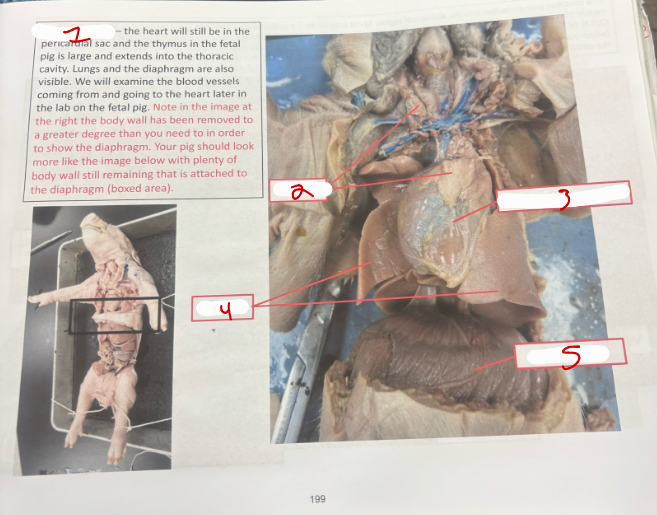
Thoracic Cavity
thoracic cavity
thymus
heart in percicardial sac
lungs
diaphragm
Abdominal Cavity
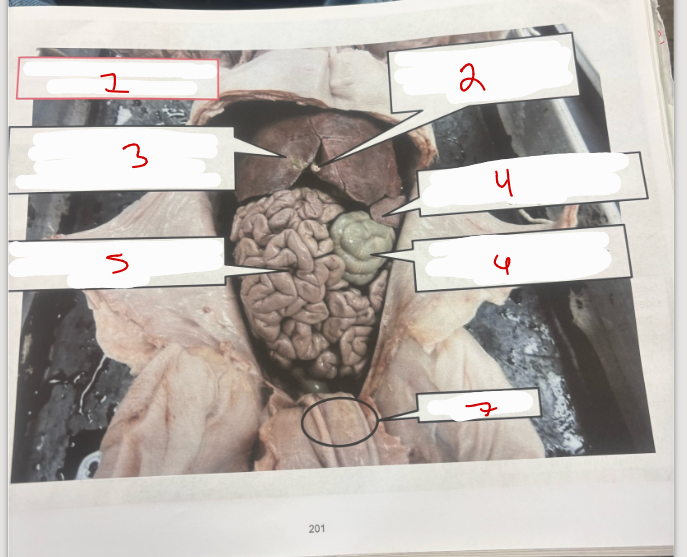
abdominal cavity - focus on the digestive system
umbilical vein
liver - large organ; site for bile production, carbohydrate storage, plasma protein production and deotxification of foreign substances
spleen - lymphoid organ (red blood cell phagocytosis and leukocyte storage)
small intestine - loosely coiled tube; major site for digestion and nutrient absorption
large intestine (colon) - tightly coiled tube; major site for water absorption
umbilical cord
Pig 1
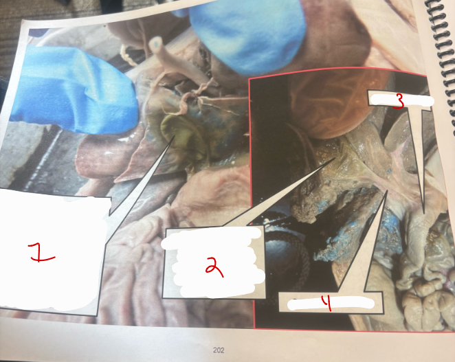
gall bladder
gall bladder
duodenum
common bile duct
Pig 2
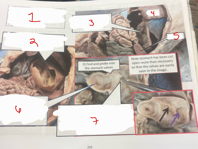
stomach - early digestion performed here; low pH dissolves extracellular matrix, kills pathogens and activates pepsinogen to pepsin to initiate protein breakdown
stomach
liver
spleen
pyloric valve - leads to the duodenum; regulates the amount of acidic chyme going from the stomach to the duodenum
cardiac valve (muscular folds) - leads to the esophagus
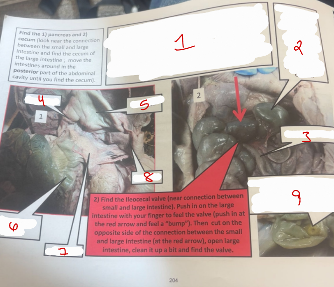
Pig 4
pancreas - soft, spongey organ between the stomach and intestine; produces hormones to regulate blood glucose levels (insulin and glucagon), digestive enzymes for the small intestine and bicarbonates to neutralize the acidic chyme from the stomach
cecum
small intestine
duodenum
stomach
large intestine (colon)
pancreas
spleen
illeocecal valve - regulates flow of chyme from small to large intestine
Pig 3
rectum and anus
rectum - short term fecal storage structure
anus (opening to the rectum)
large intestine (colon) - tightly coiled tube
kidney
rectum
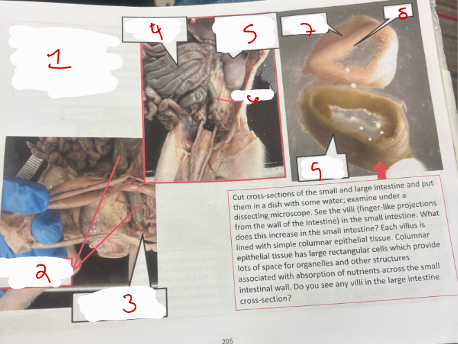
small intestine
villi
large intestine
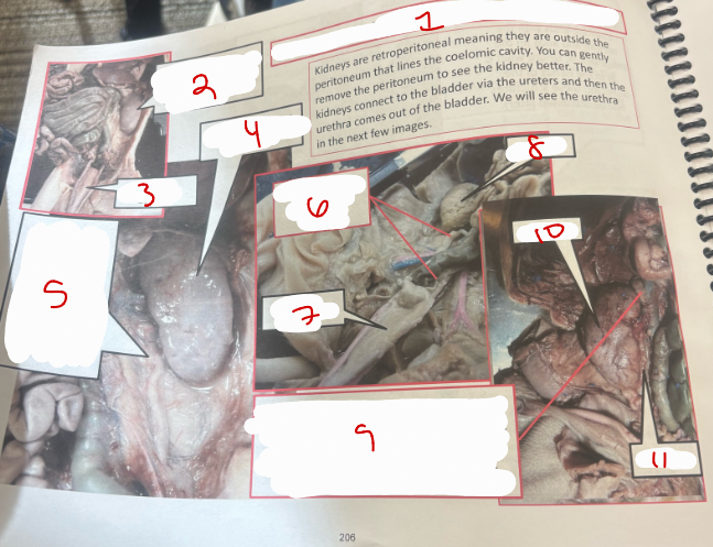
Abdominal Cavity
abdominal cavity - focus on the urogenital system
kidney
urinary bladder
kidney covered with peritoneum
ureter - tube that conducts urine from the kidney to urinary bladder
ureter leading to the urinary bladder
urinary bladder
kidney
adrenal gland - produces hormones which affect the kidney (aldosterone) and your ability to maintain the “flight or fight” sympathetic nervous system response (cortisol)
kidney
ureter
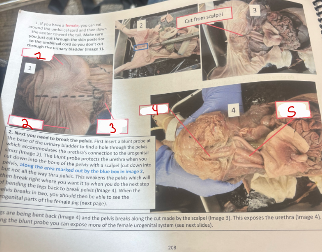
Female Pig 1
rear leg
rear leg
umbilical cord
urethra
urinary bladder
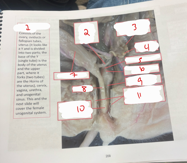
Female Urogenital System 1
female urogenital system
ureter - connect kidney to urinary bladder
ovary
horns of the uterus
body of the uterus
cervix - lower part of uterus
urinary bladder
urethra
vagina
urogenital sinus - where the vagina and urethra fuse
pelvis
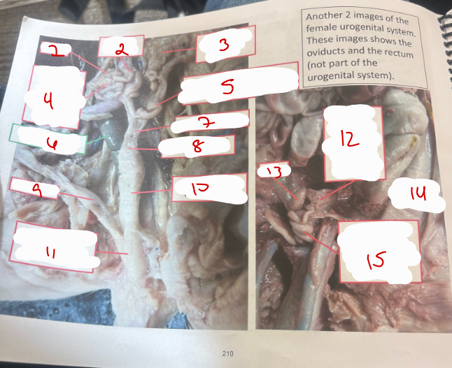
Female Urogenital System 2
ovary
horns of the uterus
ovary - eggs produced here
oviducts or fallopian tubes - tightly coiled tube adjacent to the ovary
horns of the uterus - fetal pigs develop in this part of the uterus
rectum - dorsal to the vagina
body of the uterus
cervic - lower part of the uterus
urethra
vagina - thicker than the uterus
urogenital sinus - where the vagina and urethra fuse
oviducts or fallopian tubes
ovary
large intestine
horns of the uterus
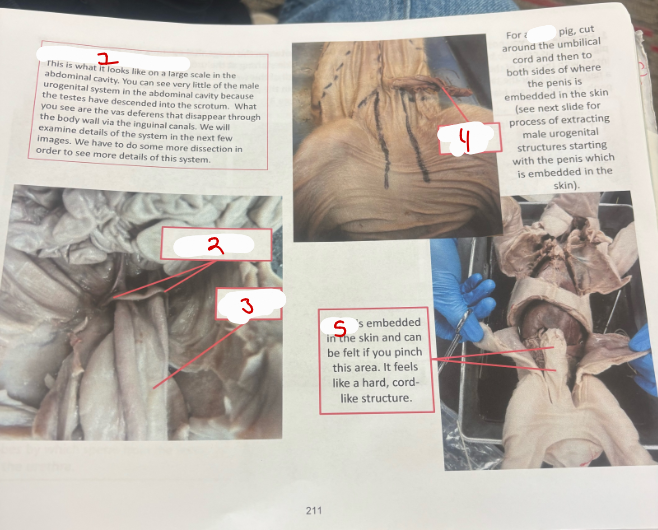
Male Urogenital System 1
male urogenital system
vas deferens
urinary bladder
umbilical cord
penis embedded in skin
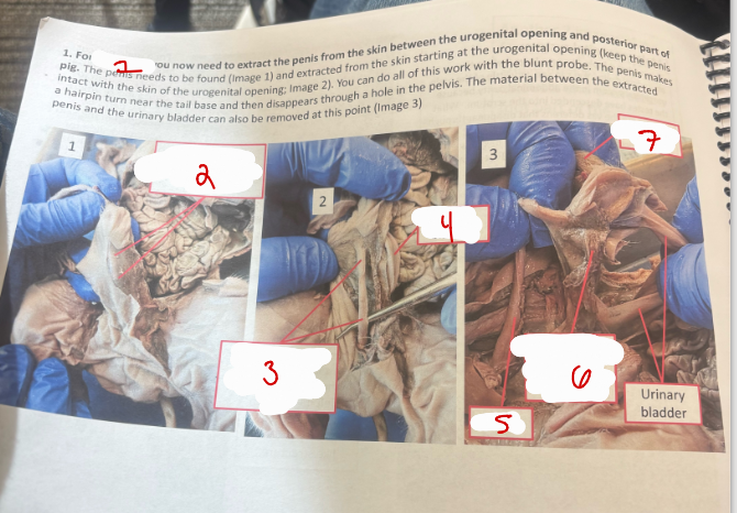
Male Urogenital System 2
male
penis
penis
urinary bladder
penis
umbilical cord
urinary bladder
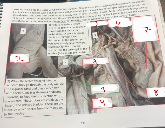
Male Urogenital System 3
scrotum with testes
vas deferens
inguinal canals
urinary bladder
vas deferens
probe is in the inguinal canal
sac with testis which has been removed from the scrotum
urinary bladder
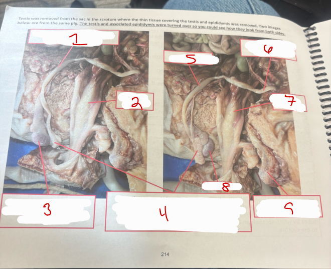
Male Urogenital System 4
vas deferens - carries sperm form epididymis to urethra
urinary bladder
testis - sperm are produced in the seminiferous tubules inside the testis
epididymis - tightly coiled tube around testis. sperm mature here. they become motile after coming in contact with fluid from the seminal vesicles
vas deferens
vas deferens coming from other testis
urinary bladder
testis
other testis still covered by thin tissue
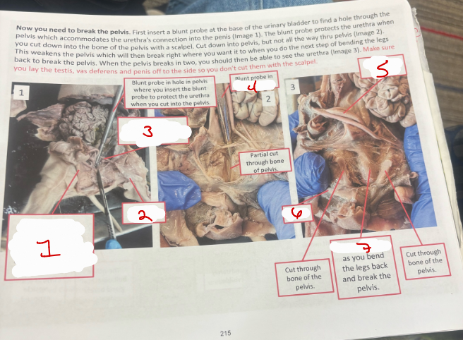
Male Urogenital System 5
penis (urethra inside0
testis in sac
vas deferens - connects to the base of the urethra
pelvis
urinary bladder
penis
urethra
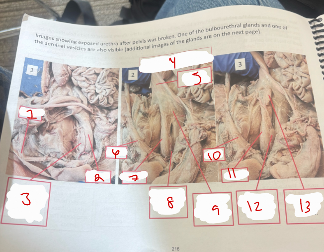
Male Urogenital System 6
penis
urethra
one of the bulbourethral glands on the side of the urethra
urethra
urinary bladder
penis
urethra
other bulbourethral glad
one seminal vesicle under this thin tissue
penis
urethra
other bulbourethral gland barely visible
one seminal vesicle exposed
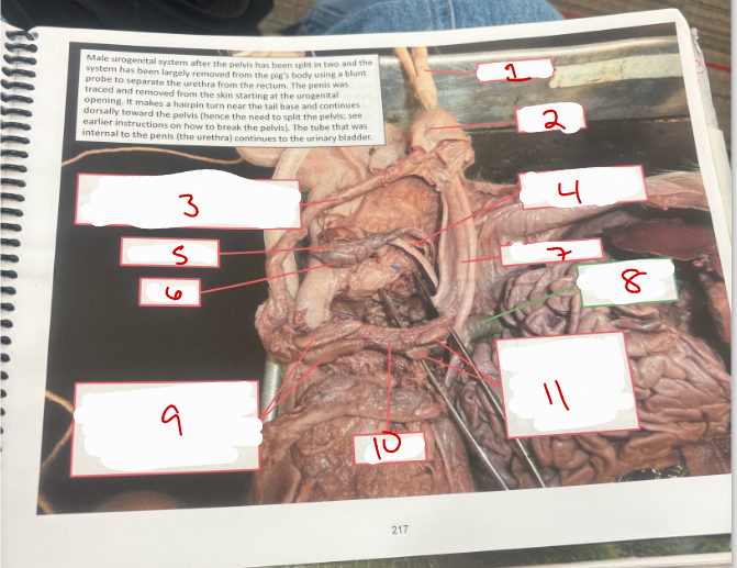
Male Urogenital System 7
umbilical cord
urogenital opening
penis
vas deferens - connects epididymis to the base of the urethra
epididymis
testis
urinary bladder
rectum (not part of urogenital system)
bulbourethral glands (on both sides of the urethra) - add mucus to the semen to lubricate the urethra for the sperm. also called cowper’s gland
urethra
seminal vesicles (at base of urinary bladder) - adds fructose to the semen that stimulate spermatozoa to become highly motile

Male Urogenital System 8
vas deferens - connects to the base of urethra
seminal vesicles (at base of urinary bladder) - add fructose to the semen that stimualte spermatozoa to become highly motile
urinary badder
prostate gland
urethra
prostate gland - adds prostatic fluid to the semen (slightly alkaline fluid)
seminal vesciles
prostate gland
urethra
vas deferens
vas deferens
prostate has been cut to follow vas deferens internally
vas deferens
ejaculatory duct closeup
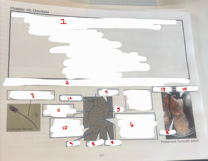
Chordata
Phylum Chordata - chordata means “cord bearing”; general characteristics of phylum include notochord, dorsal tubular nerve cord, post-anal tail, pharyngeal pouches or slits, endostyle or thyroid gland)
Subphylum Urochordata (turnicates) - adults are mainly sessile and are found in all oceans at arious depths. larvae are free-swimming and have the major chordate characteristics including notochord, post-anal tail, pharyngeal gill slits, and dorsal nerve cord
notochord - only chordate characteristic visible in slide
atrial siphon
anus
phayngeal basket - filtering structure; organic matter too large for the holes gets stuck in mucus from endostyle and then gets transported to stomach
intestine
stomach
heart
atrium - filtered water flowes through here and then out through atria siphon
endostyle - roduces ucus to trap food
oral siphon
oral siphone
atrial siphon
intestine

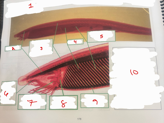
Subphylum Cephalochordata 1
Subphylum Cephalochordata (lancelets) - have major chordate characteristics
notochord
dorsal nerve with photoreceptor cells (black dots)
gill bars
gill slits (spaces between the gill bars)
oral hood with tentacles - supported by notochord
wheel organ - ciliated (provides current to draw food into mouth)
velum - vertical membrane with mouth (not visible)
enostyle - produces mucus to trap food
filter feeding - cilia on wheel organ produce a current that draws water with suspended food particles into the mouth ( a hole in the velum). food gets trapped in the mucus from the endostyle which covers the gill bars to the hyperbranchial groove to the instestine.
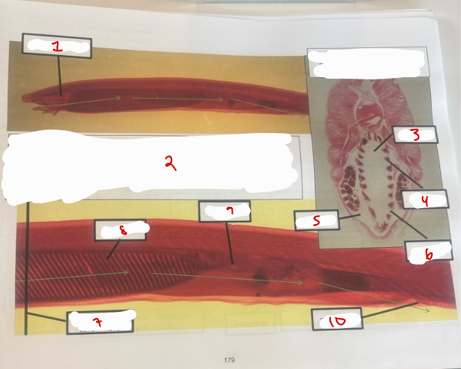
Subphylum Cephalochordata 2
wheel organ
filter feeding - cilia on the wheel organ draws water into the mouth, it moves into the pharynx, passes around the gill bars (through the gill slits) which are inside the animal to the atrium and then filtered water exits the animal via the atriopore
pharynx
gill bar
atrium
gill slit
gill bar
intestine
atriopore
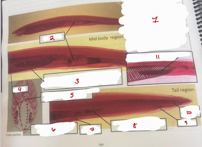
Subphylum Cephalochordata 3
Filter feeding - food suspended in the water gets trapped by mucus form the endostyle. it is then moved up the gill bars using cilia to the hyperbanchial groove, then moves back via cilia to the intestine. a diverticulum off the intestine (hepatic cecum; remember a cecum is a blind sac) phagocytizes food participles. digestion and absorption primarily occurs in the intestine and undigested food exits via the anus
intestine
hepatic cecum - blind sac, phagocytizes food particles
gill bars
hyperbranchial groove - moves trapped food to instestine
endostyle - produces mucus to trap food
atriopore
intestine - has undigested food in it (black spots)
anus
post anal tail
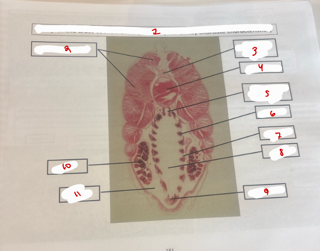
Amphioxus
Amphioxus
myotomes - muscles
dorsal nerve cord
notochord
hyperbranchial groove
gill bar
gill slit
pharynx
endostyle
gonad
atrium
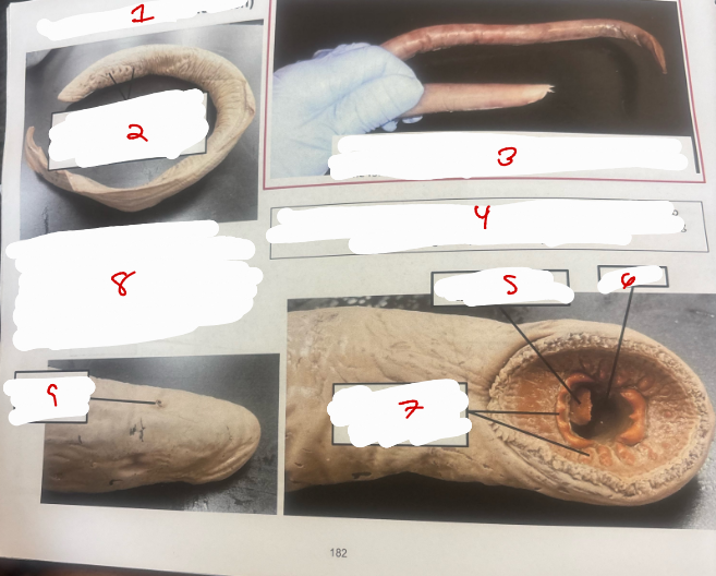
Superclass Agnatha
Sueprclass Agnatha (jawless fish)
Gill slits - water drawn into the mouth, passes over the gills (oxygen is extracted) and then passes out of the fish via the gill slits
Class Myxini (hagfishes) - marine fish that eat polychaete worms and dead and dying marine life. they produce copious amounts of slime for anti-predator purposes
Sea lamprey - lives in the Atlantic ocean and runs up streams and rivers to spawn. it invaded the great lakes wehere it has caused damage to the fishing industry. adults grow up to 1 m and parasitize fish
tongue - used to rasp hole in a fish
mouth
horny teeth 0 used to attach lamprey mouth to fish host
Class petromyzontida (lampreys) - many are anadromous (run up rivers and streams to spawn). the name petomyzon means “stone sucking” which describes how this fish, when swimming upstream, sometimes grasps a rock iwth its sucker-like mouth to hold its position in the stream. some species are parasitic and feed on the blood and body fluids of fish
nostril - single, middorsal on top of head
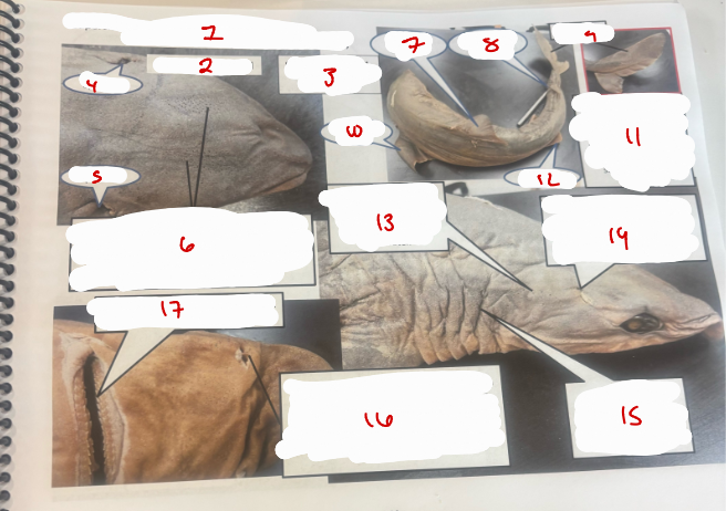
Superclass Gnathostomata
Superclass Gnathostomata (jawed vertebrates), Calss Chondrichtyes (sharks, skates, rays)
dorsal view of head
dogfish shark *squalus)
spiracle
spiracle
two clusters of holes are openings to the ampullary organs of lorenzini - these organs can detect bioelectricity produced by other living organisms when they contract their muslces, can be used to detect prey
anterior dorsal fin
posterior dorsal fin
caudal fin
pectoral fin
heterocercal - the two lobes of the caudal fin are not the same size
pelvic fin
lateral line - these tiny holes lead t o the lateral line system which detects vibrations in the water
spiracle - opening used to take in water to pass over gills when the shark is lying on the sea bottom or when buried in the sand. not all sharks have these
gill slits - after water passes over the gills (not visible) it exits the shark through the slits
nostril - has fold of skin in the middle which divides the nostril into two parts allowing water to continuously flow into one side and out through the other side of the nostril thus giving the shark continuous smell sensing of its environment
mouth 0 with multiple rows of teeth (continuously replaced)
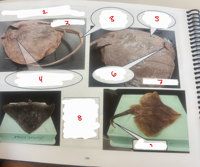
Superclass Gnathostomata 2
Superclass Gnathostomata, Class Chondrichtyes (sharks, skates, rays)
stingray - dorsal ride
barb - used to inject venom which can cause muscle cramps, swelling and rarely death
spiracle - dorsal opening used to take in water when the stingray is partiall rays have spiracles.
gill slits - water which entered via the spiracle exits here
mouth - rays eat molluscs and crustaceans
stingray - ventral side
left: smooth butterfly ray - short tail without a barb right: skate, similar to a ray, but lacks venomous barb and may have two dorsal fins on tail
dorsal fins on tail
Superclass Gnathostomata 3
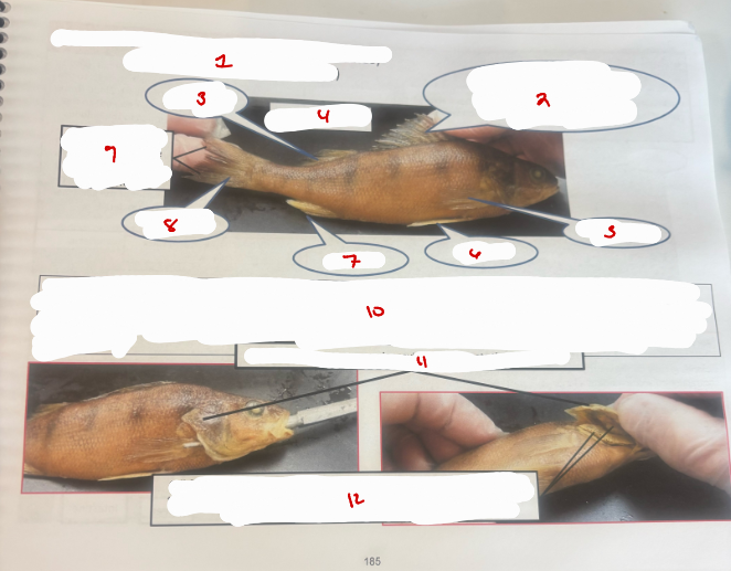
Superclass Gnathostomata (jawed vertebrate) Class Actinopterygii (bony fish)
first dorsal fin - spiny fin rays support the first dorsal fin.
second dorsal fin
yellow perch
pectoral fin
pelvic fin
anal fin
caudal fin
homocercal - 2 lobes of the caudal fin are the same size
fish breathing - mouth is continuously connected to the mouth cavity and to the opercular cavity. when the fish lowers the floor of its mouth, water is drawn into the mouth cavity through an open mouth. it then closes its mouth, raises the mouth floor and at the same time opens the opercula (this increases the volume of the opercular cavity where the gills lie) thus causing water to flow from the mouth to the opercular cavity. water then exists the fish. this process repeats itself to provide a continuous flow of water across the gills
operculum - covers opercular cavity and protects the gill filaments
gill filaments - blood flowing through the capillaries of the gills extract oxygen from the water which is moving in the opposite direction from the blood (counter current system)
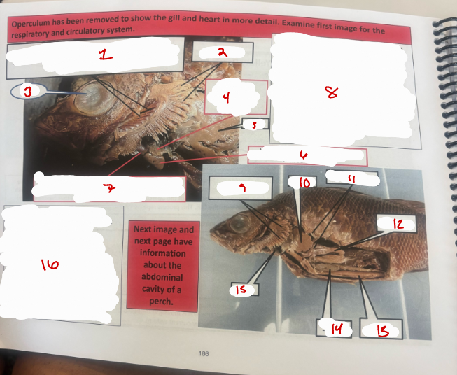
Fish 1
gill rakers - used to remove debris before water flows across gills. if the perch was a filter ffeeding fish, the rakers would be used to filter food from the wtaer.
Gill filaments
eye
Atrium - chamber with thin muscular walls
liver
ventricle - chamber with thick muscular walls
bulbous or conus arteriosus - chamber anterior to the ventricle. the aorta attaches anteriorly, leading to capillaries of the gills
fish circulatory system - single circuit. blood travels from the heart through the gills to the body and then back to the heart. blood from the body enters the atrium, descends to the ventricle where it is pumped to the bulbous arteriosus. when the ventricle relaxes (diastole(, blood is still under pressure in the bulbous anteriosus, thus maintaining even blood pressure in the aorta leading to the gill capillaries. this maintains a steady flow of the blood through the capillaries of the gills
gill filaments
liver
pyloric ceca
stomach
intestine
spleen
heart
bulbous arteriosus - acts as an elastic reservoir. expands greatly during ventricular contraction to accommodate a large amount of the blood volume from the ventricle. subsequent elastic recoil of the bulbous arteriosis gradually releases this volume to prevent gill damage due to high arterial systolic blood pressures, and to provide a more even flow of blood through the capillaries of the gill during each cycle of ventricular contraction and relaxation
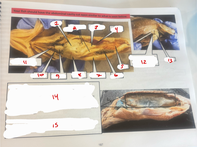
Fish 2
liver
pyloric ceca
stomach
swim bladder
ovary
spleen
intestine
gall bladder
ventricle
atrium
bulbus or conus arteriosus
gall bladder - pulled out by the forceps to make it more visible
liver
swim bladder info
Superclass Gnathostomata
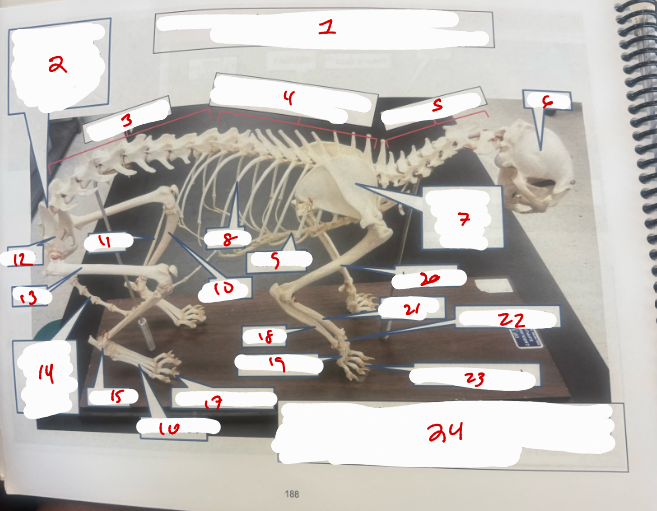
Superclass Gnathostomata, Class Mammalia (mammals) - cat skeleton
sacrium - vertebrae are fused are between the lumbar and caudal vertebrae
lumbar vertebrae
thoracic vertebrae - vertebrae with ribs attached to them
cervical vertebrae
skull
scapula (part of pectoral girdle)
ribs
sternum
tibia
fibula
pelvis
femur
caudal vertebae - vertebrae associated with the tail
tarsals
metatarsals
phalanges (toes)
ulna
metacarpals
humerus
radius
carpals (wrist)
phalanges (toes)
divisions of skeleton: 1. axial skeleton - bones that lie around the body’s center of gravity such as the skull, vertebrae of the spine, ribs and sternum and the appendicular skeleton - boens of the legs and feed including pectoral and pelvic girdles
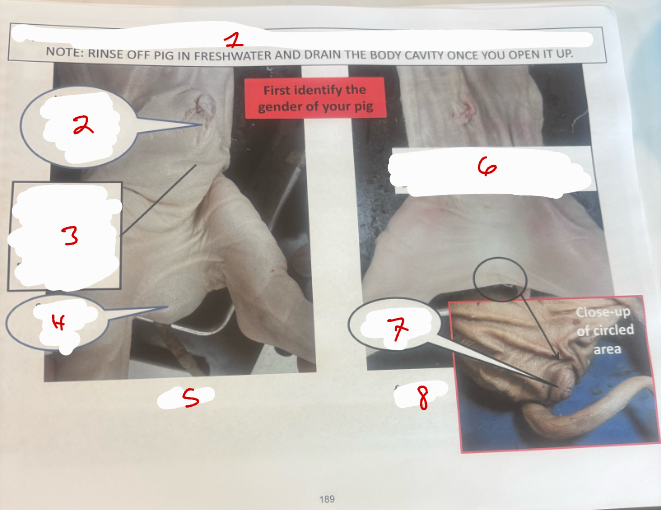
Pig 1
Superclass Gnathostomata, Class Mammalia
urogenital opening - just below umbilical cord
penis
scrotum with testes
male
gential papilla just ventral to base of tail
genital papilla
female
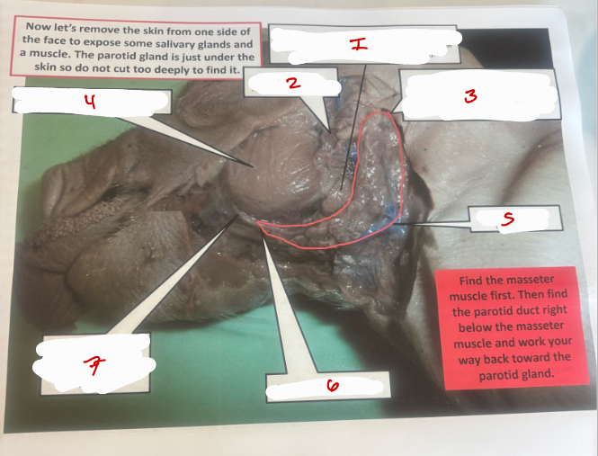
Pig 2
mandibular gland partially visible, rest under parotid gland
lymph node - partially visible
parotid gland starts here, salivary gland that adds saliva and digestive enzymes to food in the mouth
masseter muscle - helps close the jaws (pulls lower jaw up)
parotid gland
parotid gland
parotid duct - carries digestive enzymes from parotid gland to oral cavity
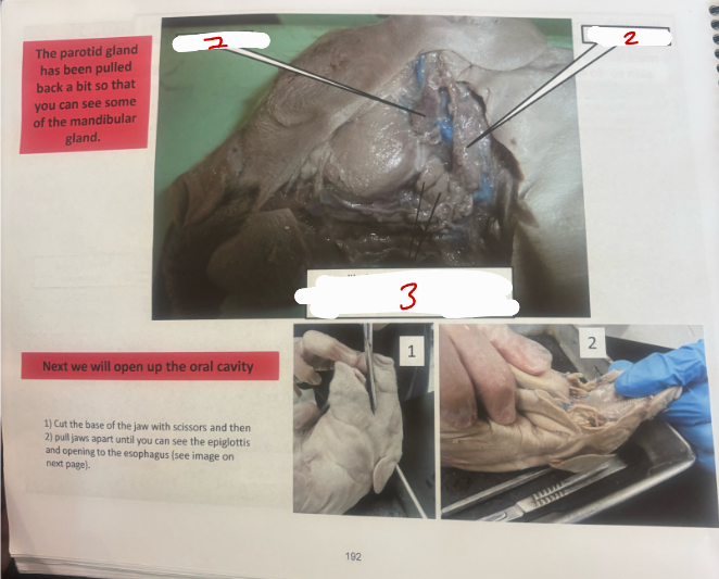
Pig 3
lymph node
parotid gland
mandibular gland fully visible since parotid gland is pulled back a bit. there is a fissure in this salivary gland
Pig 4
hard palate
soft palate
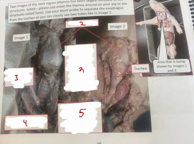
esophagus
trachea
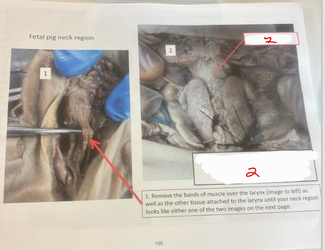
larynx
epiglottis
mouth
nose
esophagus
epiglottis - covering opening to respiratory system
opening to nasopharynx - when mouth is closed the epiglottis fits into the opening to the nasopharynx. it channels air, dorsal to the soft palate, into the glottis
hard palate
soft palate
opening to the esophagus
glottis - opening to the respiratory system
epiglottis
meconium - dark green/brown material seen here on the pigs tongue.
tongue
Fetal pig
larynx ( voice b ox)
thymus - large glandular, lymphoid tissue rich in lymphocytes ( especially T cells, cells that are part of your immunological response to antigens)
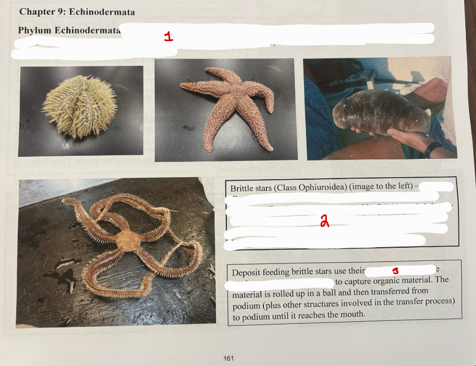
Phylum Echinodermata
(means prickly skin); examples include sea stars (starfish), brittle stars, sea urchins, sea cucumbers, and sea lilies; characteristics include water vascular system, marine, benthic, endoskeleton
(means serpent tail); widely distributed in all oceans. arms of brittle stars, as the name implies, easily autotomize (spontaneous casting off of a limb or other body part)
podia - secrete copious amounts of mucus to capture organic material
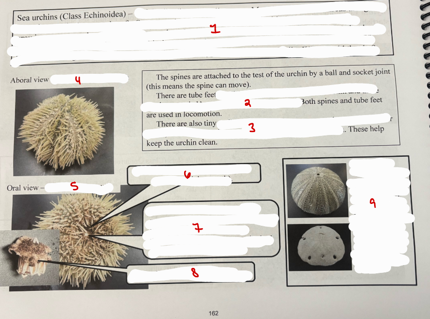
Class Echinoidea
(means spiny)
podia - in 5 major regions of the urchin and these can be extended beyond the ends of the spines
pedicellariae (jawed structures on the ends of slender stalks) between the spines and particularly around the mouth
side without the mouth
side with the mouth
pedicellariae here
mouth - it has 5 teeth. the entire mouth structure is called “Aristotle’s lantern”. Aristotle apparently thought the jaw-like structure looked like the frame of a lantern.
Internal parts of Aristotle’s lantern
Top one is the endoskeleton of an urchin, consists of fused dermal ossicles. Bottom one is from a sand dollar
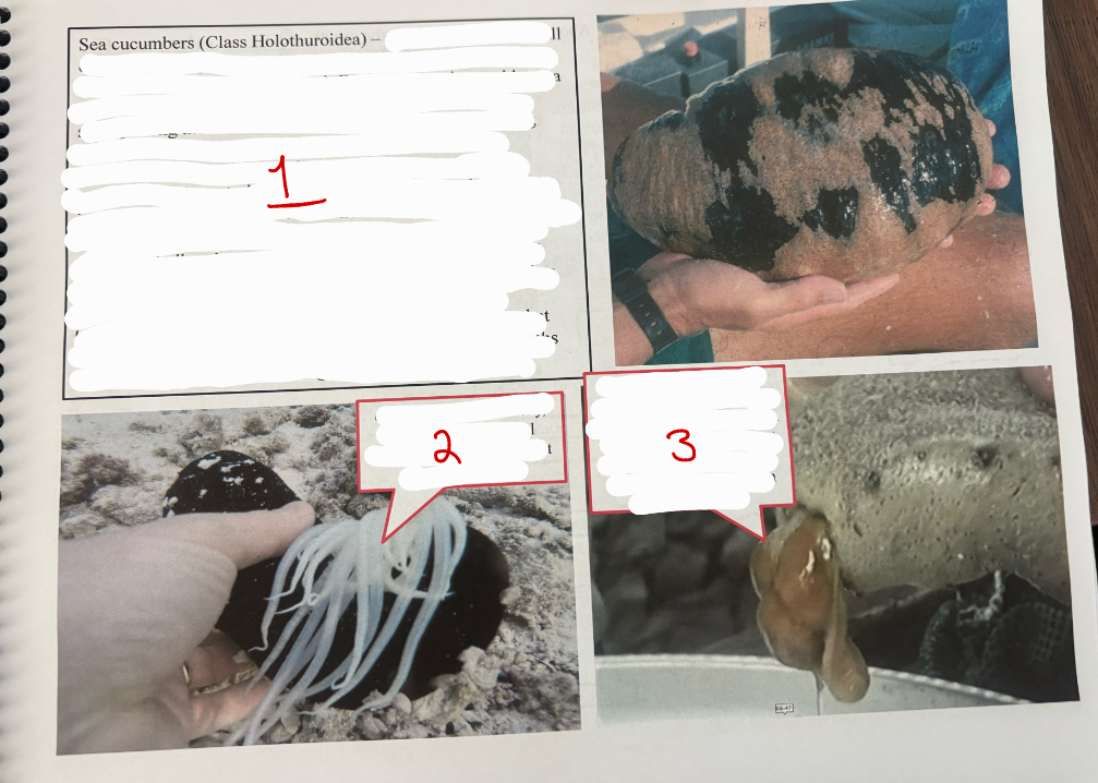
Class Holothuroidea
(means plant-like animal) deposit feeding sea cucumbers use their tentacles to push sediments from the ocean floor into their mouth. organic material is then digested and absorbed and the inorganic sand is expelled out of the anus. while sea cucumbers do not have many predators, some species have anti-predator tactics including the release of sticky, white tubular material from the anus (cuverian tubules) as well as expelling their internal organs out of the mouth or anus (evisceration)
cuvierian tubules 0 sticky, white tubular material released from the anus that deters predators
evisceration - internal organs being expelled out of the anus in response to a disturbance. this doesn’t kill the animal. internal organs are regenerated in several months.
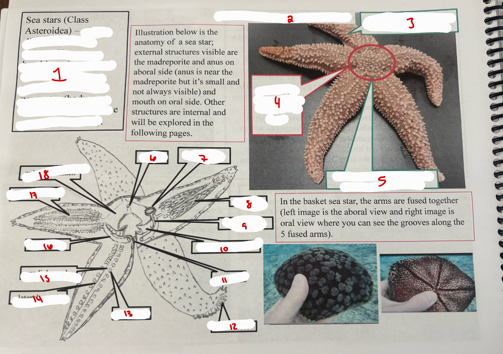
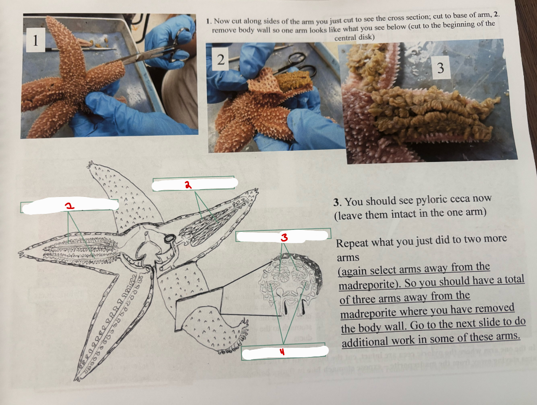
Class Asteroidea
(means star like) they are benthic and most are predators. they are pentaradial in their symmetry (body parts arranged radially in five or multiples of five)
aboral view - side without the mouth
arm - some have 5, other sea stars have more arms
central disk - arms attach to the central disk
madreporite - opening to the water vascular system
anus
madreporite
gonad
stone canal
cardiac stomach
mouth
tube feet
ampulla
lateral canal
radial canal
ring canal
pyloric ceca
pyloric stomach
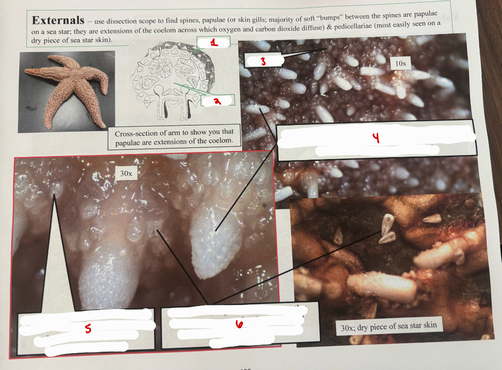
Sea Star Externals
papula
coelom
papulae
spines - the structure for which this phylum is named after
papulae - extensions of the coelom across which oxygen is obtained and carbon dioxide diffuses to the surrounding sea water
pedicellariae - pincer-like structures that help keep sea stars free from unwanted organisms that might want to attach themselves to it
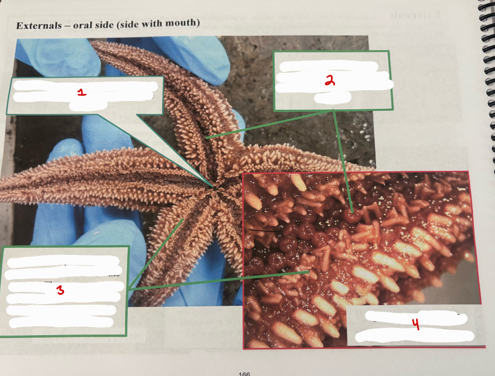
Sea Star Externals Oral Side
mouth
tube feet - externally you see the podia of the tube feet which occupy the ambulacral groove
ambulacral spines - spines that border both sides of the groove. they are movable and can interlock when the groove is contracted, thus protecting the tube feet
close up of the ambulacral groove
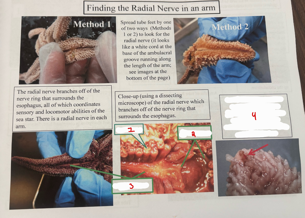
Sea Stars Finding the Radial Nerve
tube foot
nerve ring
radial nerve
light sensitive eyespot at arm tip that is connected to the radial nerve
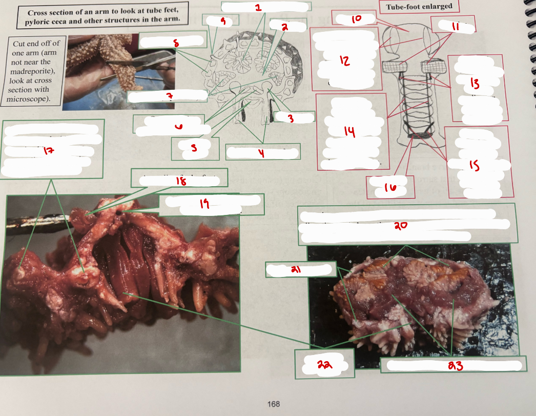
Sea Star Arm Cross Section
pyloric ceca (2 per arm)
coelom
ampulla
podia of tube feet
radial nerve
ossicles of ambulacral ridge
gonads (2 per arm)
dermal ossicles
papula
ampulla
ossicles
ampulla muscles - contracts, then water moves from ampulla into podium to elongate it
postural (orienting) muscles - to orient podium in particular direction
retractor muslces - contracts, then water moves from podium into ampulla to shorten podium
levator muscles - contract, creates suction by raising center of podium
terminal plate
dermal ossicles - white particles embedded in the dermis, this along with connective tissue make up the endoskeleton
ampulla of tube foot
canal through which the radial canal runs
pyloric ceca (2 per arm) - attached to stomach; food digestion and absorption is completed here. enzymes are produced in these blind sac (ceca) structures.
dermal ossicles
podia of tube feet
gonad of sexually mature sea star
Sea Star Pyloric Arm
pyloric ceca (2 per arm)
gonads (2 per arm)
pyloric ceca (2 per arm)
gonads (2 per arm)
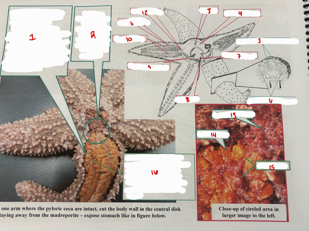
Sea Star Arm Close Up
pyloric ceca (2 per arm) - attached to pyloric stomach via pyloric duct. food digestion and absorption completed here. enzymes are produced in these blind sacs. food digested and absorbed in the ceca go into fluids filling the coelom of the sea star arm. the fluids in the coelom move throughout the arm via cilia lining the coelom.
pyloric stomach (below this and attached to it is the cardiac stomach which can be everted through the mouth)
anus
pyloric stomach
pyloric ceca (2 per arm)
eseophagus
gonads (2 per arm)
mouth
cardiac stomach
pyloric ducts
pyloric ceca
pyloric duct
pyloric stomach
pyloric ceca
pyloric ducts
pyloric ducts connect the pyloric stomach to the pyloric ceca and extend along the length of each arm (2 per arm). food moves from the stomach to the ceca via the ducts
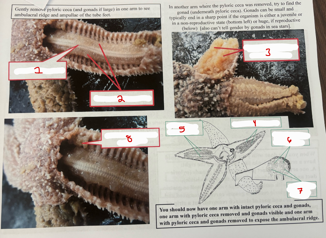
Sea Star Arm Removed Pyloric Ceca
ambulacral ridge
ampullae of tube feet
gonad of sexually mature sea star
gonads (2 per arm)
pyloric ceca (2 per arm)
pyloric ceca (2 per arm)
gonads (2 per arm)
gonad of immature sea star
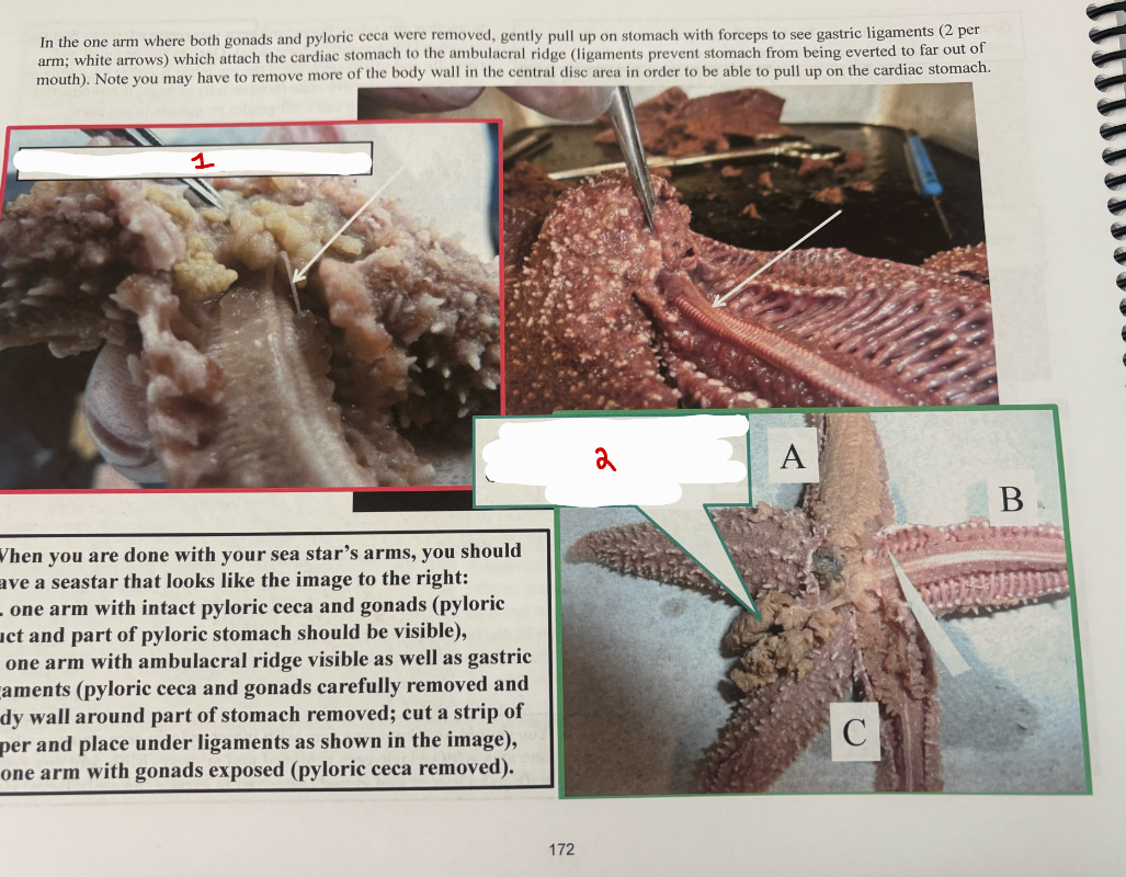
Sea Star Arm Final
gastric ligament
pyloric ceca pulled away to uncover gonads
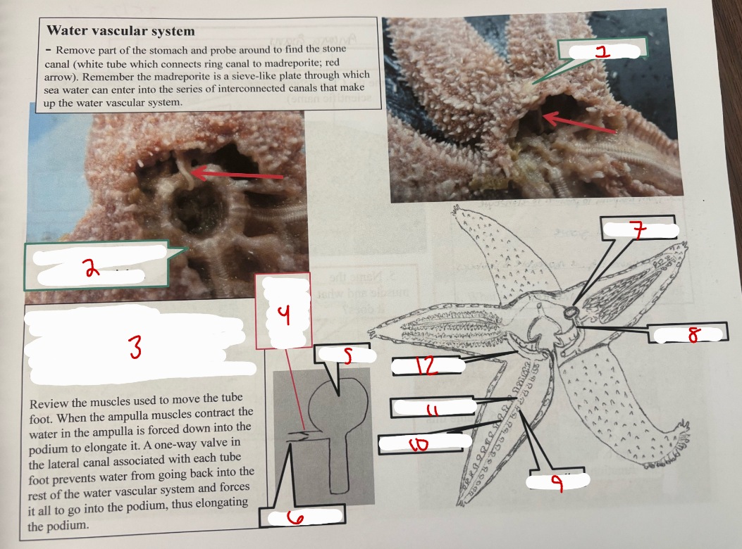
Water Vascular System
madreporite
ring canal is hidden by these ossicles
stone canal - disappears into the ring canal (ring canal hidden by ossicles), ring canal connects to the radial canal in each arm and the radial canal connects to each tube foot via lateral canals
one way valve inside lateral canal
ampulla
lateral canal
madreporite
stone canal
ampulla
lateral canal
radial canal
ring canal
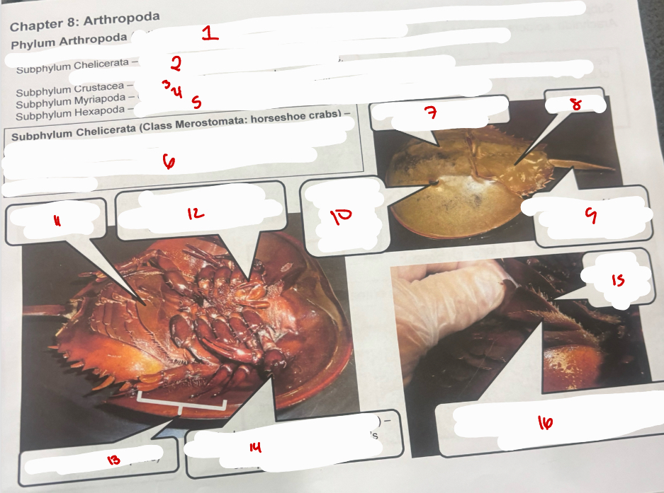
Phylum Arthropoda
Phylum Arthropoda - means “jointed food”, characteristics of this phylum include exoskeleton and jointed legs; phylum with greatest diversity or number of species
Subphylum Chelicerata - no antennae, 1st appendages are chelicerae, 2nd pair pedipalps and 4 pairs of walking legs, spiders, ticks, horshoe crabs, scorpions
Subphylum Crustacea - gills, 2 pairs of atennae, crabs, crayfish, pillbugs, shrimp, lobster
Supphylum Myriapoda - one pair of atennae and many legs (either 1 or 2 pair per beody segment); milipedes, centipedes
Subphylum Hexapoda - head, thorax, abdomen body segments, 3 pairs of walking legs, insects
Subphylum Chelicerata (Class Merostomata: horshoe crabs) _ found along the Atantic coast. They feed on molluscs and dead fish. A portion of the horshoe crabs b,ood is used by the biomedical industry to ensure that their products are free of bacterial contamination
Carapace - exoskeleton of the cephalothorax
abdomen
telson - used o help crab turn right side up when turned over
compound eye - can see lines, shapes, and borders
Gill opercula - cover gills
Chelicera (1st pair of appendages) - used to detect and manipulate food
walking legs (4 pairs)
pedipals (2nd pair of appendages) - in males, used to clasp female’s carapace during mating
gill operculum - cover gills
book gills - blood pumped through the gills contains hemocyanin instead of hemoglobbin
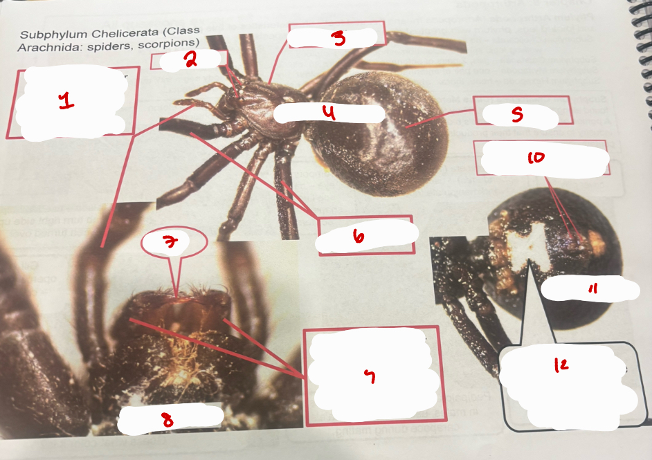
Subphylum Chelicerata (Class Arachnida)
Pedipalps (2nd pair of appendages) - used to grip prey and in males, transfer sperm
eyes
cephalothorax
dorsal view
abdomen
walking legs
fang
ventral view
chelicerae (1st pair of appendages) - terminal segment has a fang used to inject venom
spinnerets - extrude different types of silk
ventral view
Subphylum Hexapoda
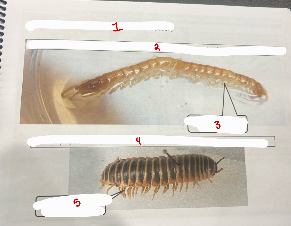
Subphylum Myriapoda (centipedes and milipedes)
Class Chilopoda - centipedes, carnivorous, active predators; 1st trunk appendage with fang
one pair of legs per body segment
Class Diplopoda - milipedes - herbivorous; some species curl up when distrubed
two pairs of legs per body segment
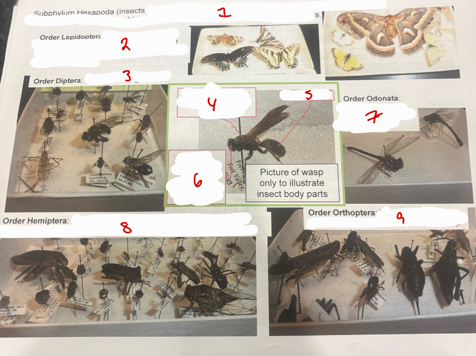
Subphylum Hexapoda
Subphylum Hexapoda (insects): here are a few insect orders; many insect orders has the word “ptera” in it (i. e. Diptera) which means organisms with wings or wing-like parts
Order Lepidoptera - wings covered with tiny scales, wings often with colorful markings, coiled tube for amouth (butterflies, moths)
Order Diptera - one pair of wings (flies)
Thorax - locomotor structures on this segment
abdomen
Head - senosry and feeding structures on this segment
Order Odonata - large eyes, large membranous wings (dragonflies)
Order Hemiptera - piercing mouthparts (like a syringe needle), wings covered with leathery-like dover (ambush bugs, wheel bugs, cicadas)
Order Orthoptera - hind legs adapted for jumping (crickets, grasshoppers, katydids)
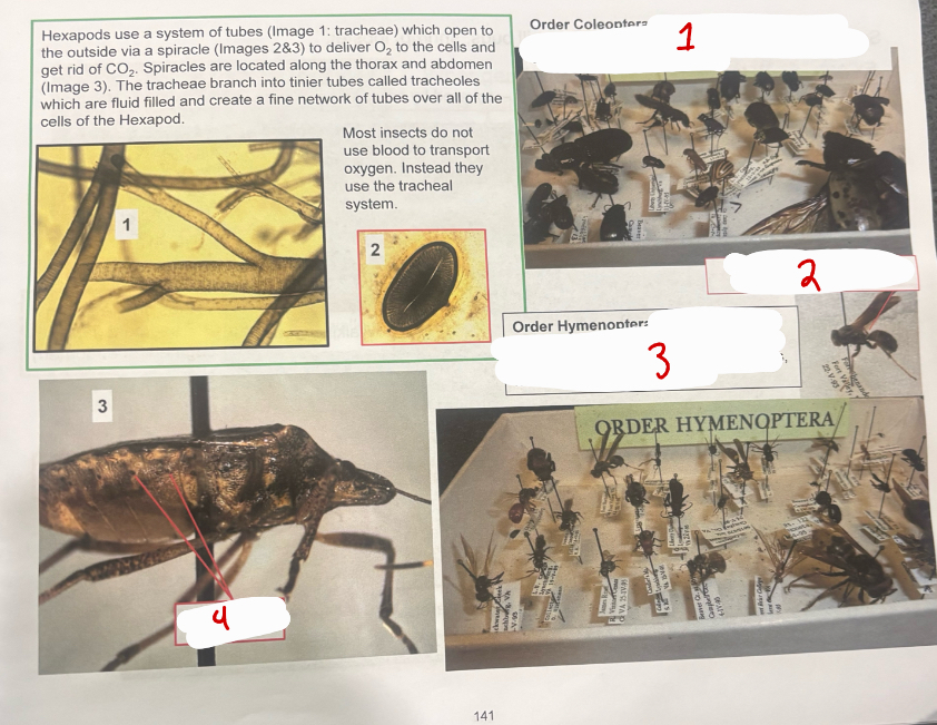
Hexapods
Order Coleoptera - chewing mouthparts, wings covered by a hard, often shiny covering (tiger beetles, dung beetles, ladybird beetles)
Order Hymenoptera - if winged, 2 pair of wings, most with thin connection between thorax and abdomen (honey bees, wasps, hornets, yellow jackets, ants)
Thin connnection between thorax and abdomen
spiracles
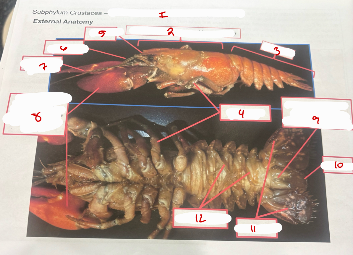
Subphylum Crustacea
Subphylum Crustacea - crabs, crayfish, pill bugs, shrimp, lobster
cephalothorax - covered by carapace
abdomen
walking legs
rostrum
antennules
antennae
chelipeds - 1st pair of walking legs; chela is the greek word for claw
anus on the telson segment (last segment of abdomen)
telson
uropods
swimmerets
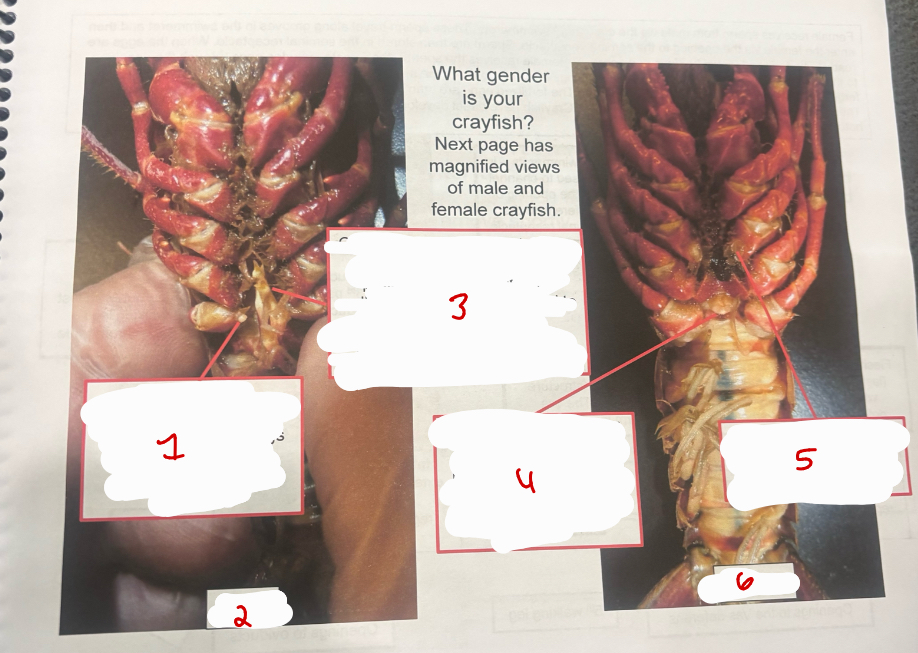
Gender
white, fleshy protuberance at base of 5th pair of walking legs (last ones) bear openings to the vas deferens
male
copulatory swimmerets (stouter than other swimmerets and project anteriorly between walking legs; in males) - used to channel sperm form the opening of the vas deferens to the females seminal receptacle
opening to the seminal receptacle - hard protuberance between last set of walking legs. receives sperm from male
opening to the oviducts at the base of the 3rd pair of walking legs
female
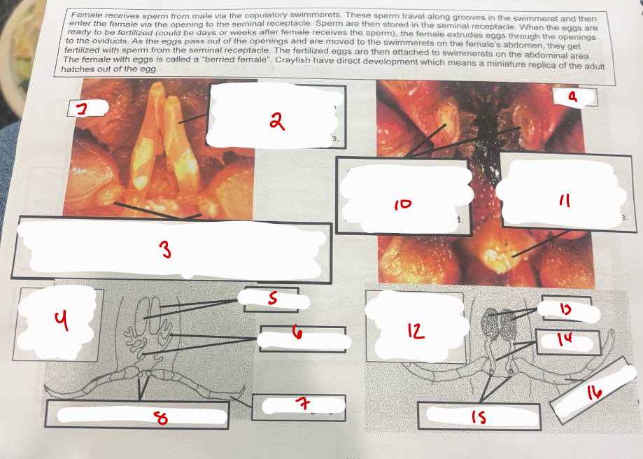
Female and Male
male
copulatory swimmerets - in males and used to channel sperm from the opening of the vas deferens to the females seminal receptacle
fleshy protuberance at the base of the 5th pair of walking legs (last ones) bear the openings to the vas deferens (structure where sperm are . the vas deferens connects to the testes where sperm is produced
internal and external view of male reproductive system
testes
vas deferens
5th walking leg
openings to the vas deferens
female
openings to the oviducts at the base of the 3rd pair of walking legs. oviducts connect to the ovaries where eggs
opening to seminal receptacle - hard protuberance between last sets of walking legs. receives sperm from male
internal and external view of female reproductive system
ovaries
oviducts
openings to oviducts
3rd walking leg
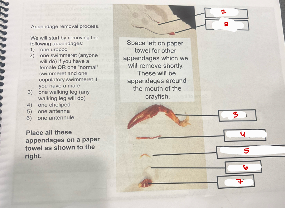
Appendage Removal
atennule
antennae
cheliped
walking leg
copulatory swimmeret
swimmeret
uropod
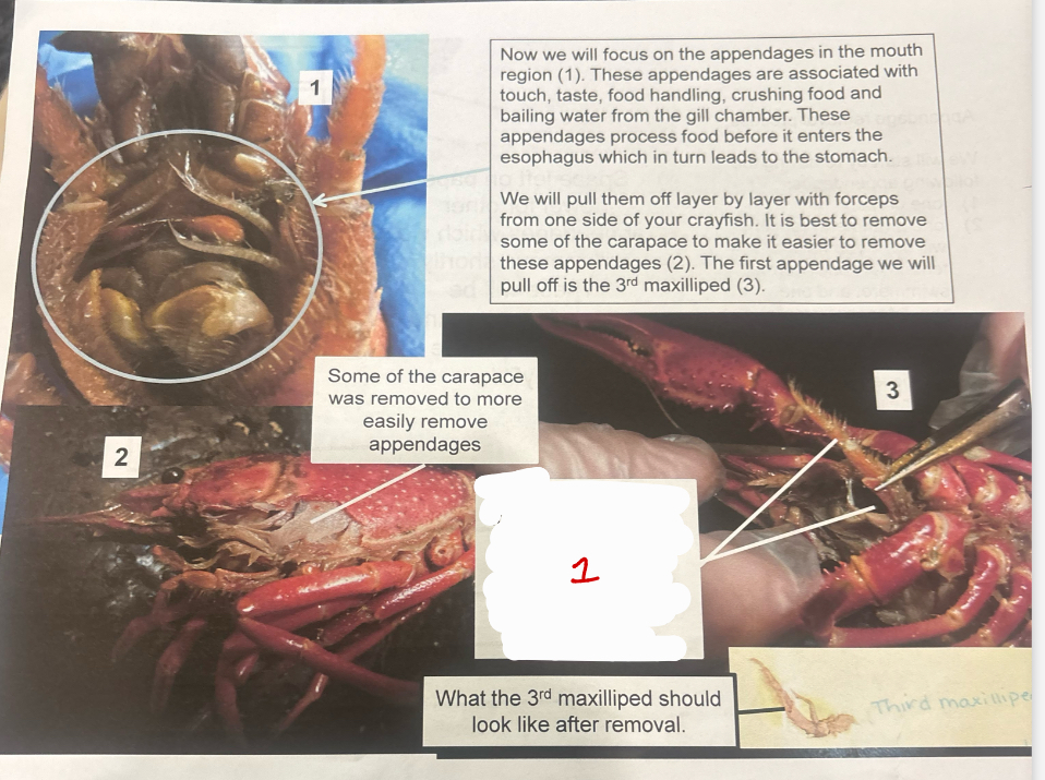
More Appendages
3rd maxilliped
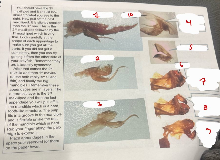
More Appendages
3rd maxilliped
2nd maxilliped
1st maxilliped
2nd maxilla with bailer; bailer moves water over the gills
1st maxilla
palp
three images of the mandible; first one is a ventral view and the palp is in a groove in mandible
here the palp has been gently pulled out of the groove in the mandible
dorsal view of mandible point out; palp has a sensory function

Carapace
3rd maxilliped
2nd maxilleped
1st maxilliped
bailer
2nd maxilla with bailer; bailer moves water over the gills
1st maxilla
palp
three images of the mandible, first one is a ventral view and the palp is in a groove in mandible
here the palp has been gently pulled out of the groove in the mandible
dorsal view of mandible with palp pointed out; palp has a sensory function
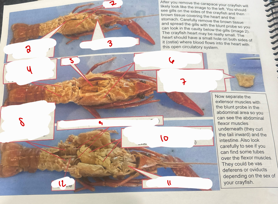
More Carapace`
gills
heart is in this location. its just anterior to the abdomen
tissue covering stomach
abdominal extensor muscles 0 muscle use to straighten out the tail after the flexors bend the tail
heart
gills have been separated with a blunt probe to expose cavity with heart
heart removed from crayfish showing shape of heart and ostium. tip of ifne tipped forceps shown for scale
abdominal flexor muscles - major force for rapid backwards swimming by flexing the tail. these muscles are underneath the extensor muscles
vas deferns (thin tubes: this is a male crayfish)
hepatopancreas (yellow, soft structure that wraps around the stomach and extends into the abdomen)
tissue covering stomach
intestine
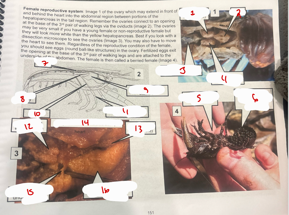
Female Reproductive System
ovary (with eggs visible)
heart
pyloric portion of the stomach
ovary (with eggs visible)
berried female crayfish
lots of eggs attached to underside of abdomen
heart
ovary
pyloric portion of stomach
oviduct
3rd pair of lwalking legs
ovary with eggs visible
ovary with eggs
oviduct
hepatopancreas in abdominal area
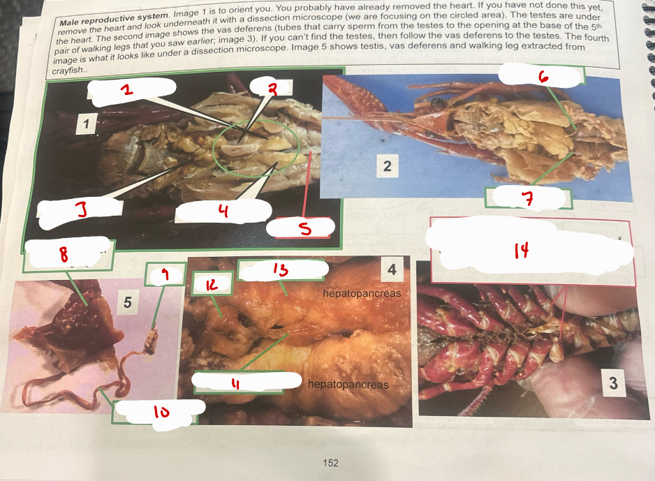
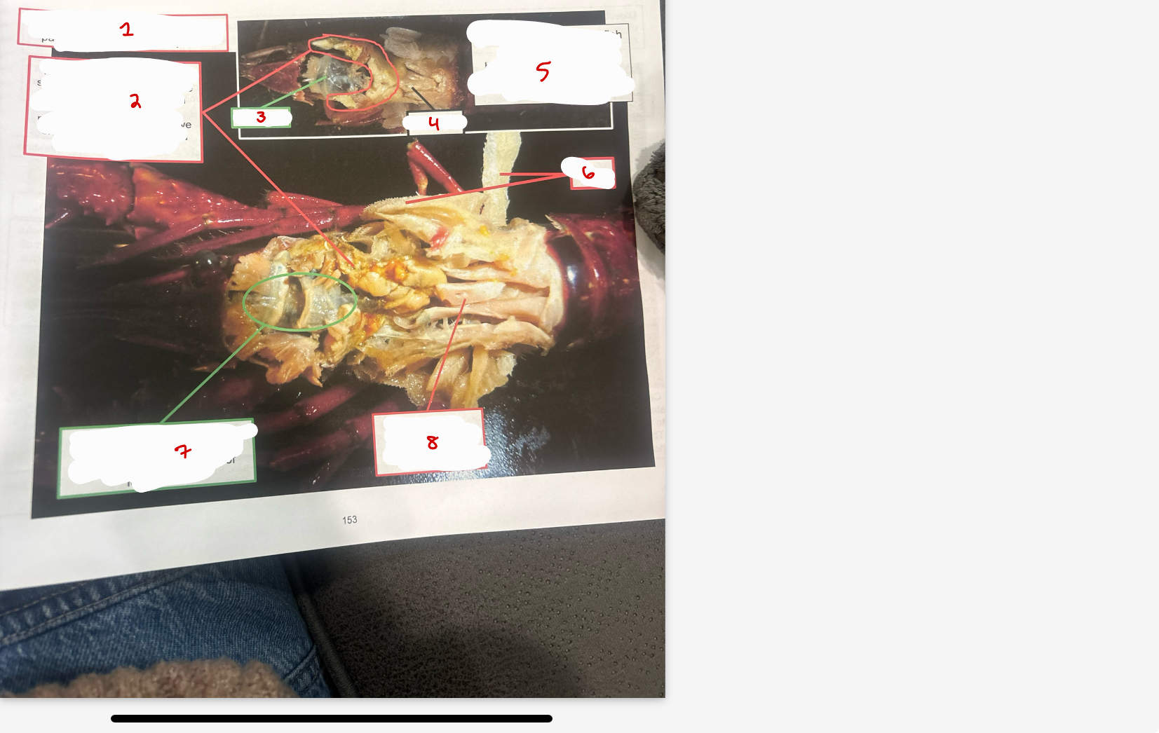
Male Reproductive System
hepatopancreas
heart
heaptopancreas
hepatopancreas
intestine
vas deferens
vas deferens
base of 5th pair of walking leg
testis
vas deferens
vas deferens
testis
vas deferns
white fleshy protuberance at base of 5th pair of walking legs (last oness) bear openings to the vas deferens
Digestive System
Digestive system
hepatopancreas 0 serves as both the liver (hepato) and pancreas. secretes digestive enzymes to the stomach and absorbs fine particles
stomach
heart
gills
stomach - initial breakdown and digestion of food occurs
heart
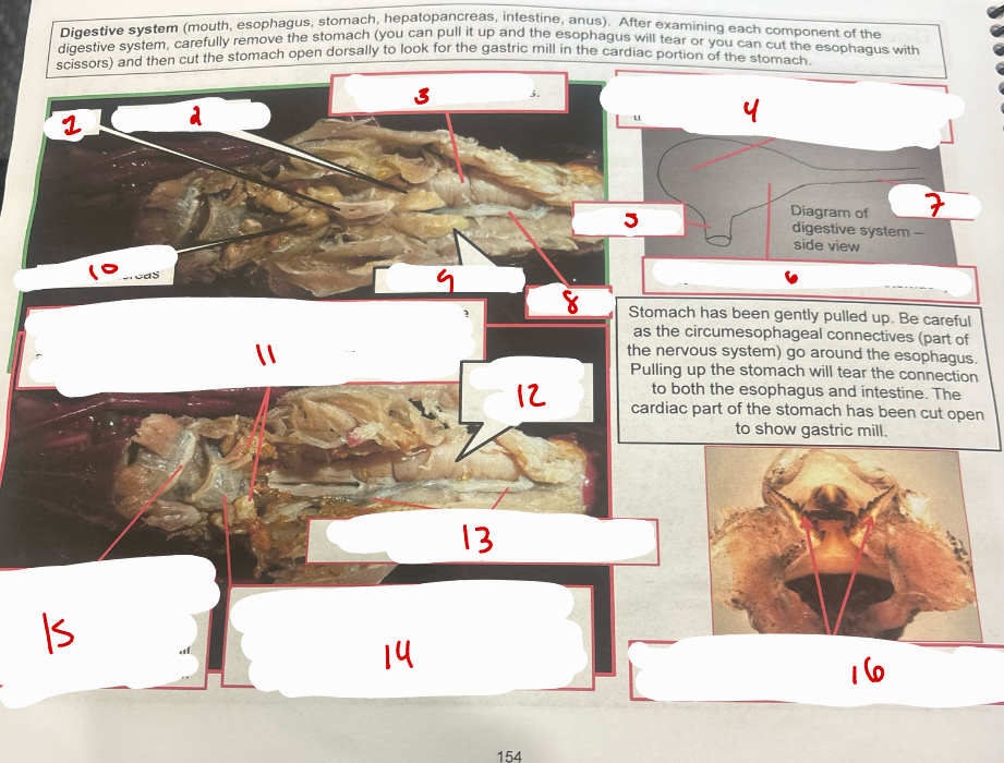
Digestive System
heart
hepatopancreas
abdominal flexor muscles
cardiac stomach (1st part of the stomach) which is connected to the esophagus. this part has the gastric mill (teeth) used to grind up food
esophagus
pyloric stomach (2nd part of stmoach)
intestine
intestine
hepatopancreas
hepatopancreas
hepatopancreas - secrets digestive enzymes to the pyloric stomach and absorbs fine partciles for digestion and absorption
abdominal flexor muscles
intestine - digestion and absorption of food is completed here
pyloric stomach (2nd part of stomach_ which is directly connected to the cardiac stomach. it receives enzymes from the hepatopancreas to further digest food
cardiac stomach (1st part of stomach - which is connected to the esophagus. this part has the gastric mill which grinds up food
cardiac stomach cut open to show gastric mill
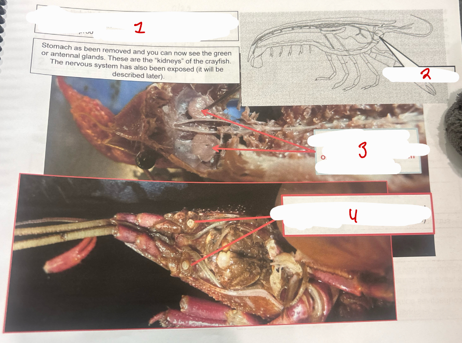
Excretory System
antennal gland
green or antennal glands - excretory organ, forms urine from the hemolymph
opening to the green glands (ventral side of animal at base of antennae)
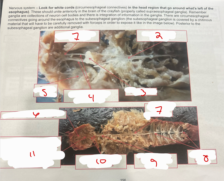
Nervous System
supraesophageal ganglia
subesophageal ganglion
esophagus
circumesophageal connectives
green gland
supraesophageal ganglia
thoraic ganglion
abdominal ganglia
subesophageal ganglion
circumesophageal connectives
esophagus was here
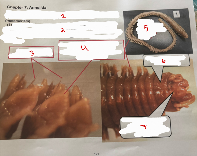
Annelida
Phylum Annelida (little ring) - have serial repetition of body parts both externally and internally (metamerism)
Clade Errantia or Class Polychaeta (bristle worms) - (poly long hair) - can have many setae
setae - used in locomotion and defense
parapodia (pair per body segment) - foot-like structures used in locomotion and respiration (these are vascularized)
Nereis (sandworms and clamworms in this genus)
Peristomium - segment around mouth
Prostomium (segment before mouth) with sensory structures (eyes and tentacles)
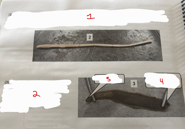
Worms
Clade Sedentaria (Class Oligochaeta) - aquatic and terrerestial worms (oligo (few) long hair (chaite)
Class Hirudinea (leeches) - many are parasitic
oral sucker with mouth
caudal sucker - bigger than the anterior sucker
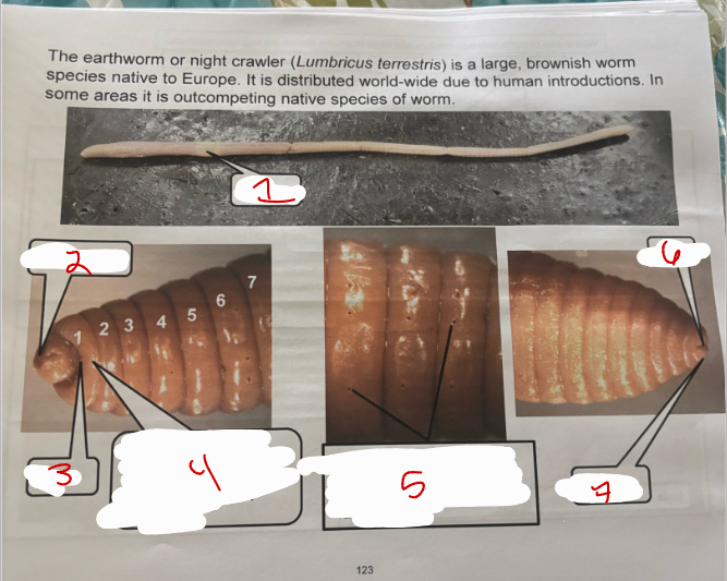
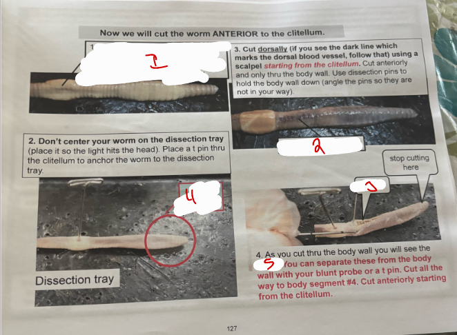
Lumbricus Terrestris
clitellum
prostomium
mouth
peristomium - first segment
setae - help worm hold onto a surface its crawling along
anus
pygidium
Earthworm Ventral Side
ventral side
seminal grooves
male pores
openings to the seminal receptacles
female pores
male pores
male pores - spermatozoa exit here and travel along seminal grooves to another earthworms seminal receptacles
seminal grooves
male pores (2)
earthworms are hermaphroditic (have male and female organs). when they mate, they exchange spermatozoa (mutual insemination)
female pores
female pores - oviducts discharge eggs from these pores (2)
male pore
openings to seminal receptacles
opening to seminal receptacles
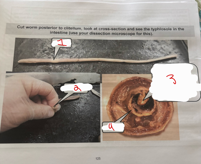
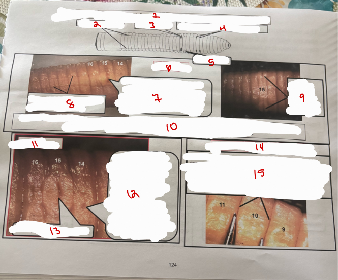
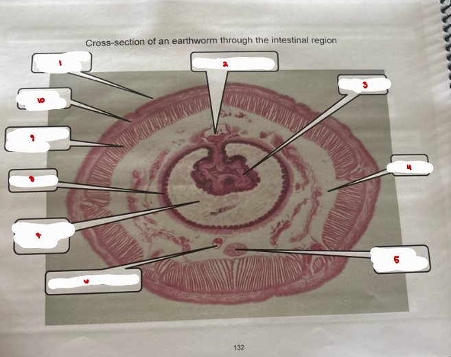
Earthworm Cut
clitellum
typhosole
typhlosole - fold in intestine that inreases surface area available for digestive enzyme production and food absorption
intestine
Earthworm Stuff
dorsal blood vessel
intestine - where enzymatic digestion and absorption occurs
intestine
intestine pushed to the side to expose the ventral nerve cord and blood vessel
ventral blood vessel
ventral nerve cord (white in appereance)
remnants of septa
metanephrida - function like a kidney nephron; take in coelomic fluid via ciliary action and reabsorb glucose and salts while ammonia and excess water are excreted through a pore (nephridiopore) to the outside
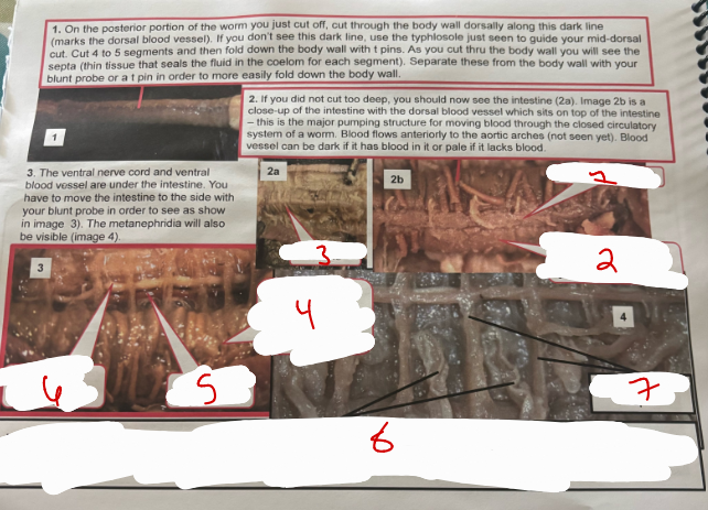
Anterior Cut Earthworm
ventral side
dark mid-dorsal line
septa
lit area
Dorsally Cut Earthworm
dorsal blood vessel
seminal vesicles
pharynx
aortic arches
seminal receptacles
esophagus
crop
gizzard
intestine
intestine
crop - gray in color and soft; temporary storage structure for food
seminal receptacles (2 on each side) - spermatozoa from another worm stored here
“hairy” pharynx dilator muscles (cerebral ganglia embedded)
aortic arches
seminal receptacles (2 on each side)
seminal vesicles 0 spermatozoa are produced in the testes
gizzard - white and firm, muscular grinding organ
intestine
magnified view of aotric arches
dorsal blood vessel
aortic arches - carry blood from dorsal to ventral blood vessel
ventral blood vessel
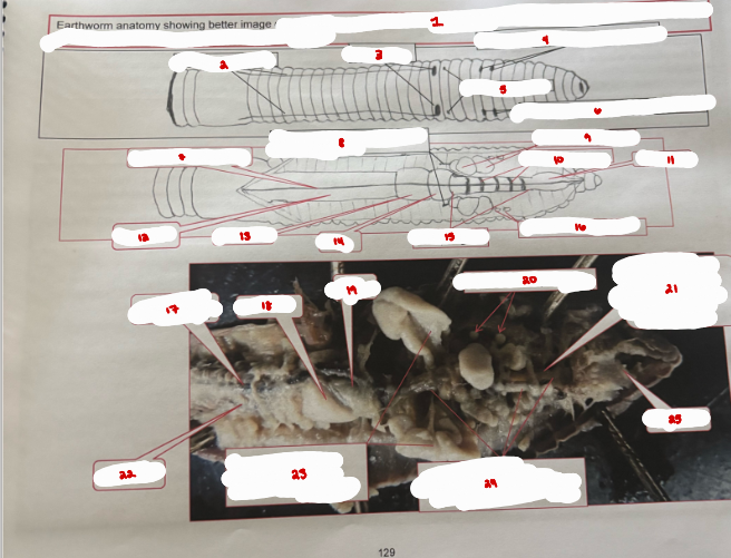
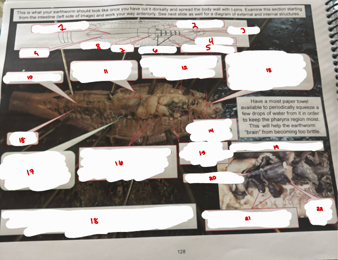
Earthworm Anatomy
esophagus - conducts food between pharynx and crop and diagrams showing both external openings and associated internal structures
seminal grooves
male pores
openings to the seminal receptacles
female pores
openings to the seminal receptacles
dorsal blood vessel
tube connecting spermatozoa to male pores
seminal vesicles
aortic arches
pharynx
intestine
gizzard
crop
esophagus
seminal receptacles
dorsal blood vessel
gizzard
crop
seminal receptacles
aortic arches would have been here (removed to see esophagus)
intestine
seminal vesicles - pulled to the side to show the esophagus
esophagus - tube connecting pharynx to crop
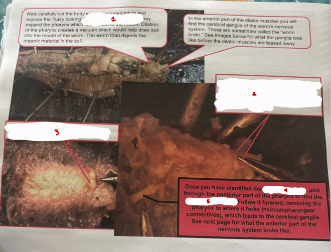
Earthworm Stuff
pharynx dilator muscles
body wall to the prostomium
cerebrial ganglia imbedded in the pharynx dilator muscles
cerebrial ganglia
ventral nerve cord
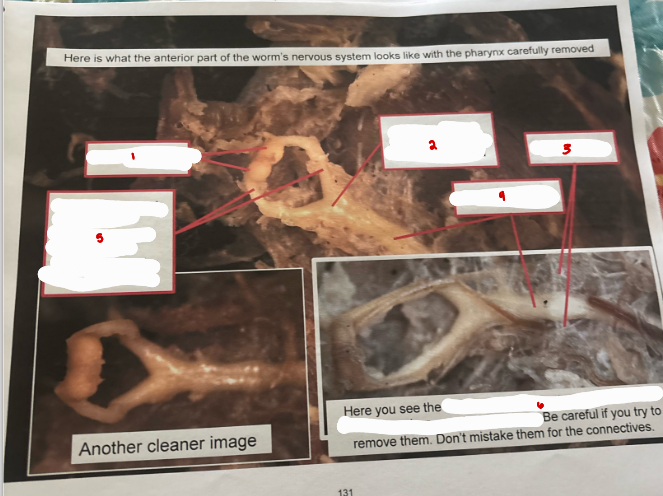
Earthworm Nervous System
cerebrial ganglia
subpharyngeal ganglian (1st part of ventral nerve cord)
lateral nerve
ventral nerve cord
circumpharyngeal connectives - connect cerebral ganglion along the ventral nerve cord
blood vessels around the circumpharyngeal connectives
Earthworm Cross Section
cuticle and epidermis
dorsal blood vessel
typhosole
coelom
ventral nerve cord
ventral blood vessel
lumen of intestine
intestine
longitudinal muscles
circular muscles
Mussel Overview
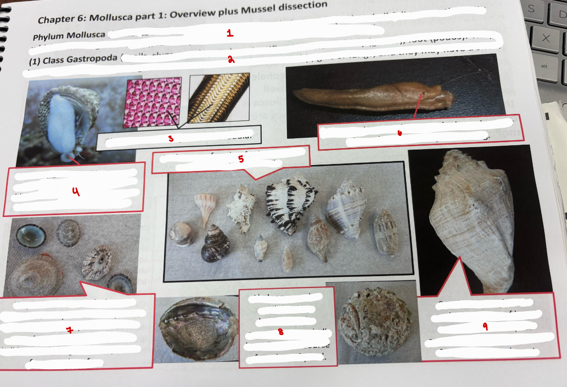
Phylum Mollusca - Mollusca means soft; characteristics include mantle (makes shell & lines mantle cavity which contains gills or lungs), radula (subset of classes in this phylum examined)
Class Gastropoda (snails, slugs, conchs, etc.) - scientific name means belly (gaster), foot (podos). They have a radula (rasping tongue-like structure with chitinous teeth), gills or lungs, and they may have a shell
Chitinous teeth or plates of a radula
Snail radula - tongue-like structure that extents out of a snail’s mouth (this snail is scarping algae off glass in an aquarium using it radula
Wide range of marine, freshwater, and terrestrial gastropoda shells
Pneumostome - air enters the lung chamber through this opening in this slug
Limpets - marine snails that are herbivorous. They remove competitors like mussels and limpets of another species by using the shell rim like a bulldozer to dislodge the competitor from a rock surface
Abalone - marine snail that is an herbivore. the nacreous inner shell layer (left) is a source of mother-of-pearl
Conch - name given to multiple species of large marine snails. They are omnivores and are harvested for their shell and meat
Mollusca Classes
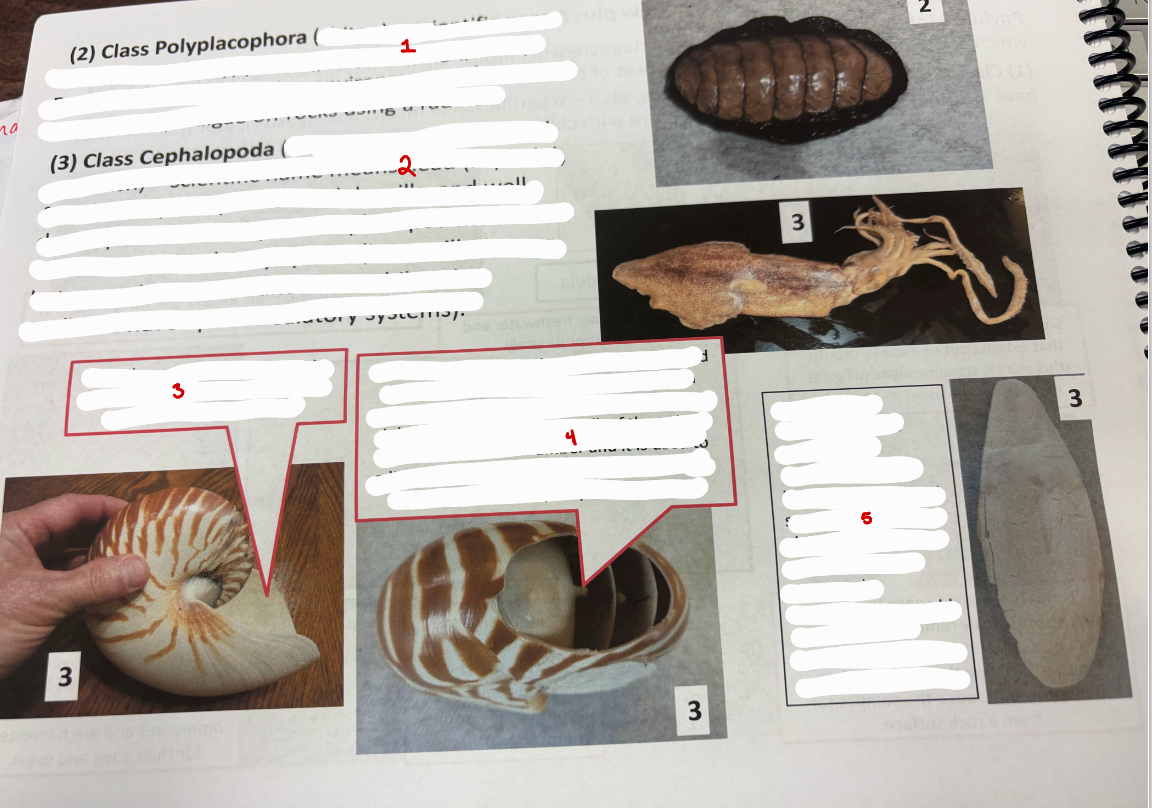
Class Polyplacophora (chiton) - scientific name means many (poly) plates (plax), bearing (phora). Found in intertidal marine environments where most species scrape algae off rocks using a radula
Class Cephalopoda (octopus, nautiloids, squid, cuttlefish) - scientific name means head (kephale), foot (podos). They have a radula, gills, and well developed arms. Unique to cephalopod mollusca is their closed-circulatory system.
Nautilus - this cephalopod adds chambers as it grows and it lives in the last chamber created
Nautilus - shows the siphuncle which connects to the bulk of the animal in the last created chamber and it is used to adjust the amount of gas in each chamber to adjust the amount of gas in each chamber to adjust the total buoyancy of the animal.
Cuttlebone - internal support structure for cuttle fish
Class Bivalvia
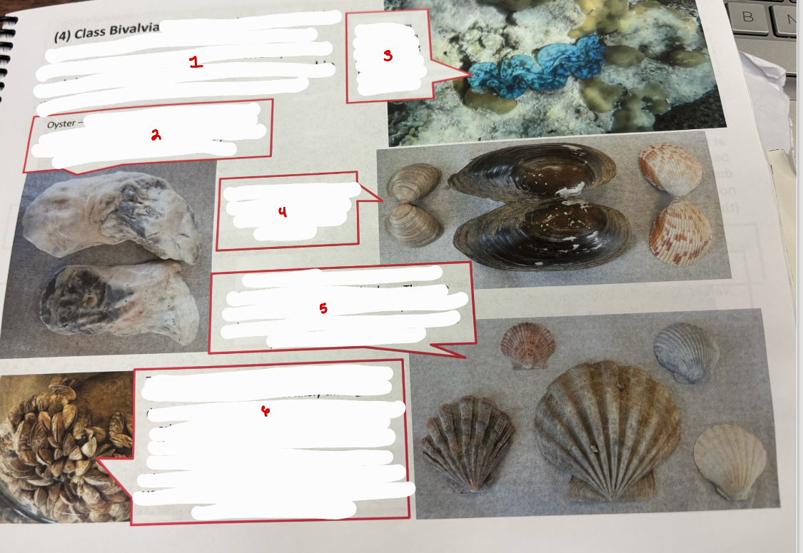
Class Bivalvia (clams, oysters, mussels) - scientific name means two (bi), valves (valva). They have 2 valves or shells, lack a radula and most are filter feeders. Found in marine and freshwater environments.
Oyster - common name for several families of marine bivalves. Several kinds of oysters are consumed raw or cooked. Pearl oysters produce pearls from their nacreous shell layer.
Giant clam embedded in coral showing blue pigments in the mantle of the clam
Several bivalves showing the two valves attached together via a hinge ligament
Scallop - marine bivalves that have characteristic fan shaped valves. they are prized for thier flesh. They can clap their two valves together quickly to swim away from a predator such as a sea star
Zebra mussel - accidentally introduced into the Great Lakes of the US most likely through the discharge of larvae in ballast water from ships originating in Europe. .these small striped bivalves have caused major damage to industry along lakes and large rivers in the eastern part of the US by clogging water intake pipes used to carry water to cool machinery
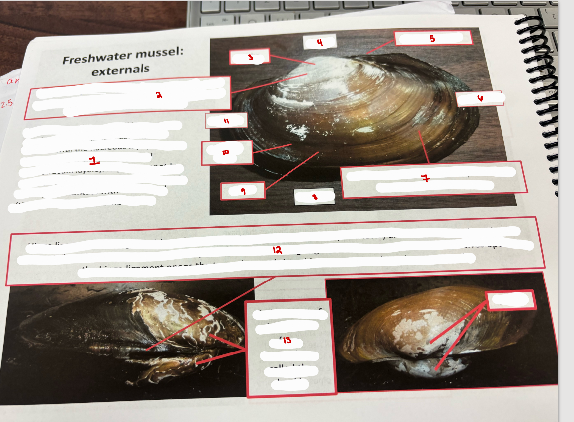
Freshwater Mussel: Externals
Valve (or shell) layers - all layers made by the mantle.
Prismatic layer - white in appearance because its made of calcium carbonate; visible hear because the periostracum has worn off
umbo
dorsal
hinge ligament
posterior
periostracum - outermost layer of valve; protects other valve layers
ventral
valve
growth rings
anterior
Hinge ligament - this ligament is under tension when adductor muscles are contracted to close the two valves
umbo - area of shell first to form (beak)
umbo
Mussel
anus
mantle
excurrent aperture
mantle
incurrent aperture - its the ventral opening made by the mantle
anus
excurrent aperture
gills
suprabranchial chamber
incurrent aperture
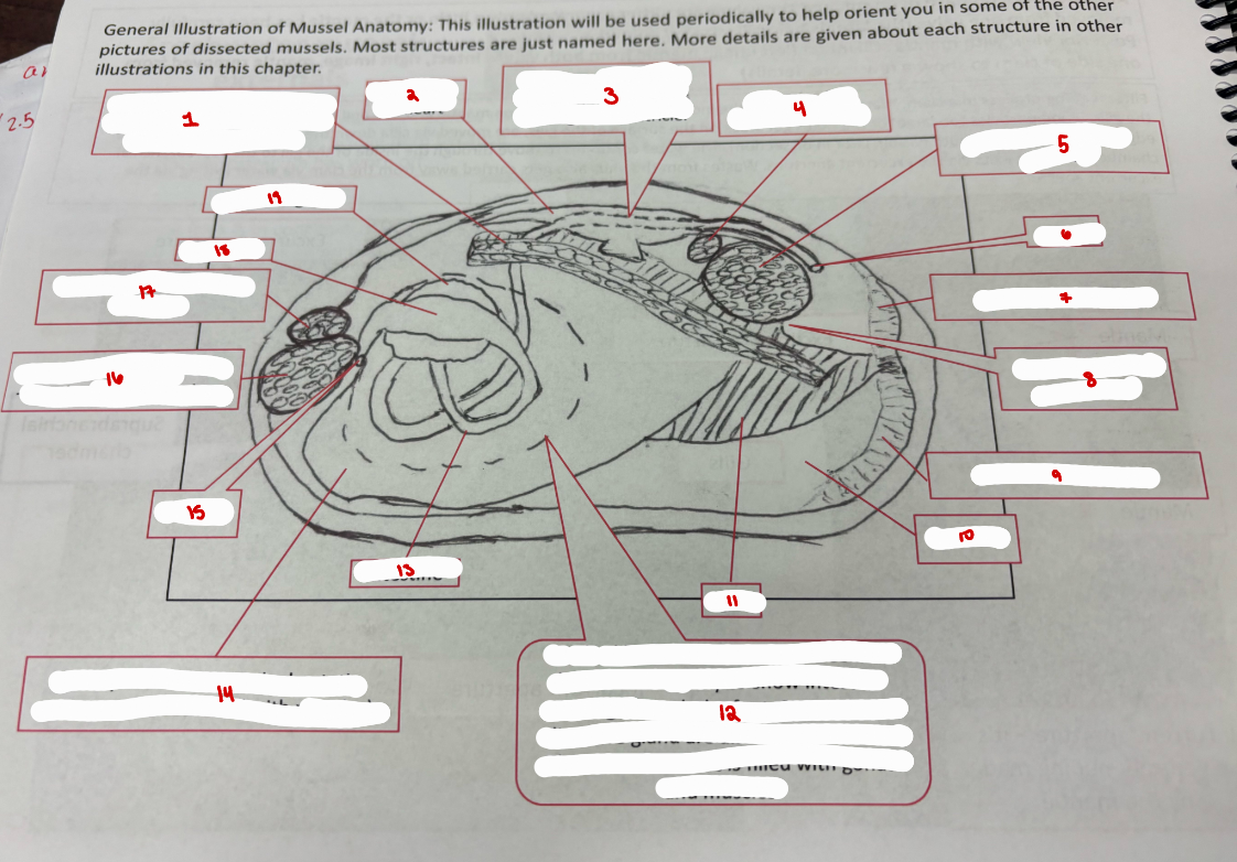
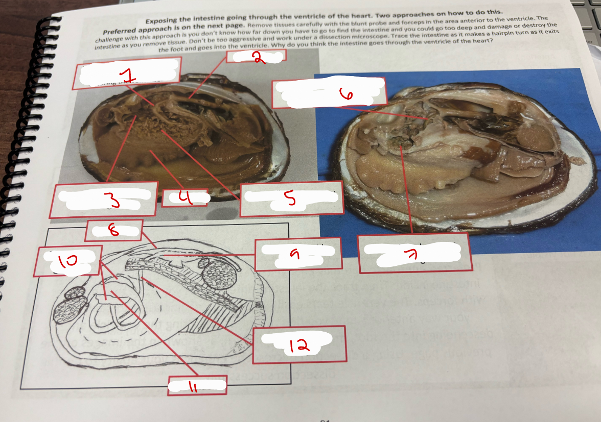
Mussel Anatomy
gills (two layers on each side of foot)
ventricle of heart
intestine - dashed line showing where it goes through the ventricle
posterior foot retractor muscle
posterior adductor muscle
anus
excurrent aperture
suprabranchial chamber
incurrent aperture
mantle
gill
dashed line is an area of the foot that has been cut away to show intestine winding through the foot. stomach and digestive gland are also inside the foot. the rest of the foot is filled with gonad and muscles
intestine
foot
mouth
anterior adductor muscle used the close the valves
anterior foot retractor muscle
stomach
digestive gland
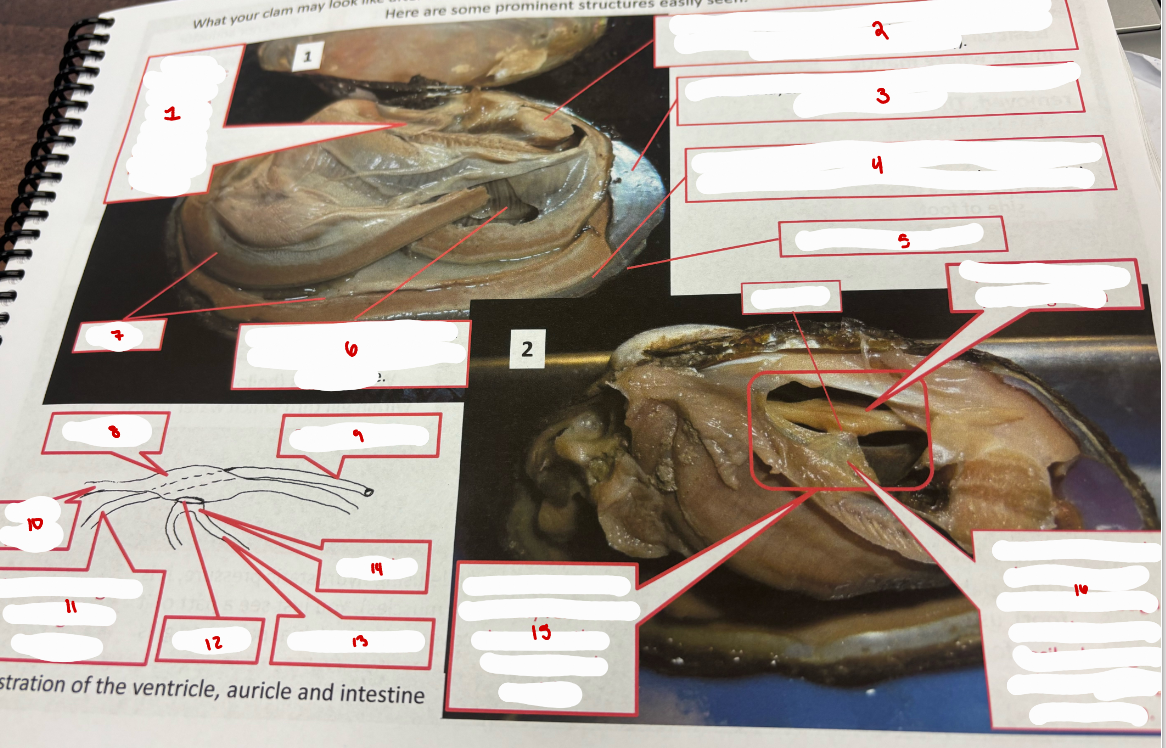
Mussel
pericardial sac
posterior adductor muscle - used to close the valves
nacreous layer (mother-of-pearl)
region where new periostracum and prismatic layer is being layed
newest periostracum
gills
mantle
ventricle
rectum & anus
blood vessel
intestine
ostium
blood vessel
auricle
pericadrial sac removed to expose the ventricle and auricle
auricle - channels blood into the ventricle through an ostium
ostium
ventricle
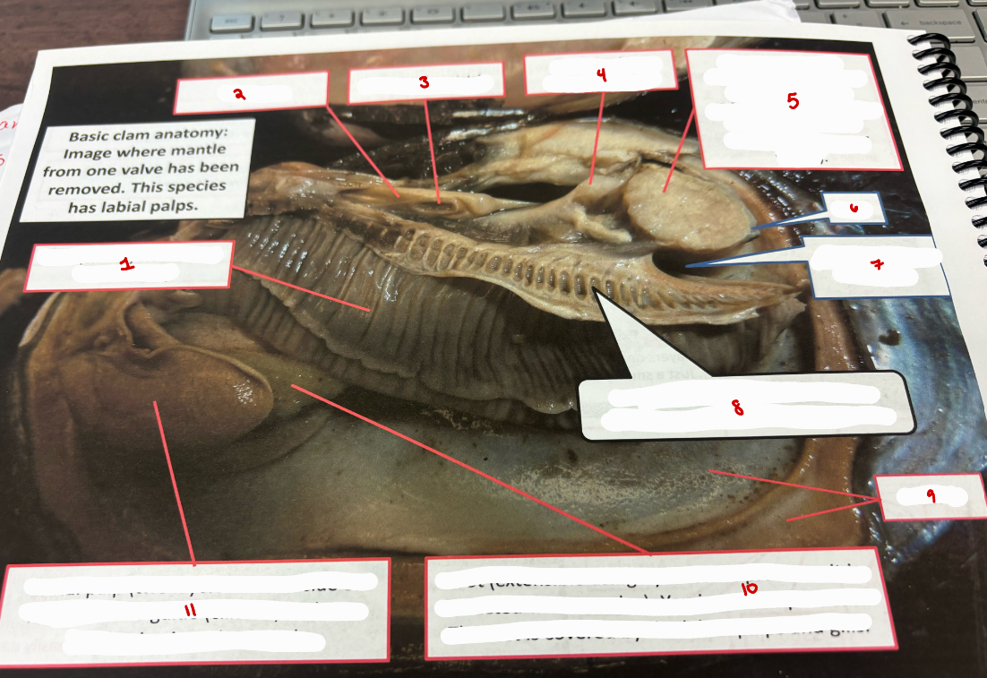
Basic Clam Anatomy
gills
ventricle
ostium of ventricle
posterior foot retractor muscle
posterior adductor muscle
anus
suprabranchial chamber
water tubes
mantle
foot
labial palp
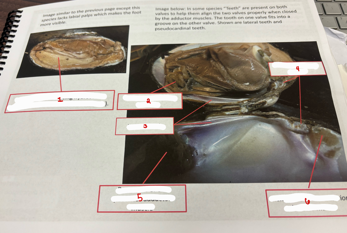
Clam Anatomy
foot
posterior adductor muscle
lateral teeth
pseudocardinal teeth
remnants of the posterior adductor muscle
remnants of the anterior adductor muscle
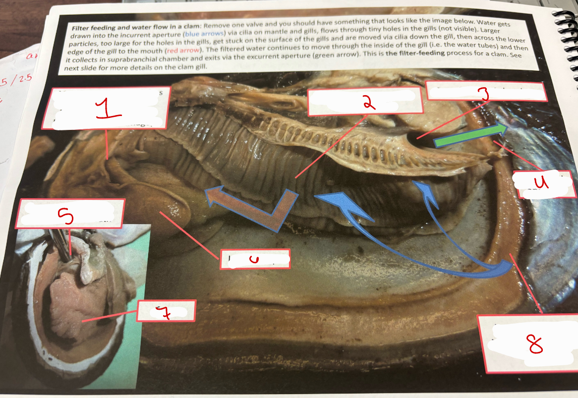
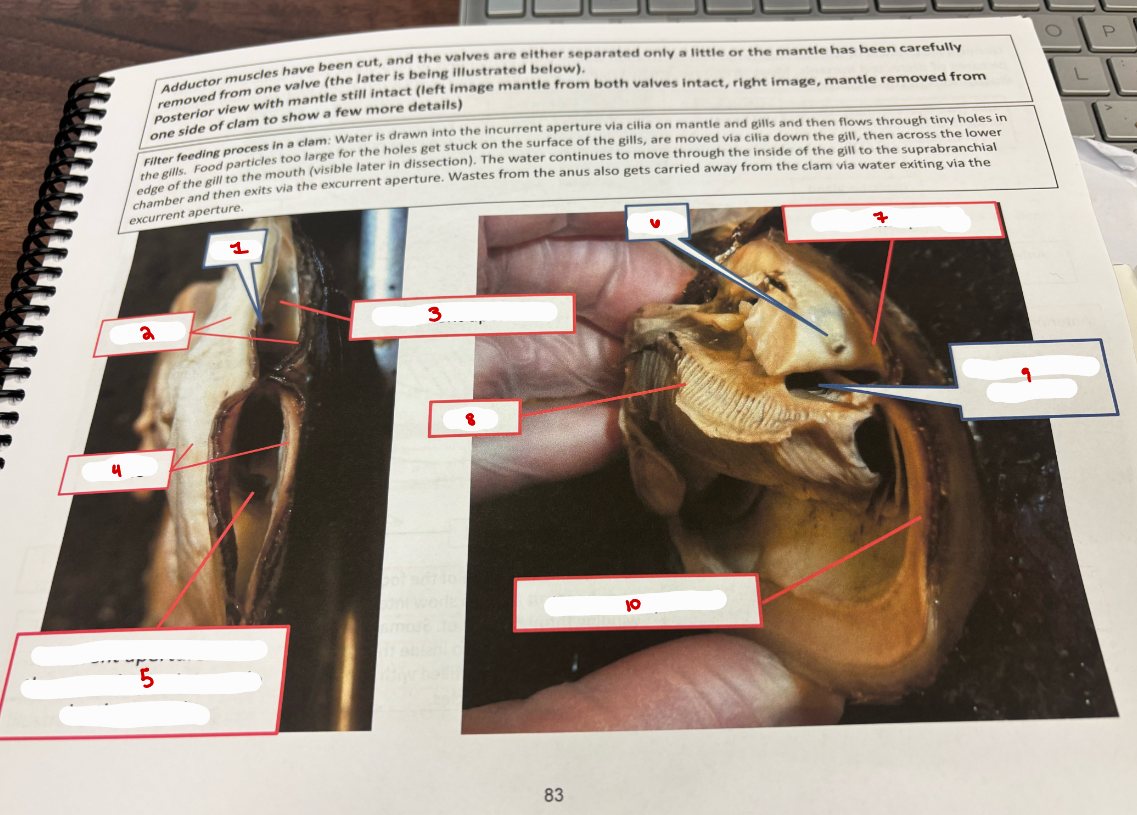
Water Flow Clam
mouth
gills
suprabranchial chamber
excurrent aperture
mouth
labial palps
foot
incurrent aperture

Clam: Close-up of Gills & Foot
ventricle
intestine
mantle
gills
mantle
gills
foot
intestine
gill filaments
water tube
intestine
gill filaments
water tube
ostia (pores on gill surface)
cilia
Clam
stomach and digestive glands
gonad
intestine
digestive gland
intestine inside foot
digestive gland
intestine
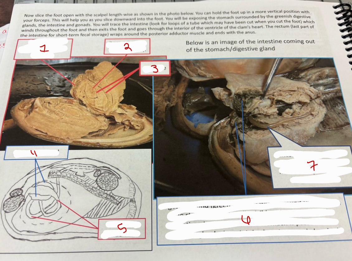
Clam Exposing Intestine
intestine coming out of foot
ventricle
stomach and greenish digestive glands
foot
gonad
intestine
stomach and greenish digestive glands
ventricle
intestine inside ventricle
digestive gland
stomach
intestine inside foot
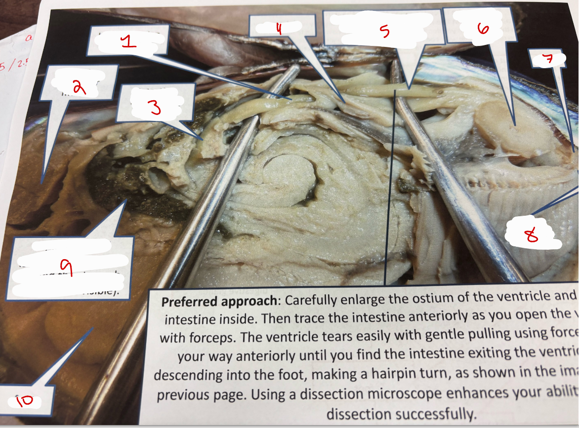
Clam 4
intestine going through ventricle
anterior adductor muscle
intestine descending into foot
ventricle
intestine exposed after the ventricle was removed
posterior adductor muscle
anus
gill
digestive gland
foot
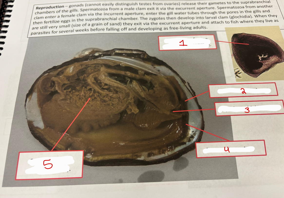
Clam Reproduction
glochidium
excurrent aperture
suprabranchial chamber
incurrent aperture
gonad
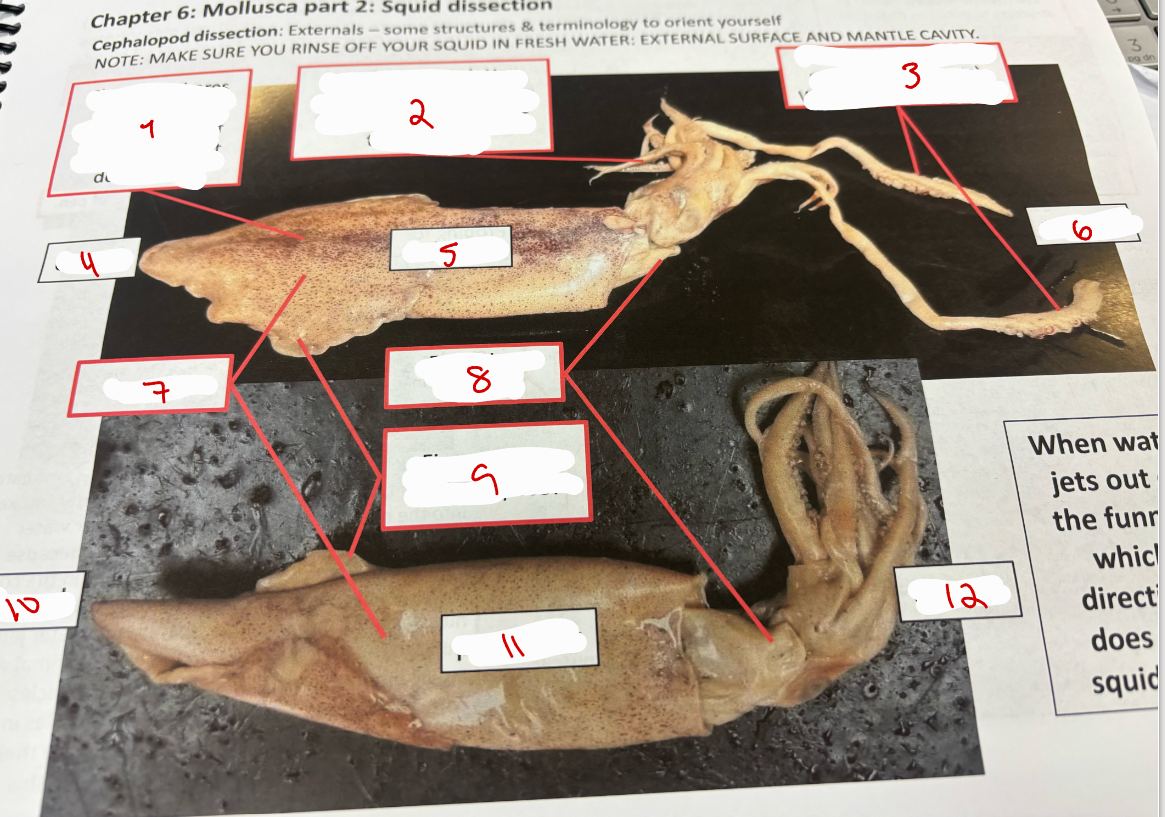
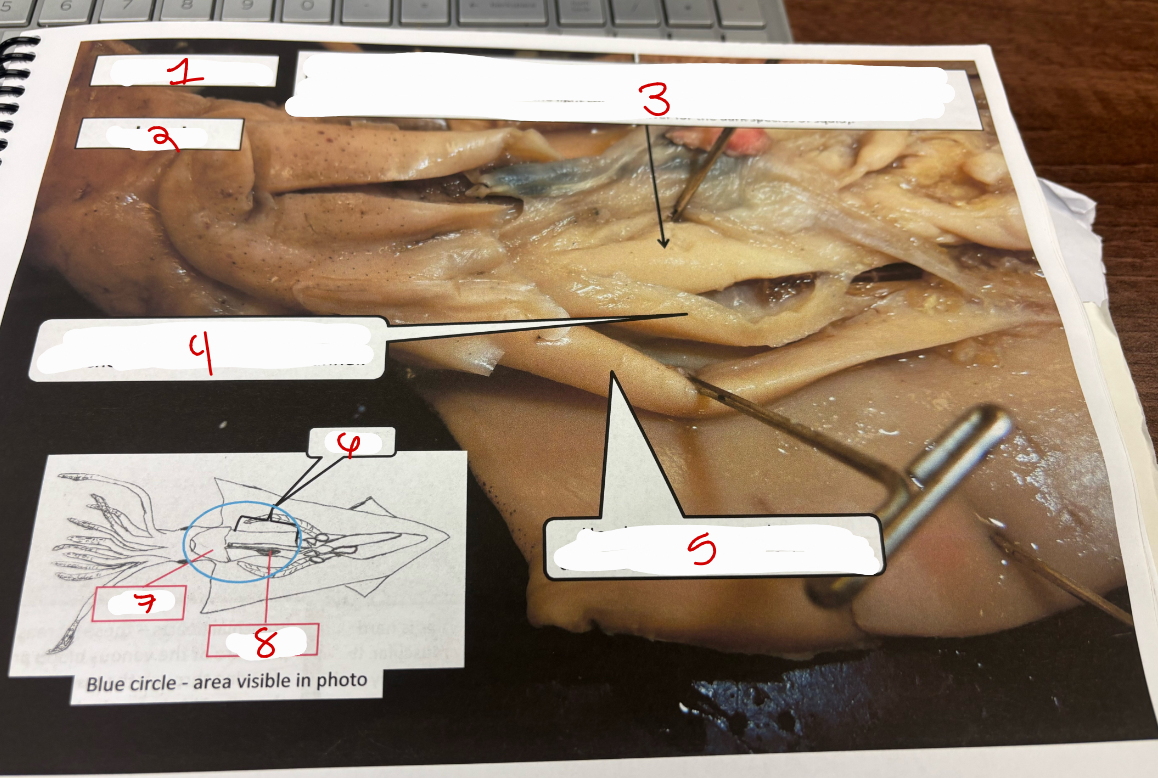
Squid 1
chromatophores -m enable rapid color change for camouflage, communication, and thermoregulation
short arms - 8, suckers along length
tentacles - 2, suckers at tip, used for catching prey
dorsal
anterior
ventral
mantle
funnel or siphon
fins - used for stability, maneuvering, and efficient low-speed locomotion
dorsal
posterior
ventral
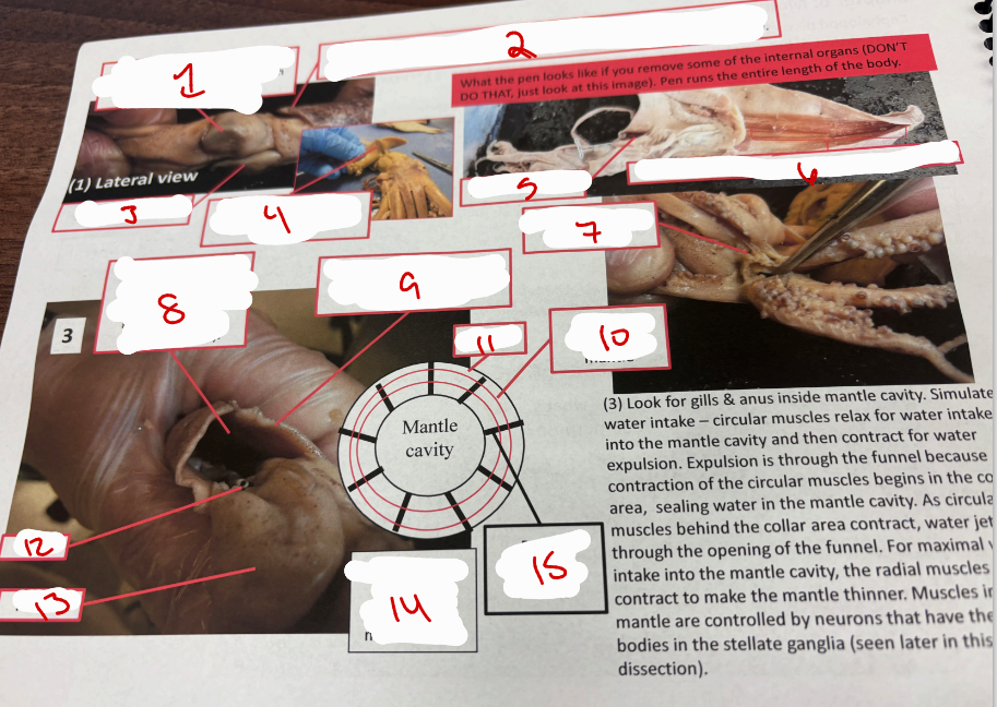
Lateral View Squid
eye
extension of pen (an internal, chitinous support structure that runs the length of the squid’s body)
funnel or siphon
mantle
extension of pen
pen
probing for the beak
mantle cavity
collar of mantle - part of mantle that encircles the mantle cavity
circular muscles in mantle
mantle
anus
funnel
cross-section through mantle and mantle cavity
radial muscles in mantle
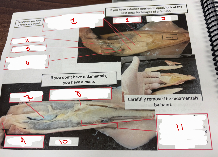
Squid 2
ink sac
male reproductive structures
funnel
male
testis - cream colored, soft, oval shaped organ under cecum. cecum is on top of the cream colored testis and its a thin-walled sac that is part of the squids digestive sytstem
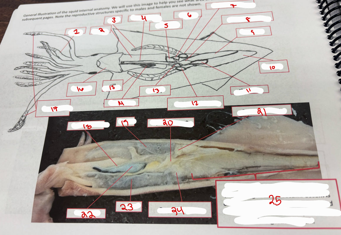
Squid General Anatomy
arms
liver
gill
intestine
pancreas
kidney
branchial heart
systemic heart
cecum
gonad
stomach
branchial heart
kidney
ink sac
gill
funnel
tentacles
intestine
gill
systemic heart
branchial heart
ink sac
gill
branchial heart
testis
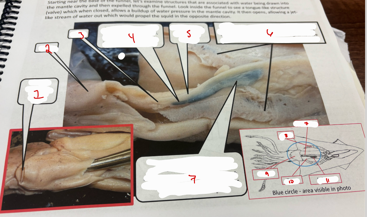
Squid 3
funnel with valve pointed out
funnel
anus
ink sac
rectum - short term fecal storage
gills
ink sac with sepia - sepia can be expelled through the funnel if the animal is disturbed
rectum
gill
funnel
liver
ink sac
Squid 5
light species
head
liver
funnel retractor muscles
head rectractor muscles
liver
funnel
ink sac