Neuronal Signaling
1/25
There's no tags or description
Looks like no tags are added yet.
Name | Mastery | Learn | Test | Matching | Spaced | Call with Kai |
|---|
No analytics yet
Send a link to your students to track their progress
26 Terms
Morphogenesis:
Organogenesis
Organs of the body develop from specific portions of the three embryonic germ layers
cells from 2 or 3 germ layers participate in forming a single organ
ex: neurulation
Morphogenesis:
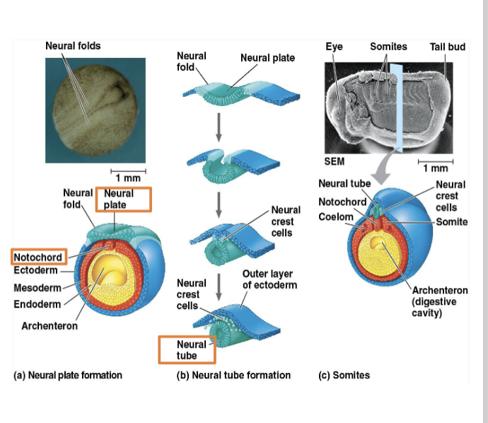
Neurulation
• Neurulation-formation of the
brain and spinal cord in vertebrates
• Mesoderm cells form notochord
• Notochord secretes signaling
molecules causing ectoderm to
form neural plate
• Neural plate cells change shape
to form the neural tube which
becomes the central nervous
system (brain and spinal cord)
• Notochord disappears before
birth, but some parts persist as
disks
What disability occurs when a portion of the neural tube doesn’t develop or close properly?
Neural tube defects (NTDs) are the disabilities that occur when the embryonic neural tube fails to develop or close properly, typically within the first 28 days of pregnancy. The most common types are spina bifida (spine/lower tube issues) and anencephaly(brain/upper tube issues), which can lead to lifelong physical disabilities or death
Contraception is preventing pregnancy by preventing
The release of gametes, implantation, and fertilization
Neuron Structure
Neuron
Fundamental unit of the nervous system
Specialized to receive and transmit electrical (long distance) and chemical (short distance) signals
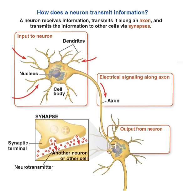
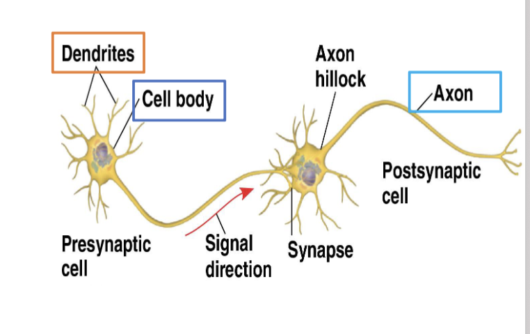
Neuron structure
• Cell body
• Contains nucleus and
organelles
• Dendrites
• Branched extension of
cell body that receive
signals from other
neurons
• Axon
• A single long extension
that transmits signals
to other cells
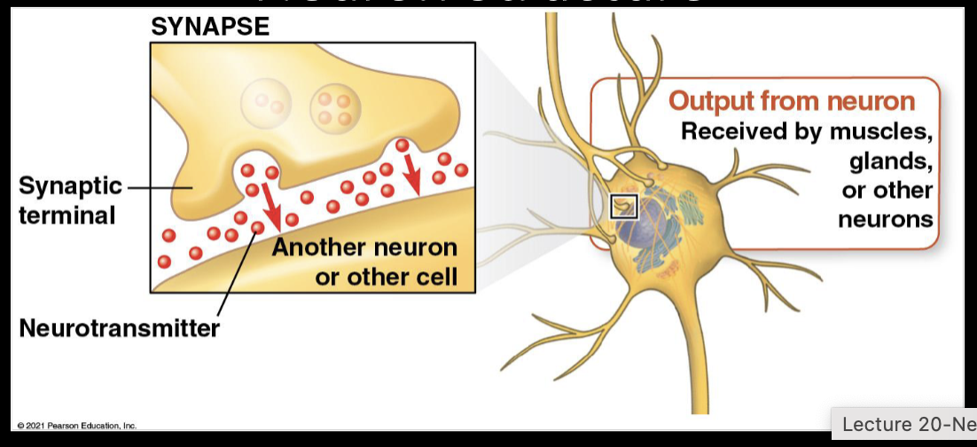
Neuron Structure
Synapse-junction between neuron and another cell
Neurotransmitters- chemical messengers that pass information
from the transmitting neuron to the receiving cell
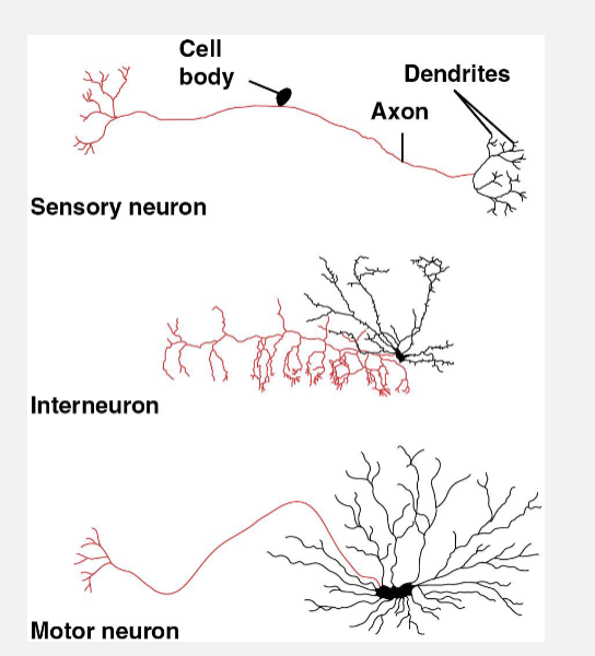
Neuron
Structure
• 3 types of neurons involved in
information processing
• Sensory neurons-transmit
information about external stimuli
• Interneurons-integrate the
information
• Motor neurons-transmit signals to
muscle cells causing them to
contract
• Shape of a neuron varies widely
depending on its role in information
processing
Neuron Structure
• When grouped together, axons of
neurons form nerves
• Central nervous system (CNS)
• Neurons that carry out
sorting, processing,
integration
• Peripheral nervous system (PNS)
• Neurons that carry
information into and out of
the CNS
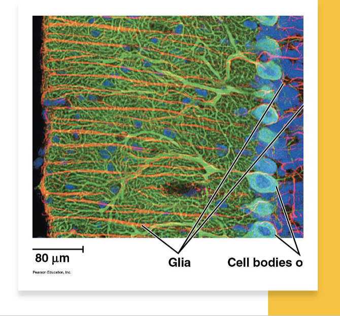
• All neurons are supported by glia
The central nervous system in vertebrates is formed from the
Ectoderm
Resting potential
• Membrane potential
• voltage (difference in electrical charge)
across a cell’s plasma membrane
• Inside of cell negatively charged relative
to surrounding fluid
• Resting potential
• membrane potential of a neuron that is
not sending signals
• Typically between -60 and -80 millivolts
(mV)
• Action potentials
• changes in membrane potential
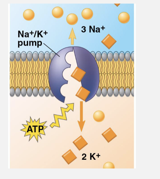
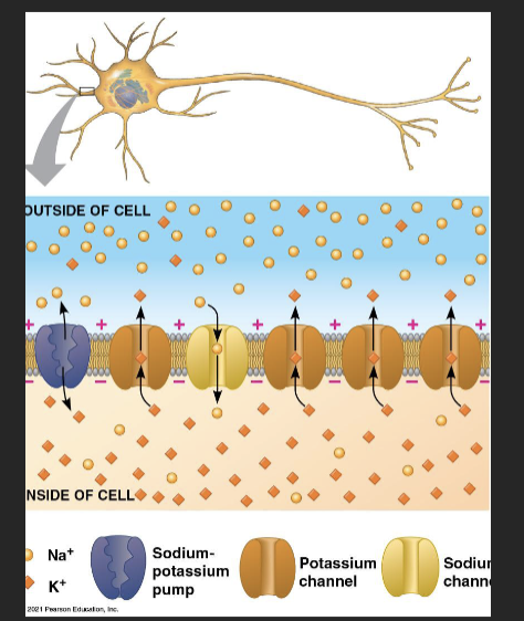
Resting potential: formation
• Ions with essential role
• Sodium (Na+ )
• Potassium (K+ )
• K+ higher inside cell; Na+
higher outside cell
• Sodium-potassium pump
maintains gradients
• Uses ATP to pump 2 K+ in
for every 3 Na+ out
Resting Potential
• Ion channels
• Allow ions to diffuse back and
forth generating the resting
potential
• Many open potassium channels;
few open sodium channels
• K+ diffuses out of cell and
negative charge builds up within
neuron
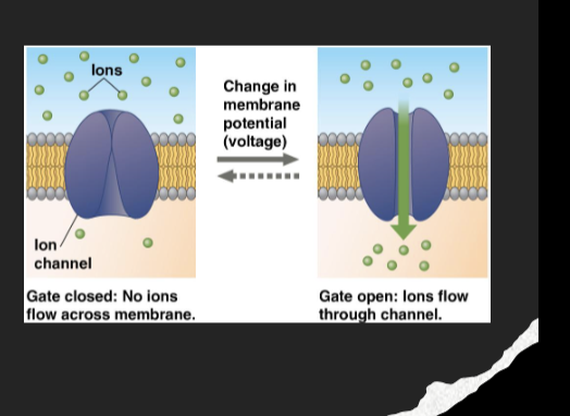
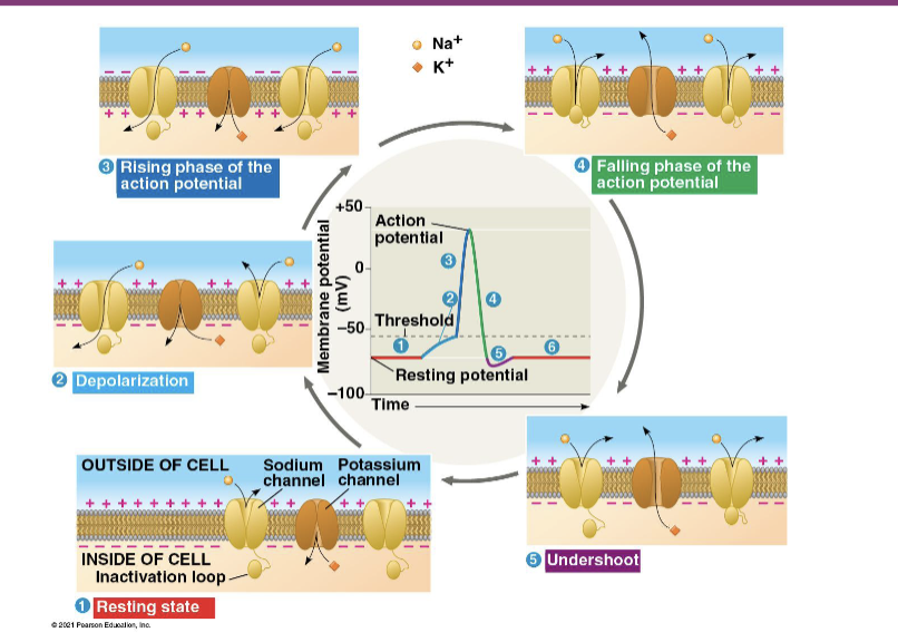
Action Potential
Gated ion channels
open or close in response to stimuli leading to changes in membrane potential
Alters permeability of membrane to particular ions
Ex: voltage gated ion channels
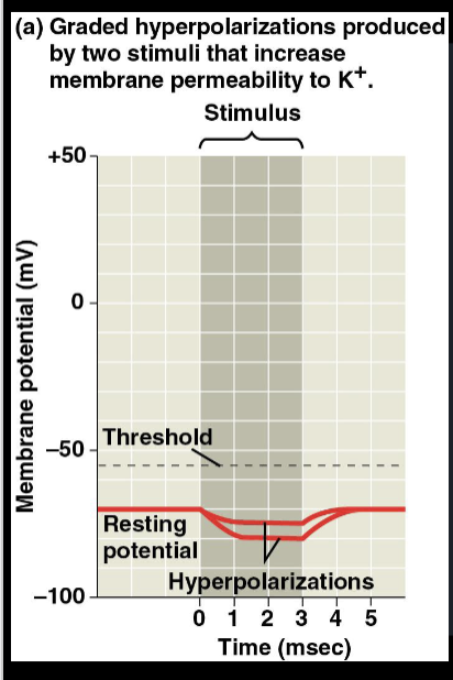
Action potential
• Hyperpolarization-increase
in the magnitude of the
membrane potential
• Depolarization-decrease in
the magnitude of the
membrane potential
• Graded Potentials-changes
in the membrane potential
that vary with the strength
of stimulus
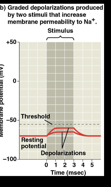
Action potential
• Action Potential
• Brief, all-or-none depolarization of a
neuron’s plasma membrane
• Generated when a graded depolarization
shifts the membrane potential to threshold
(-55mV)
• Voltage-gated sodium channels open
• Na+ flows into the neuron
• Repolarization occurs when sodium
channels are inactivated and voltage-gated
potassium channels open

Action potentials are transmitted to synaptic terminals by the
Axon
The opening of potassium channels as the membrane potential becomes positve is a form of
Negative feedback
Action potentials: Conduction
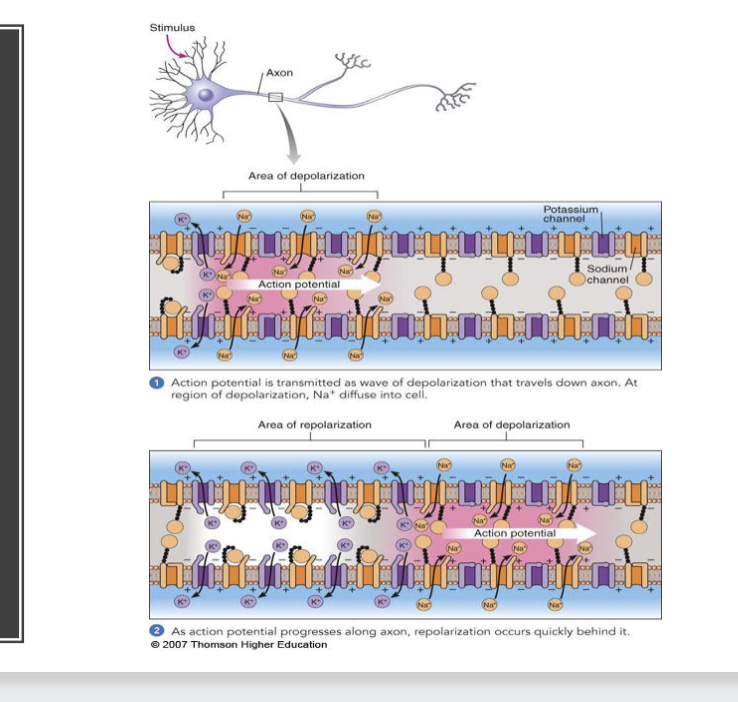
Can an action potential travel back toward the cell body?
Under normal physiological conditions, an action potential does not travel back toward the cell body; it propagates in only one direction, from the cell body (axon hillock) to the terminal. This unidirectional flow is guaranteed by the refractory period, during which the recently activated sodium channels are inactive and cannot reopen
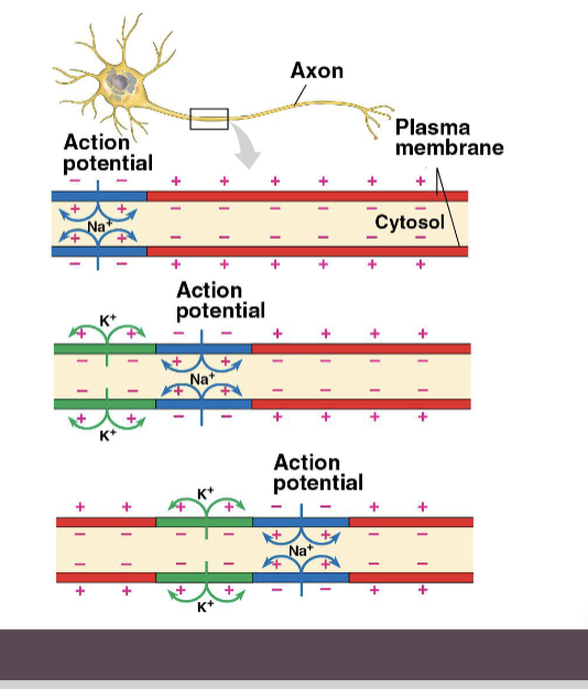
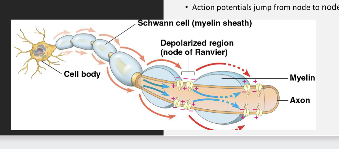
Action potentials: conduction
• Speed of conduction increases
with diameter of the axon
• Vertebrate axons have
electrical insulation-myelin
sheaths-that allow fast
conduction
• Made by glia
(oligodendrocytes in the CNS
and Schwann cells in the PNS)
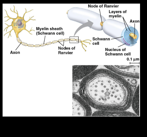
Action Potentials:
Conduction
• Nodes of Ranvier
• Gaps in myelinated axons where voltage-
gated sodium channels are restricted to
• Saltatory Conduction
• Action potentials jump from node to node
Which axon features would result in slower communication with other cells
Nonmyelinated axons,thin axons
Synapses
junctions where neurons communicate with other neurons or cells
Electrical
Contain gap junctions that allow electrical current to flow directly from one neuron to another
Chemical
A chemical neurotransmitter carries information between neurons
Most are chemical
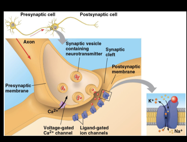
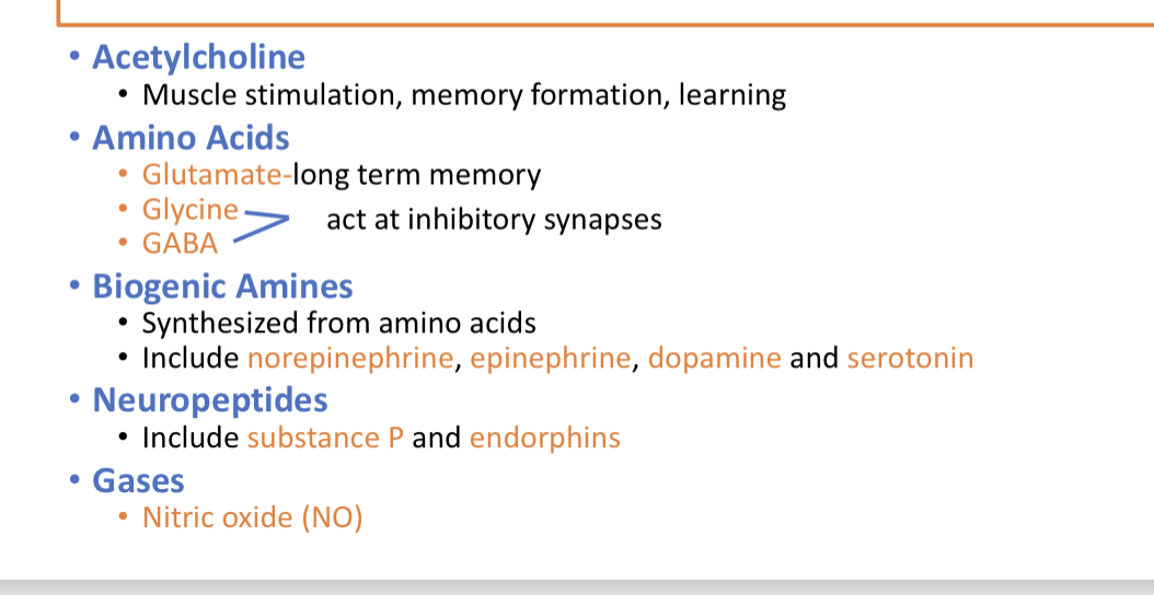
Synapses: neurotransmitters