Osteology Quiz 3
1/59
There's no tags or description
Looks like no tags are added yet.
Name | Mastery | Learn | Test | Matching | Spaced | Call with Kai |
|---|
No analytics yet
Send a link to your students to track their progress
60 Terms
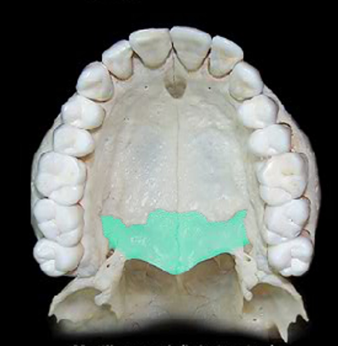
palatine bones
form the posterior part of the hard palate
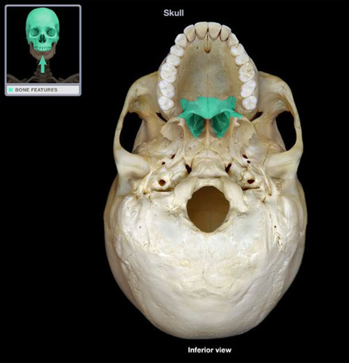
horizontal plate of palatine
forms the posterior portion of the hard palate
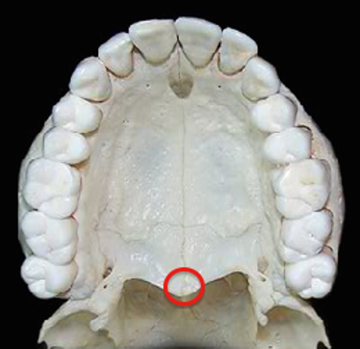
posterior nasal spine
superior surface; middle triangle on end of horizontal plate
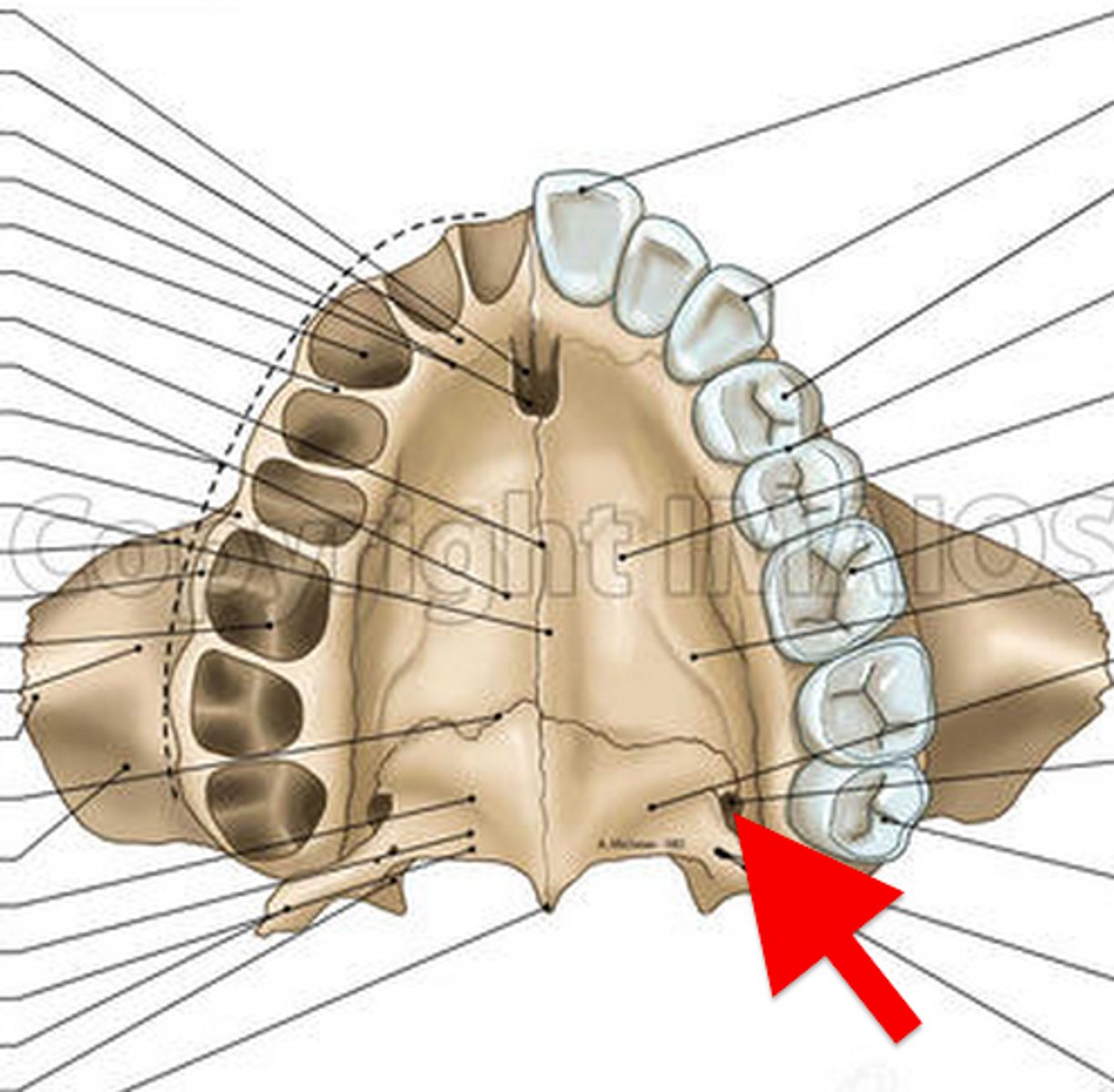
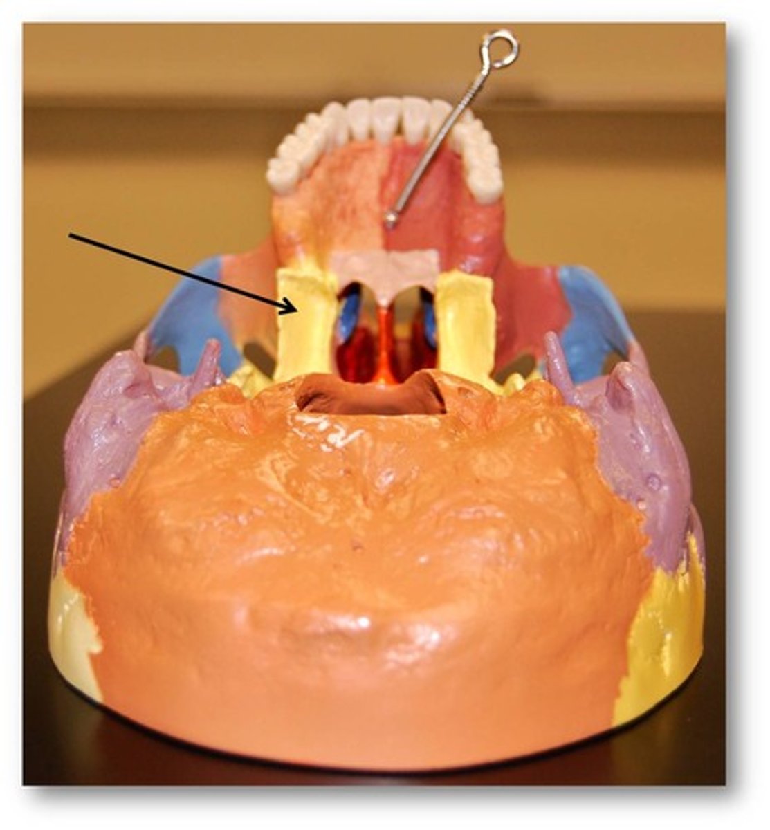
greater palatine foramen (canal)
big hole by back of molars
Vomer
"plow shape" midline bone dividing nasal cavity
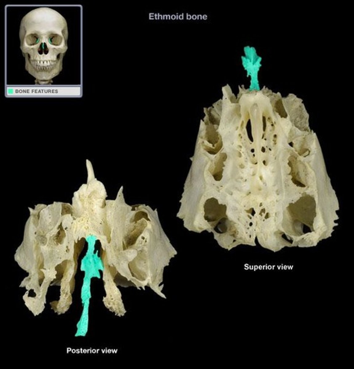
ethmoid bone
forms part of the posterior portion of the nose, the orbit, and the floor of the cranium
Cribiform plate
roof of nasal cavities (little drain holes)
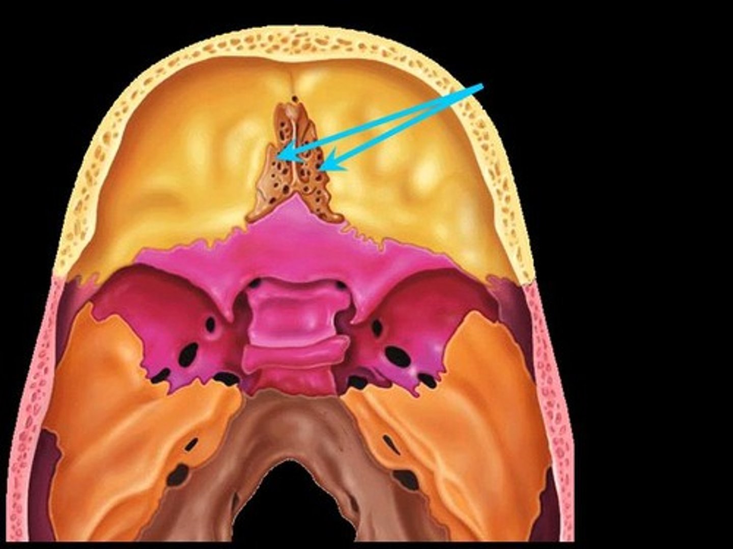
crista galli
perpendicular projection into endocranial cavity
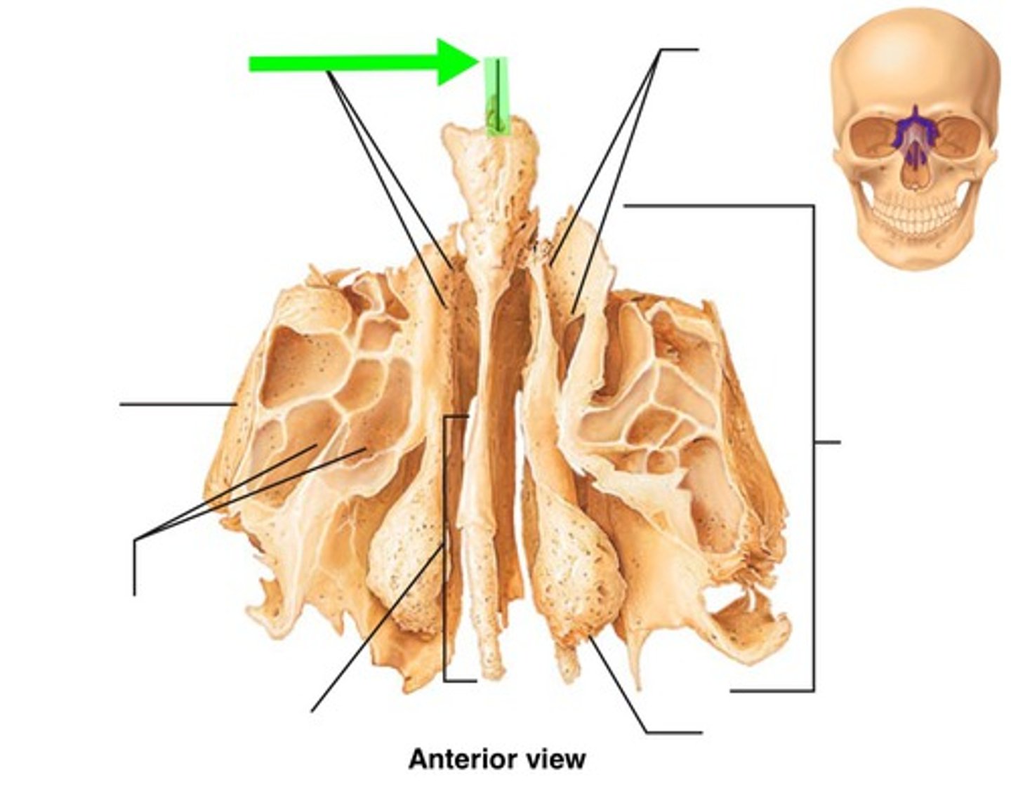
perpendicular plate
midline projection bellow cribriform plate
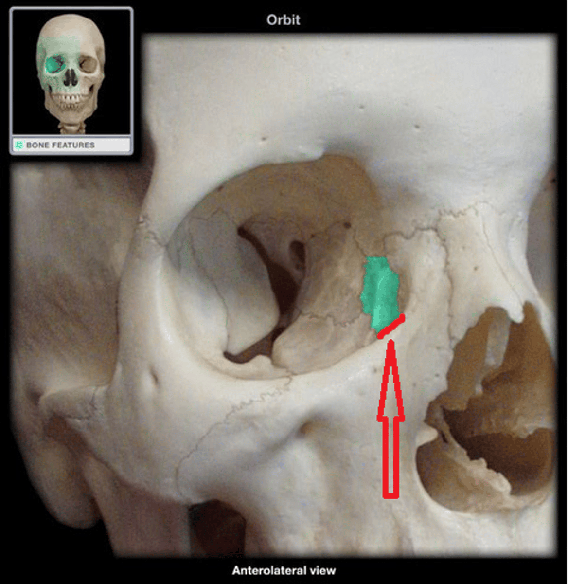
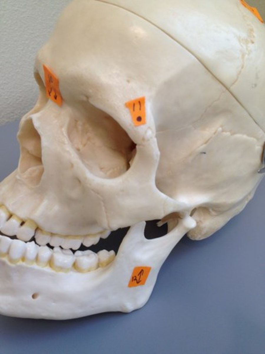
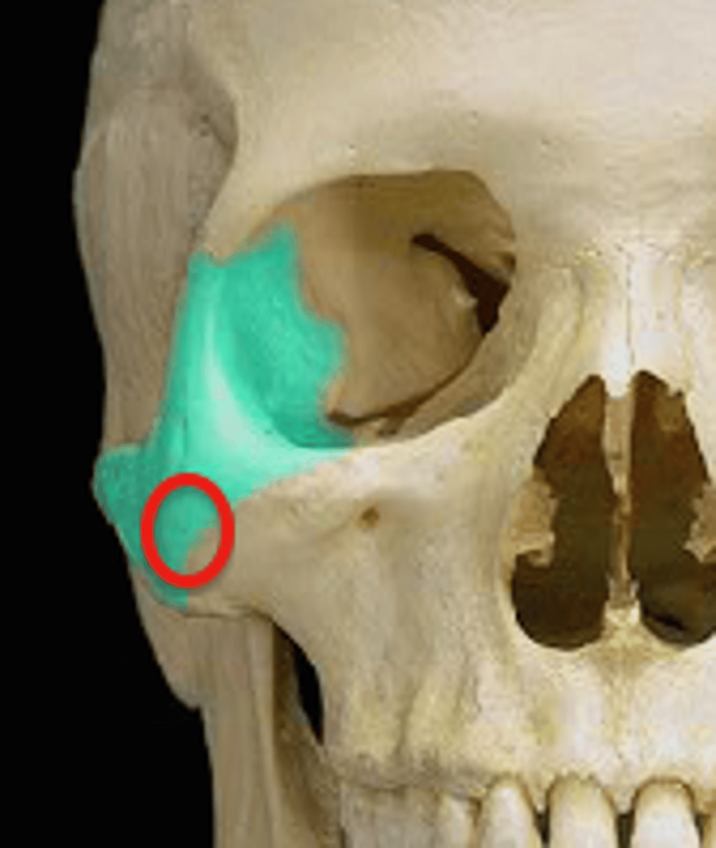
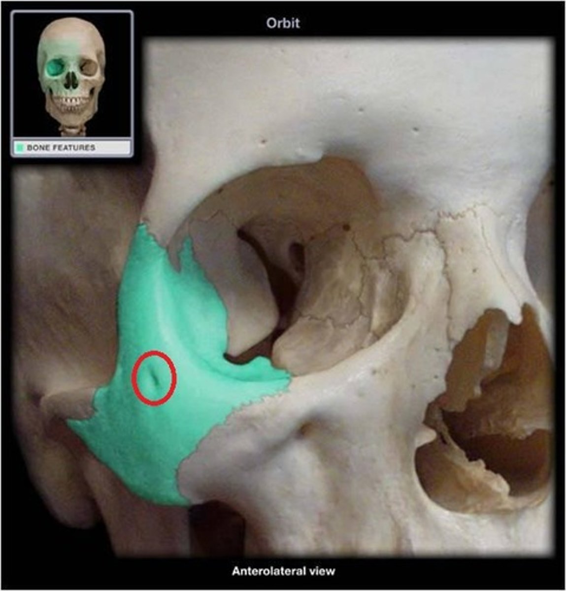
lacrimals
square outlined by sutures in lacrimal fossa
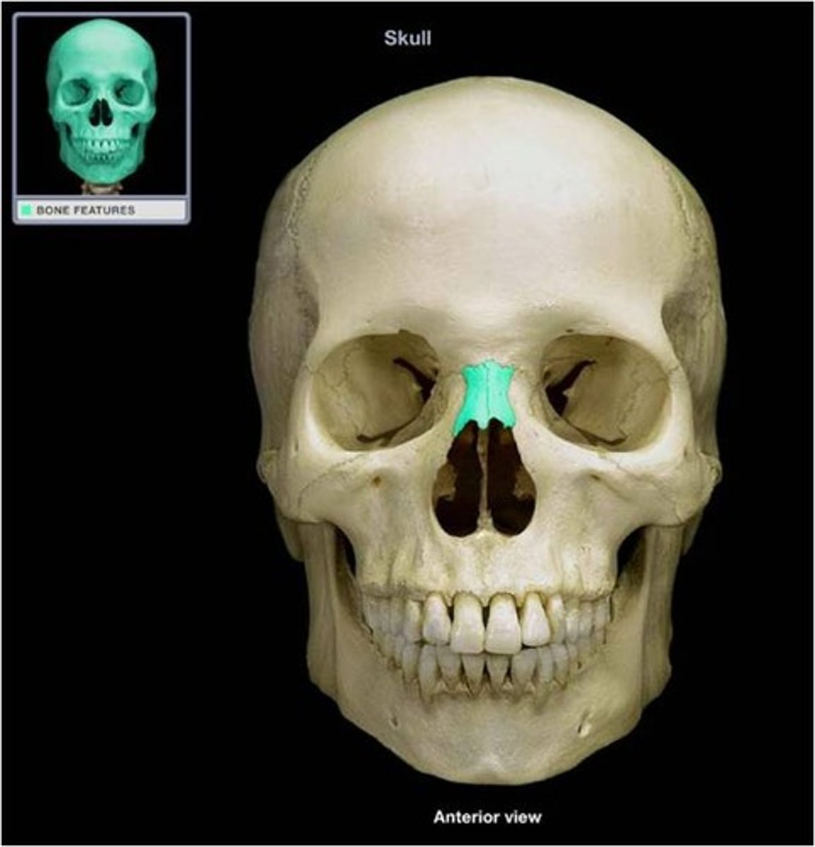
nasal bones
small thin rectangular bones bellow glabellar region (make up "nose ridge"
frontal process of zygomatic
articulates with frontal bone
temporal process of zygomatic bone
articulates with the temporal process of zygomatic bone to form zygomatic arch
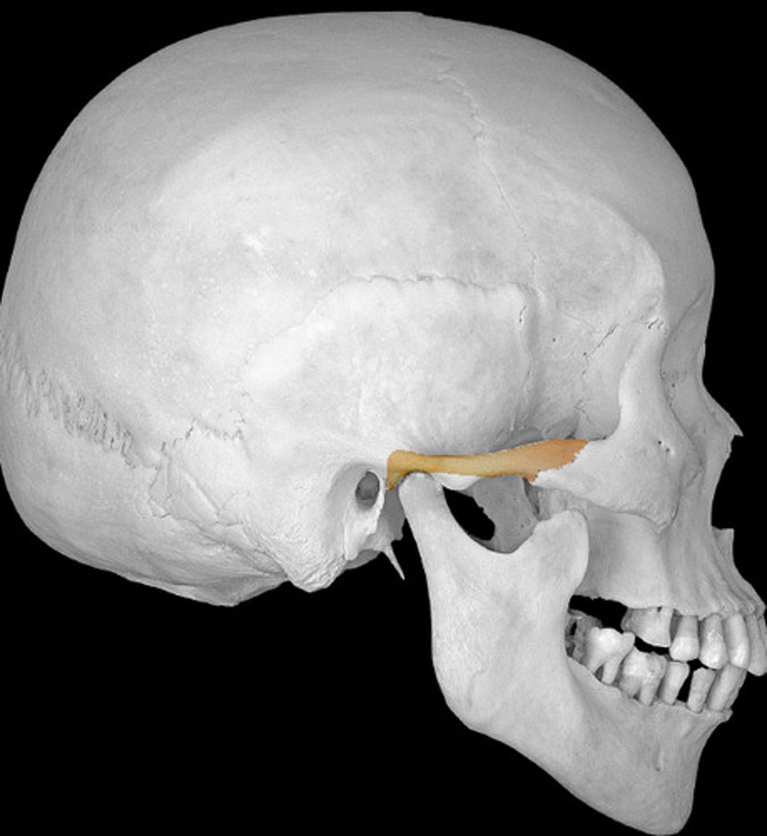
maxillary process of zygomatic
articulates with zygomatic process of maxillary bone
zygomaticofacial foramen
an opening in the zygomatic bone on lateral surface
masseteric origin
roughened, expanded inferior edge of the bone, extends from the zygomaticomaxillary to the temporozygomatic suture
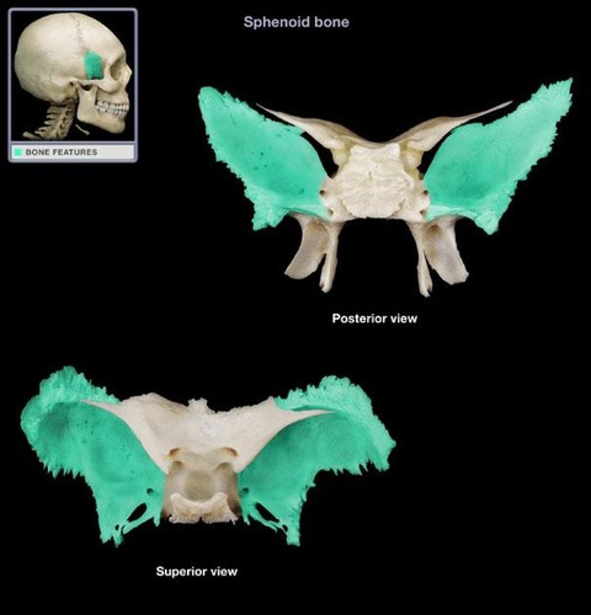
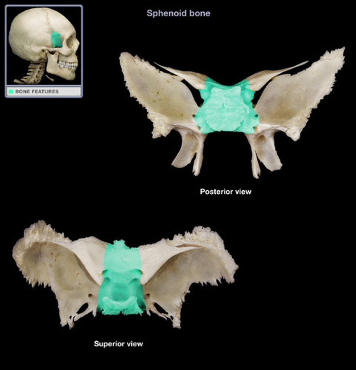
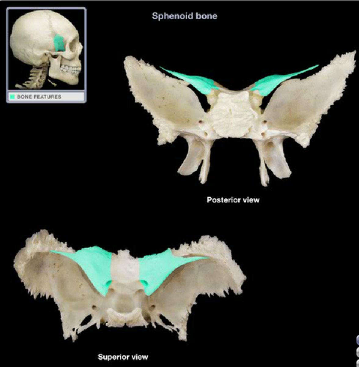
sphenoid bone
Bone that joins all of the bones of the cranium together
body of sphenoid bone
central section of sphenoid
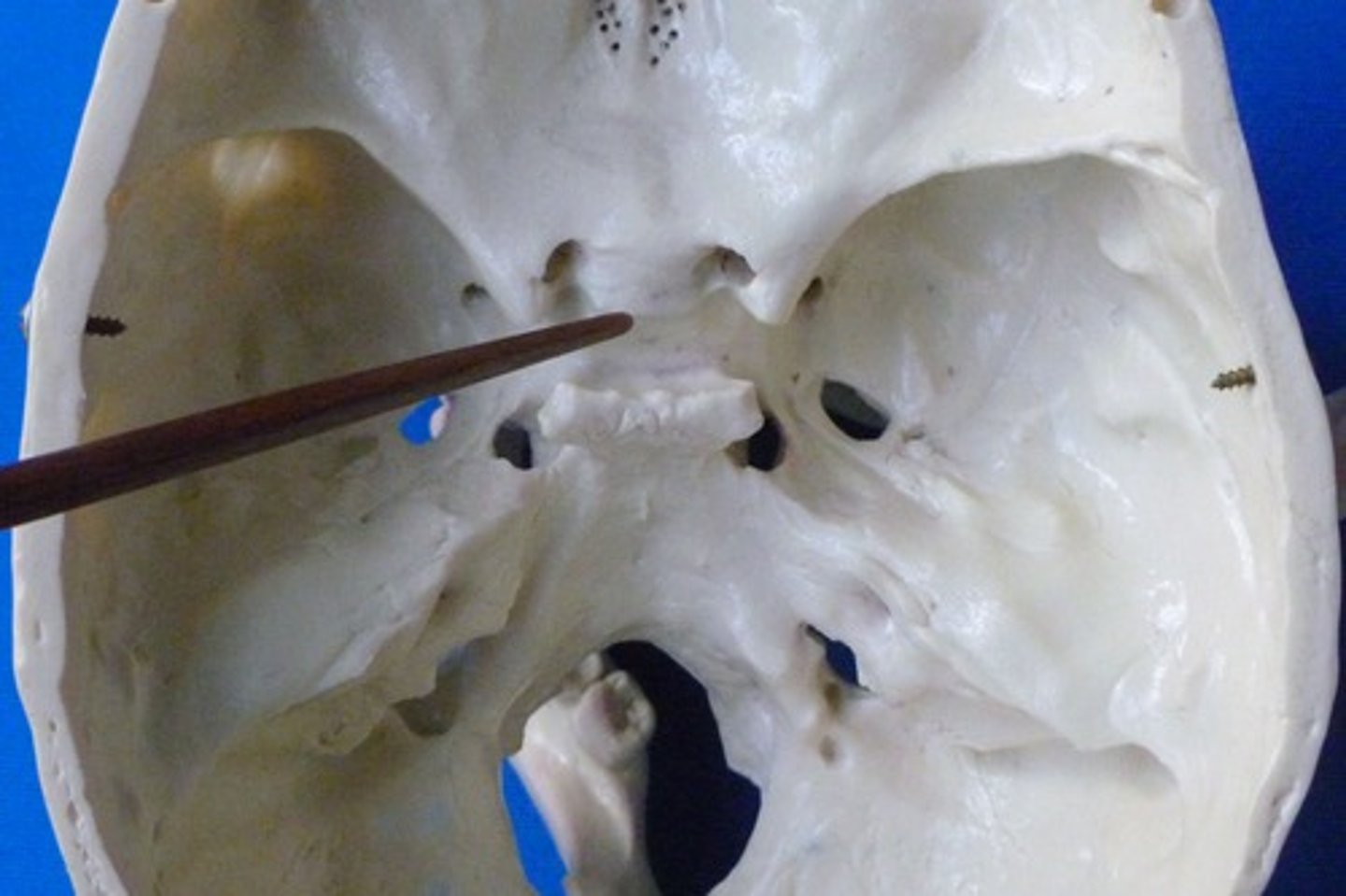
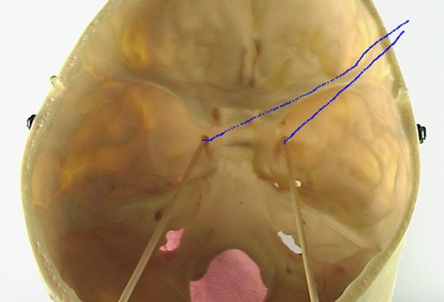
optic canals
openings in the bases of the lesser wings through which the optic nerves enter the orbits to serve the eyes

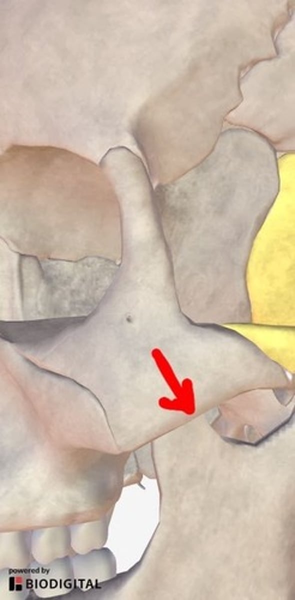
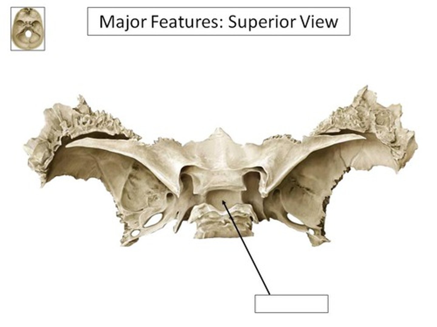
sella turcicia
"turkish saddle" grove in body of sphenoid
pituitary fossa
deep pit in "saddle"/sella turcicia
dorsom sellae
"back of saddle"
clinoid process (anterior and posterior)
knobs on hands and back of saddle
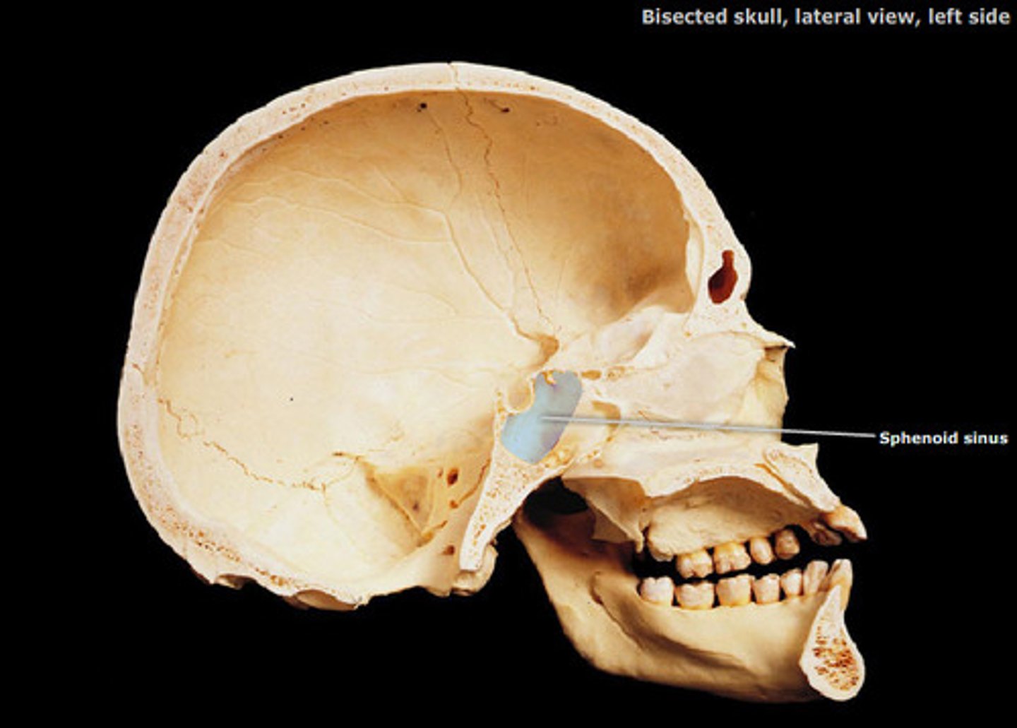
sphenoidal sinuses
Central part of sphenoid bone is riddled with air cavities.
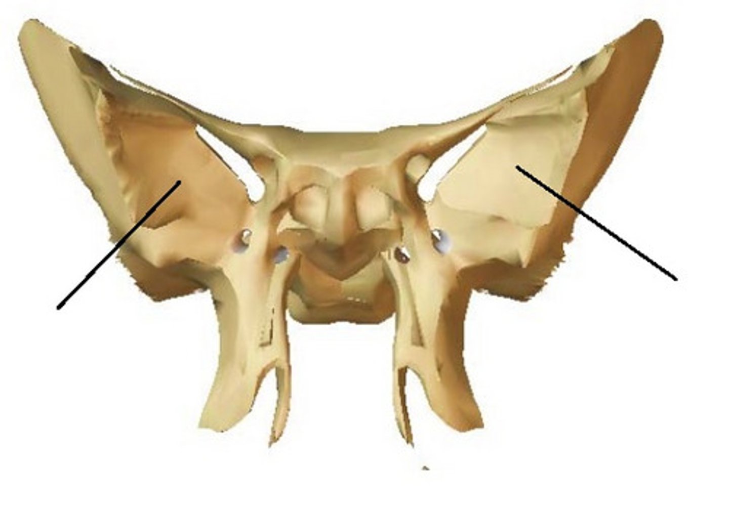
greater wings of sphenoid
project laterally from the sphenoid body, forming parts of the middle cranial fossa and the orbits
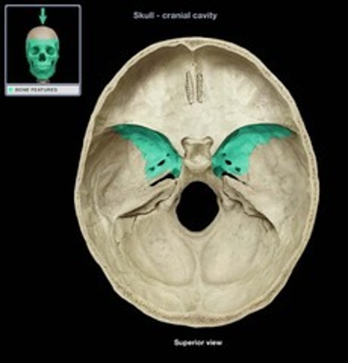
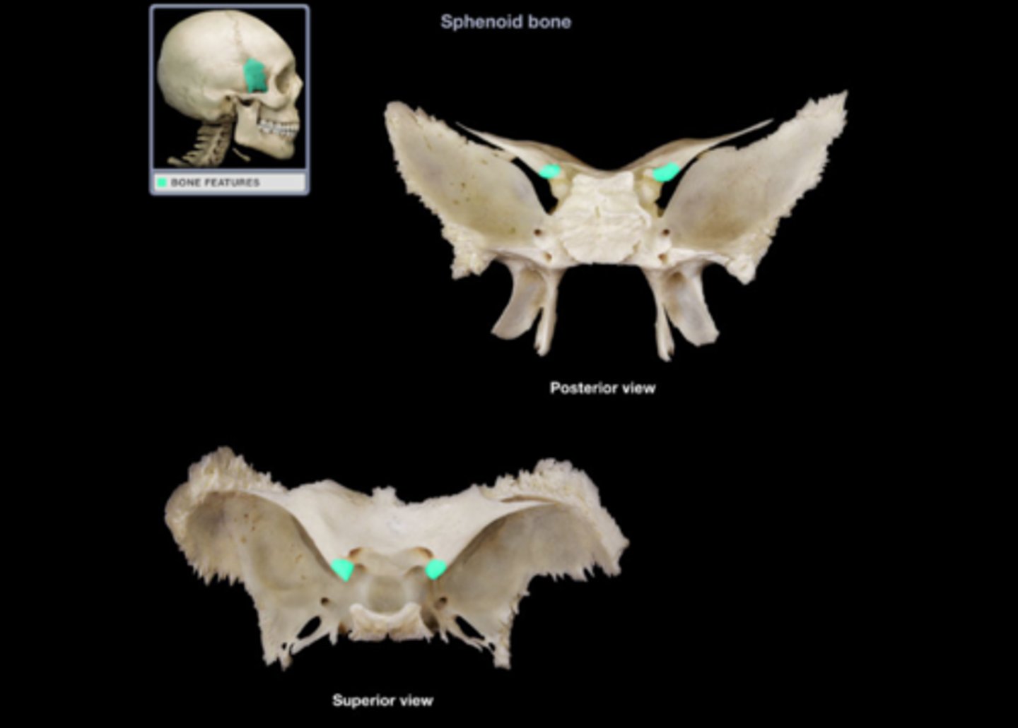
lesser wings of sphenoid
the smaller , upper wings from the posterior edge of the anterior cranial fossa
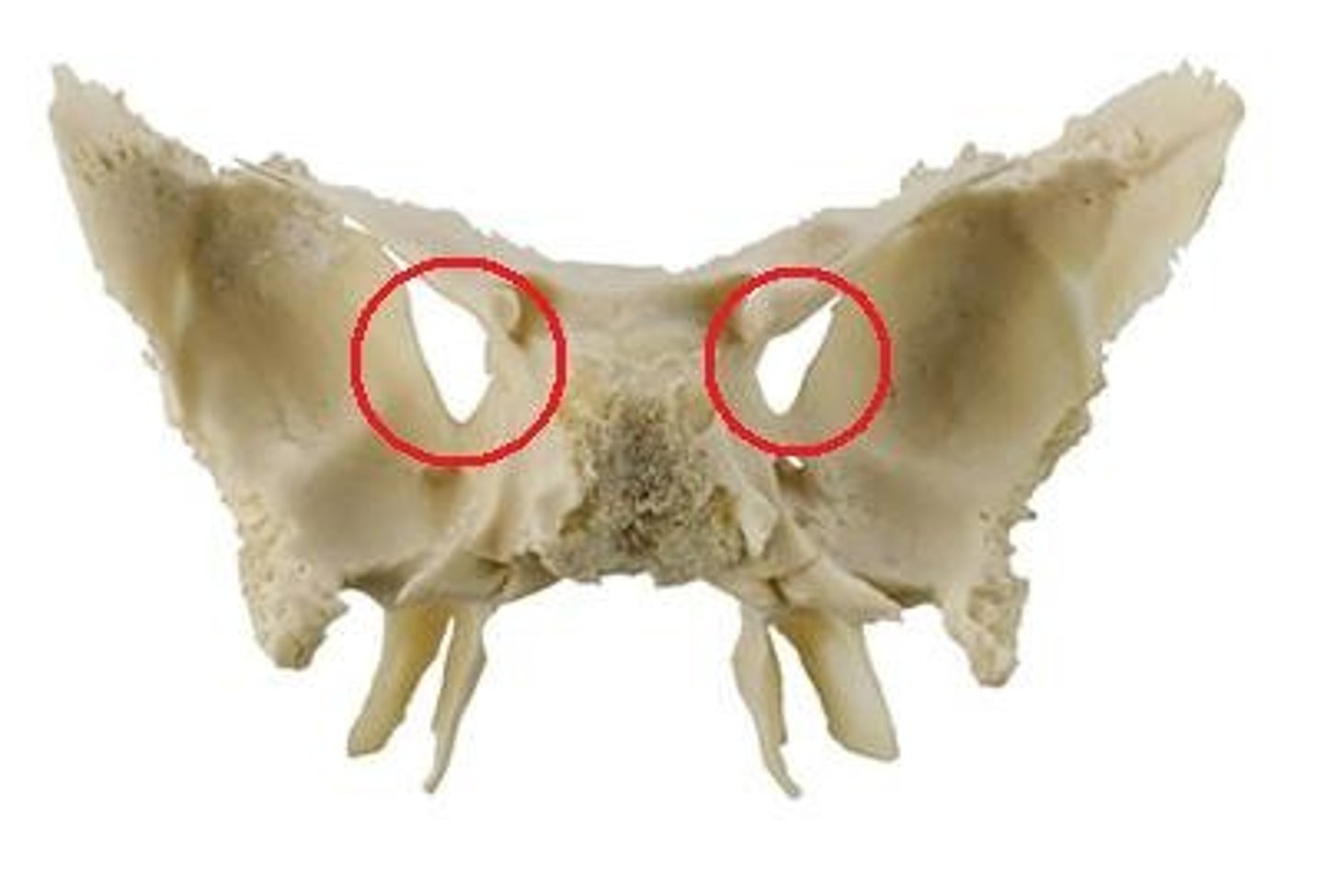
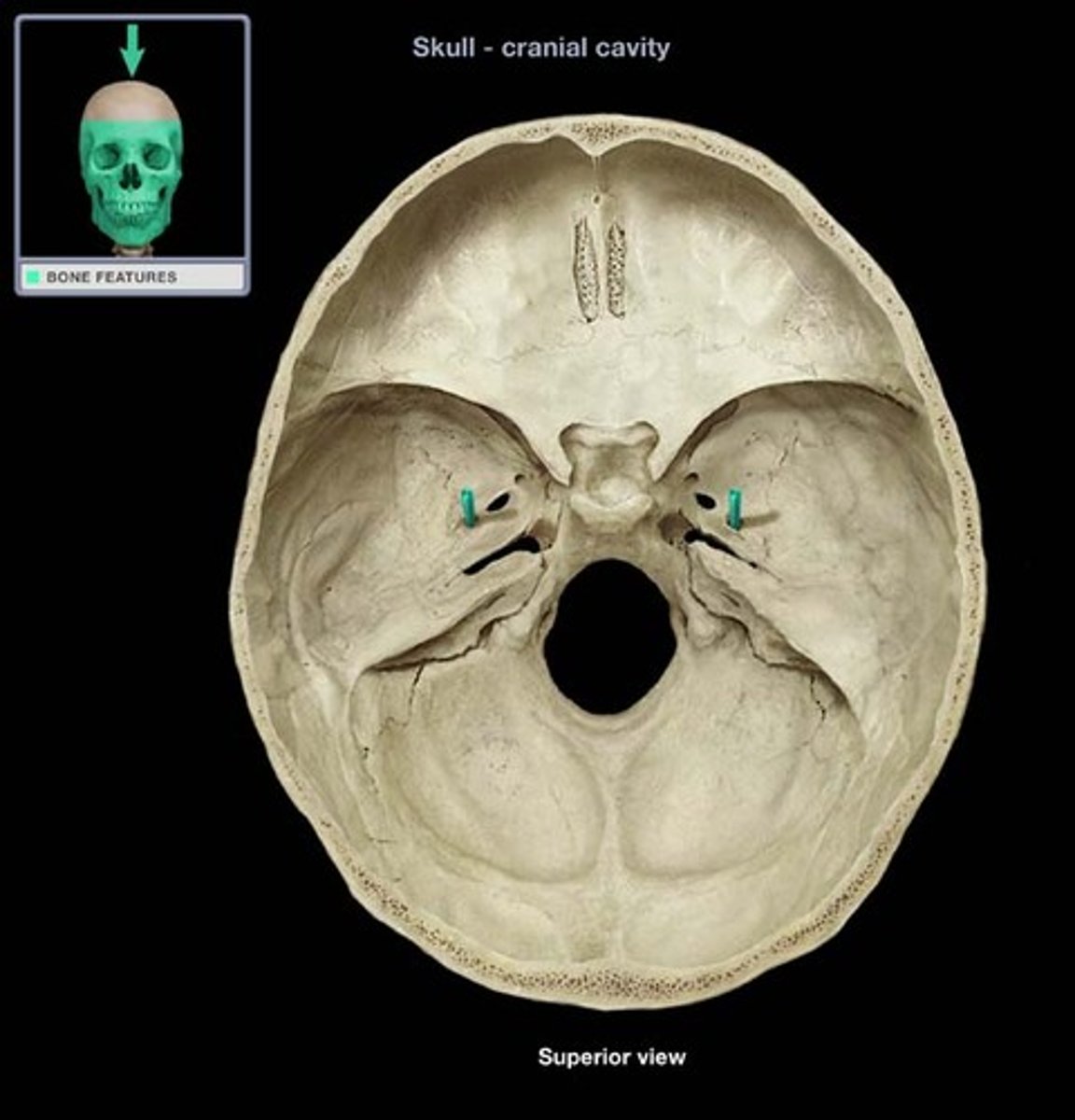
superior orbital fissure
gap/slit/fissure under lesser wing shelf endocranially (can see in orbit as long slit)
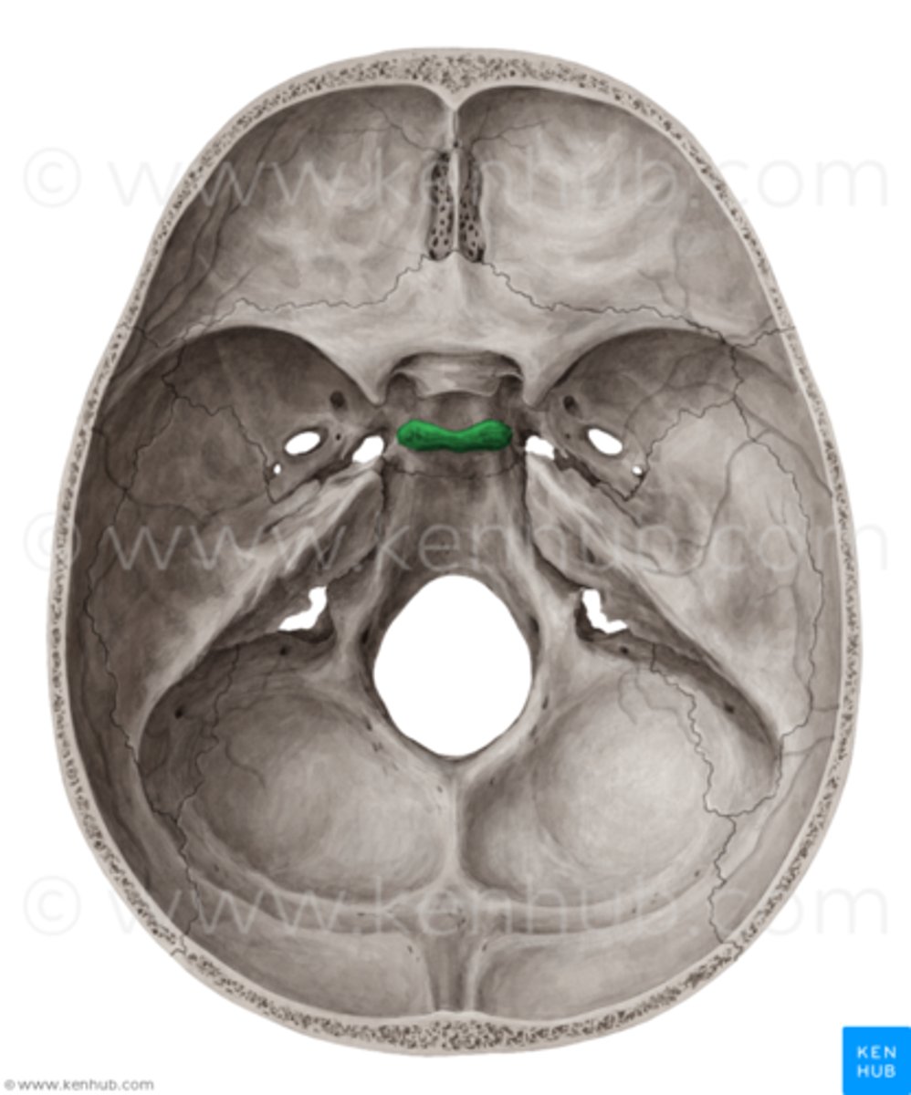
foramen rotundum (sphenoid)
hole under superior orbital fissure
foramen ovale
oval shaped hole under foramen rotundum
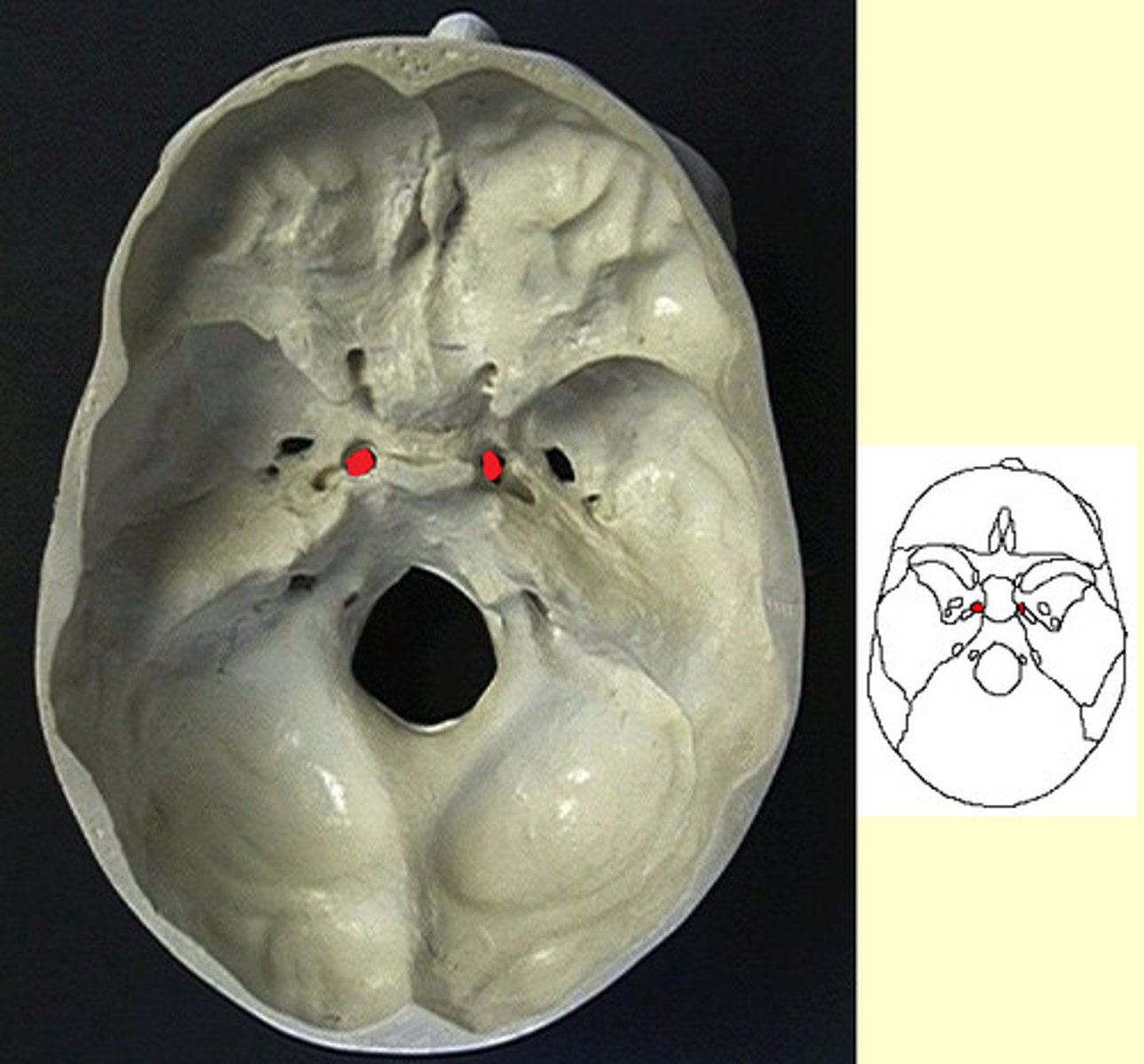
foramen spinosum
hole lateral/posterior to ovale (super tiny)
orbital surface of sphenoid
the portion of the sphenoid behind each eye socket/orbit
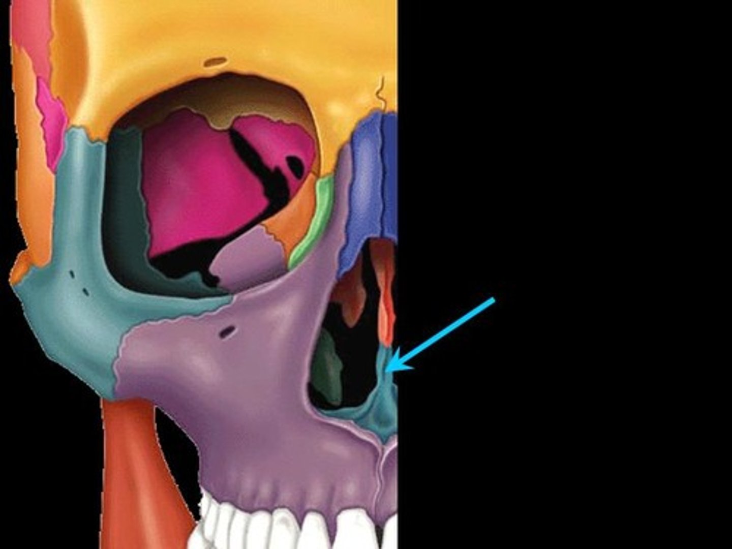
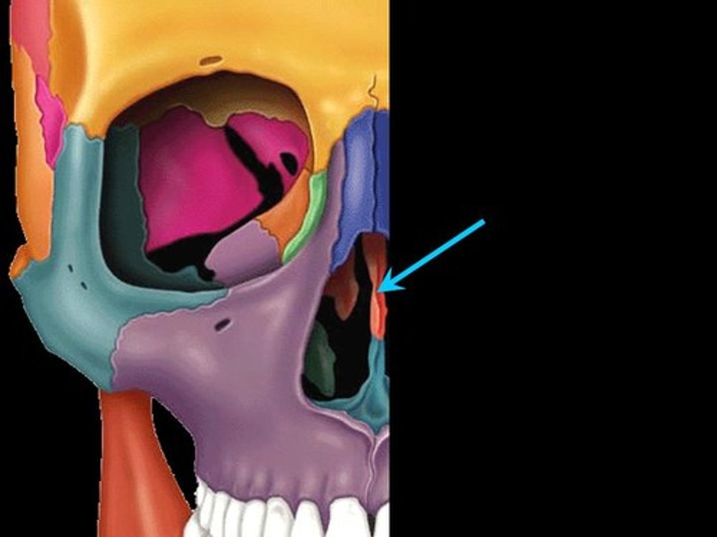
pterigoid processes
inferior "legs" of spenoid split into two plates (later sides of posterior nasal apature
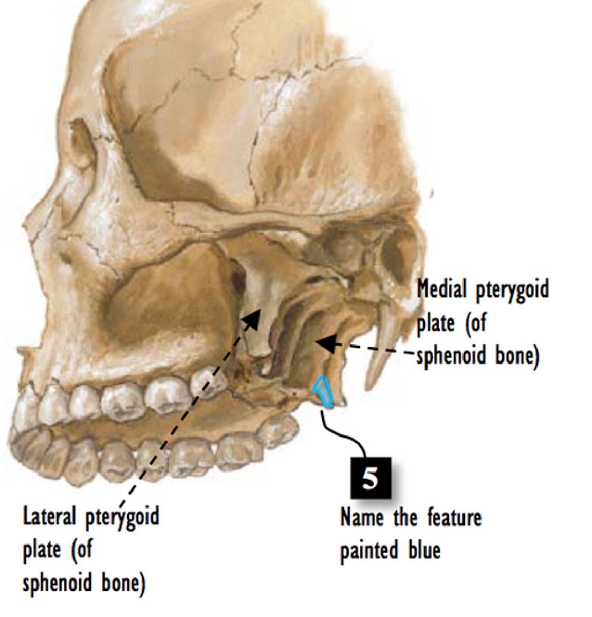
lateral/medial pterygoid plates
thin vertical plates making up pterigoid processes
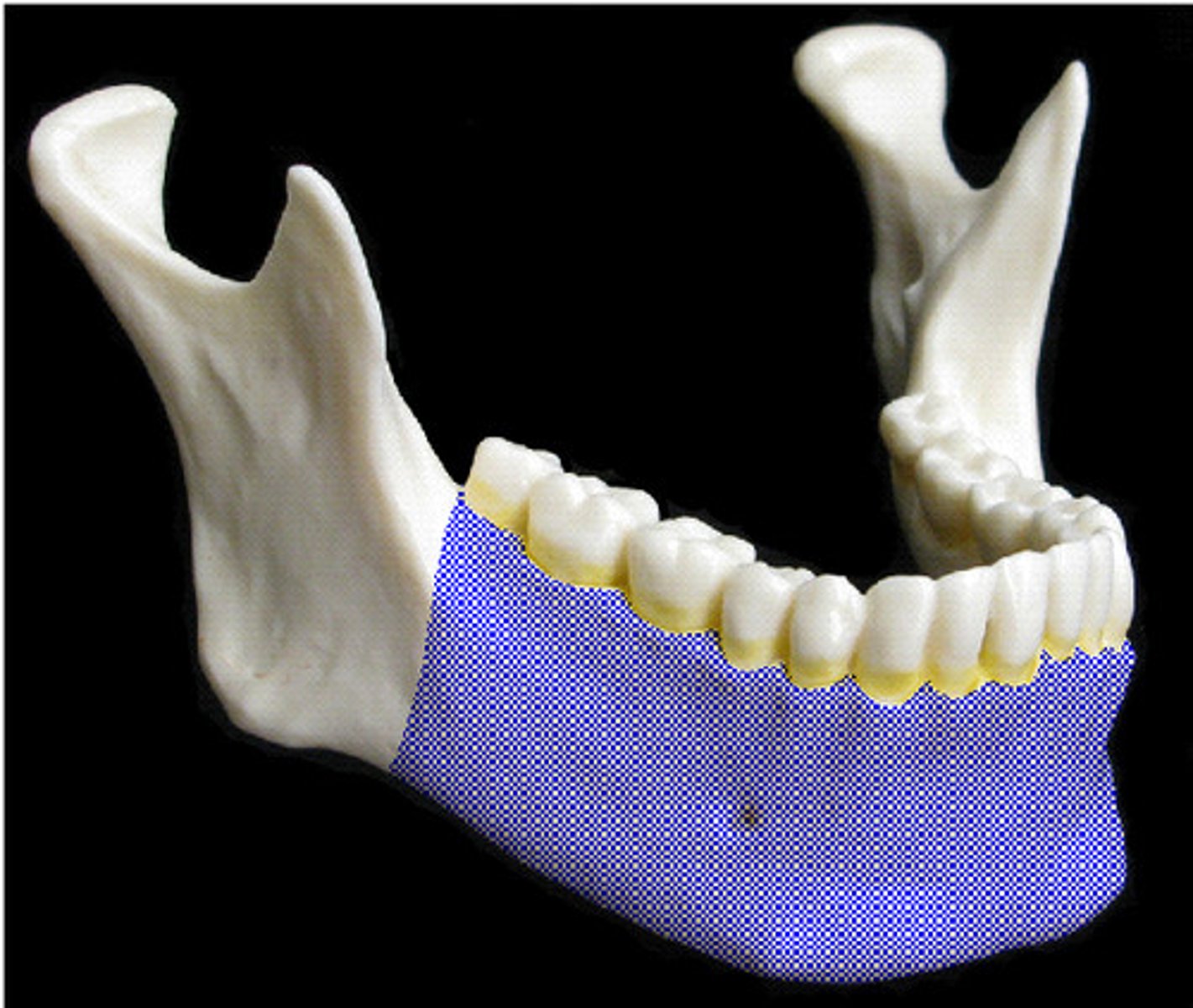
body of mandible
the horizontal portion of the lower jaw
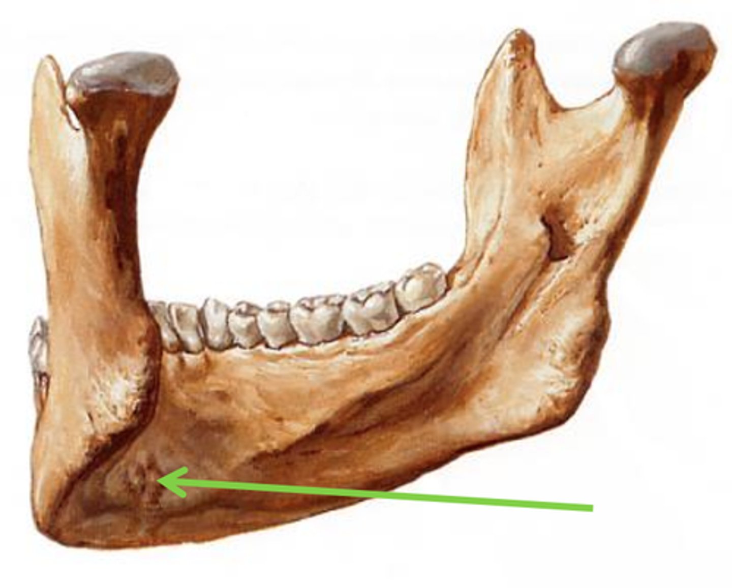
mental foramen of mandible
one of two holes located on the anterior surface of the mandible. It permits passage of the mental nerve and vessels.
mylohyloid line
Attachment for mylohyloid muscle, separates sublingual/submandibular fossae
submandibular fossa
hollow portion on medial corpus, inferior to mylohyloid line in premolar region
sublingual fossa
superior to mylohyoid line, occupied by sublingual gland
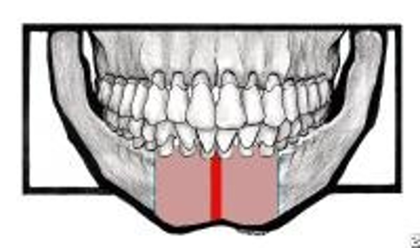
mandibular symphysis
where mandible formed by fusion of right and left processes
mental spines
are small ridges on the inner surface of the mandible; serve for attachment of certain chin muscles
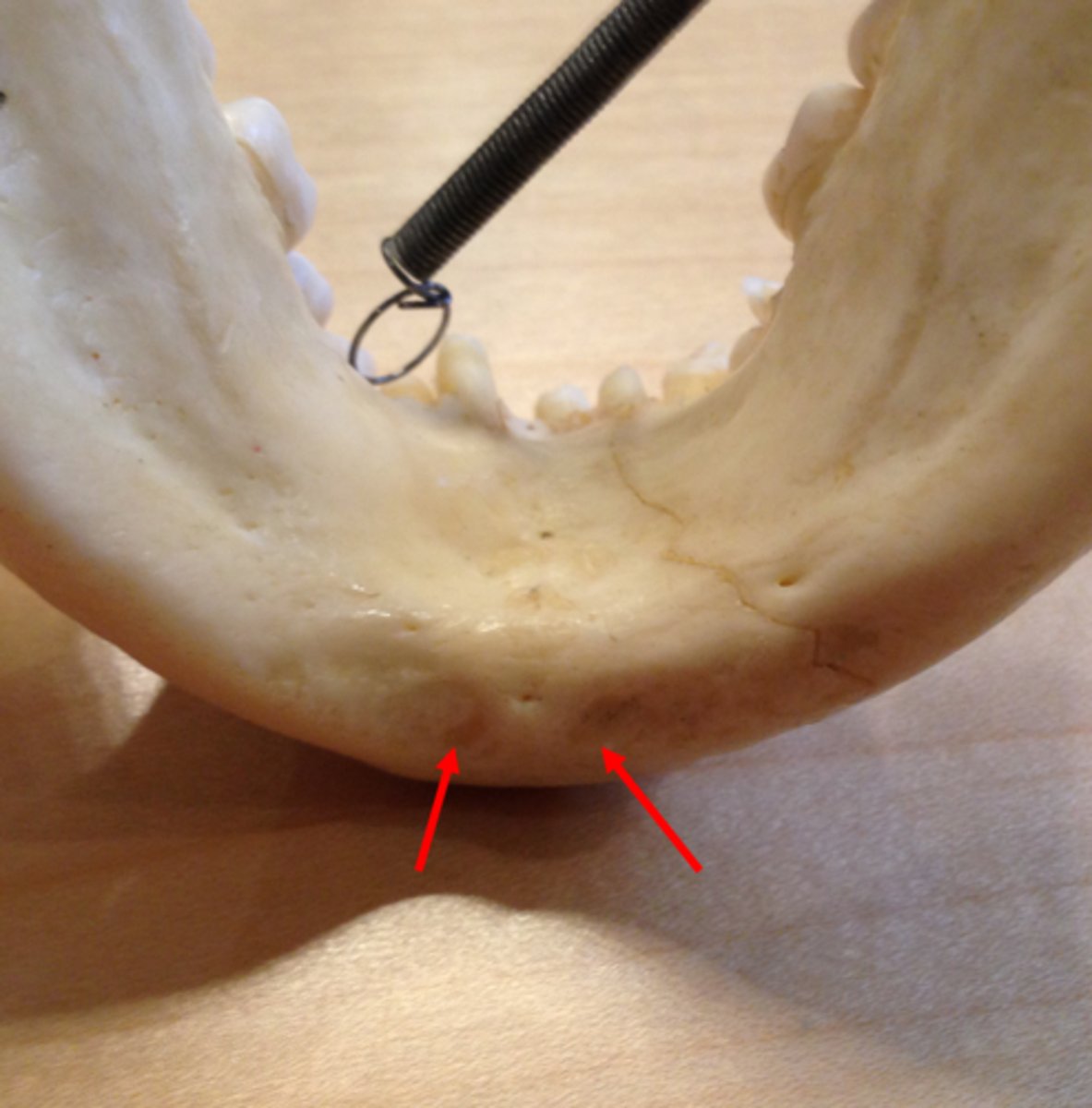
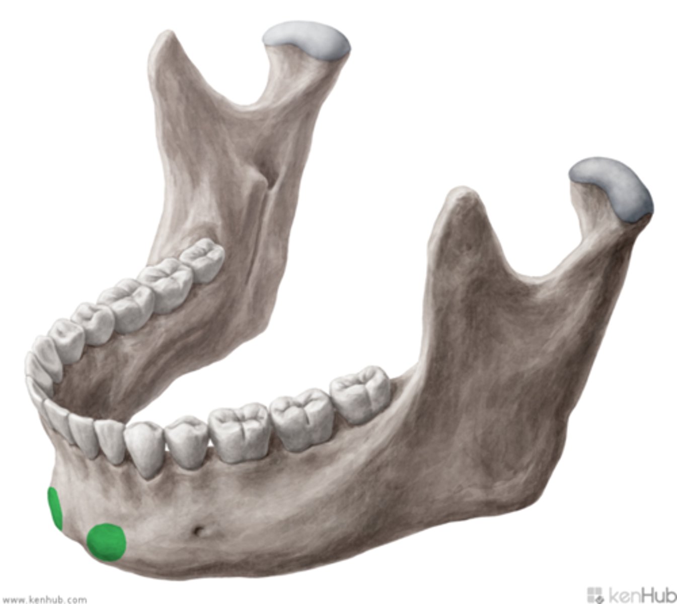
digastric fossae
points of attachment for anterior digastric muscle
mental protuberance/eminence/tubercle
chin at base of body
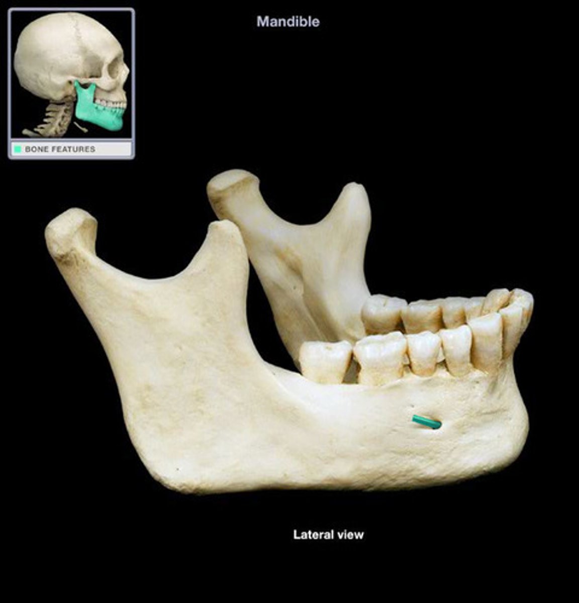
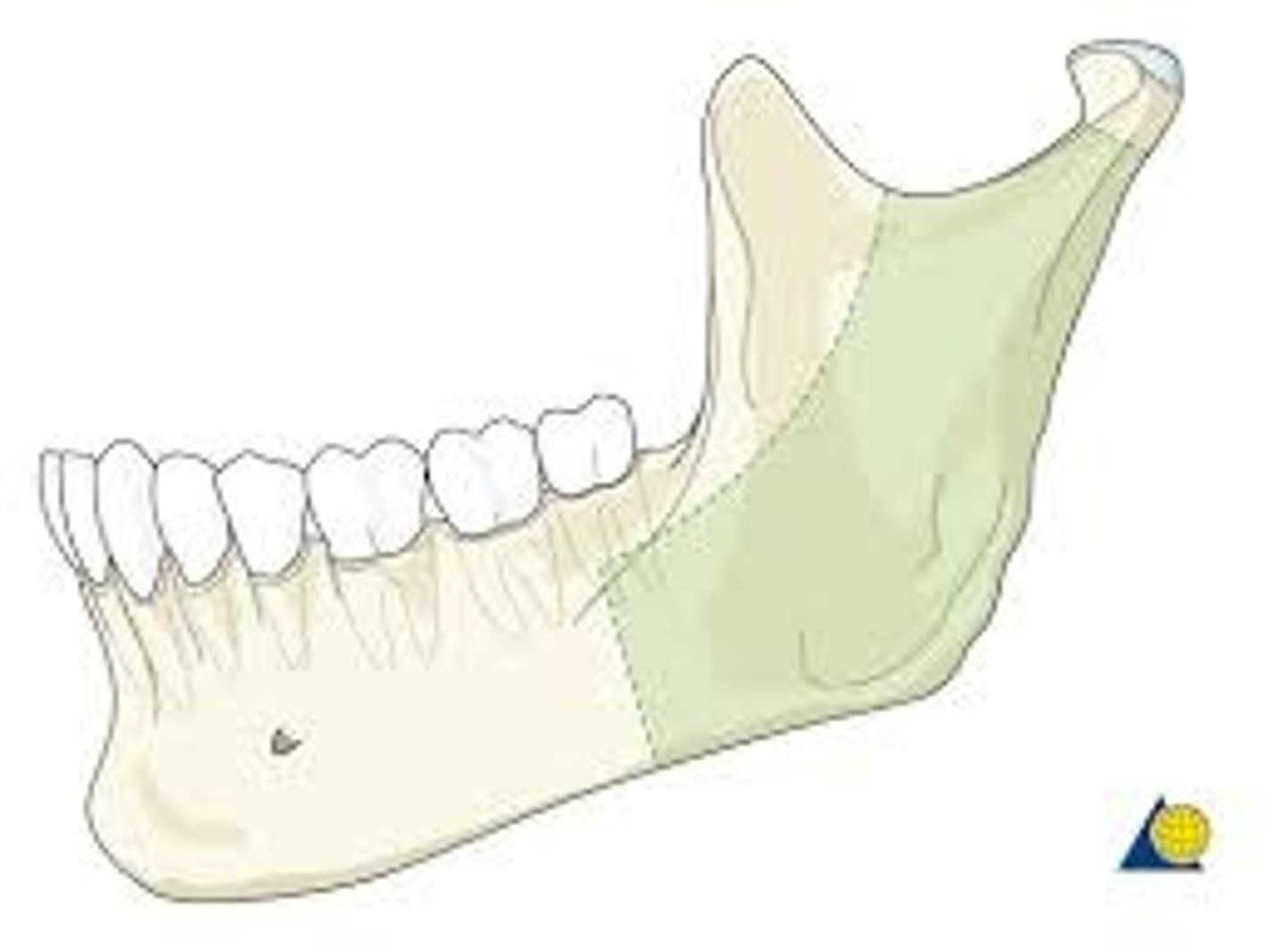
ascending ramus
Upright portion of the jaw bone
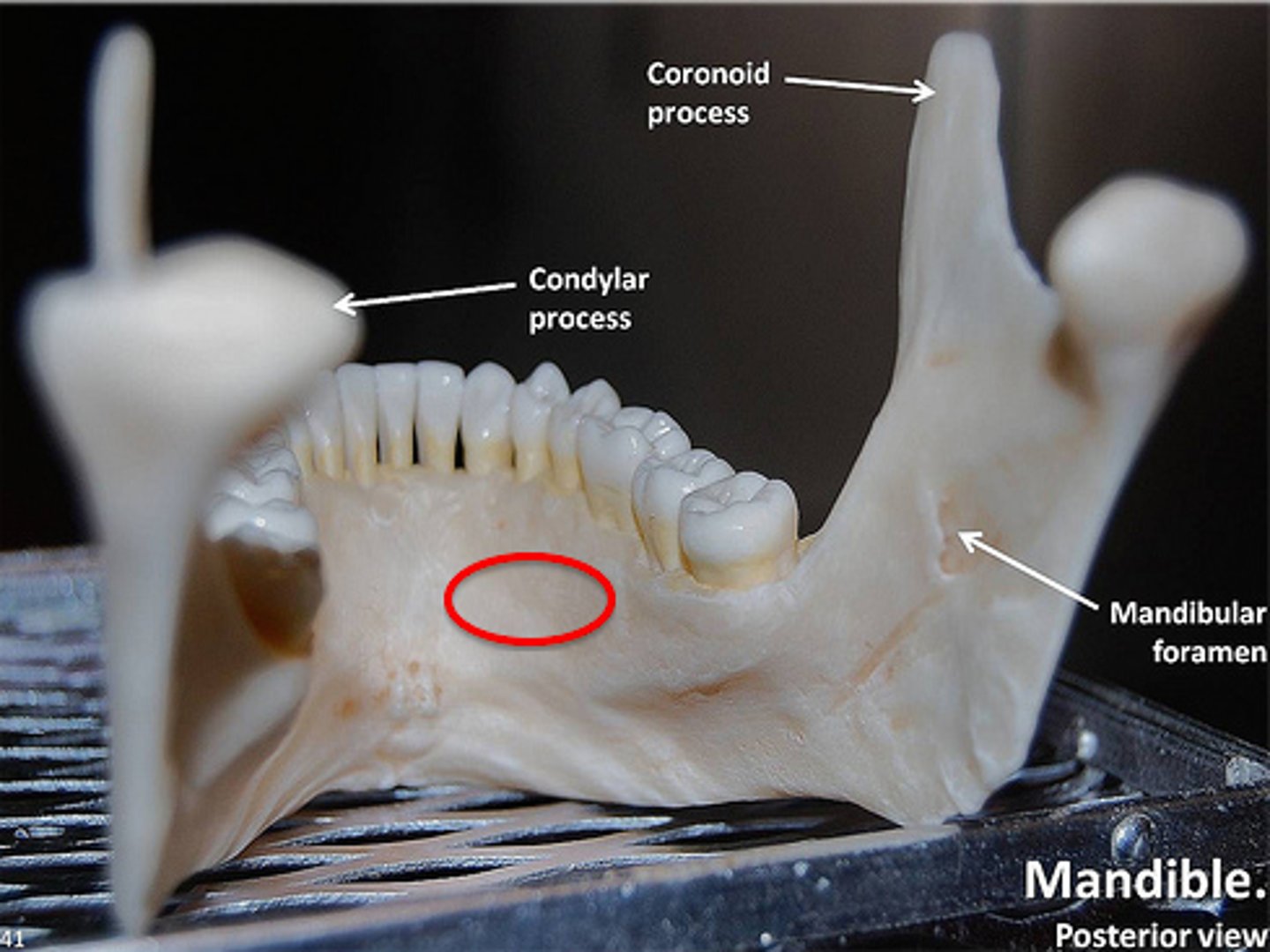
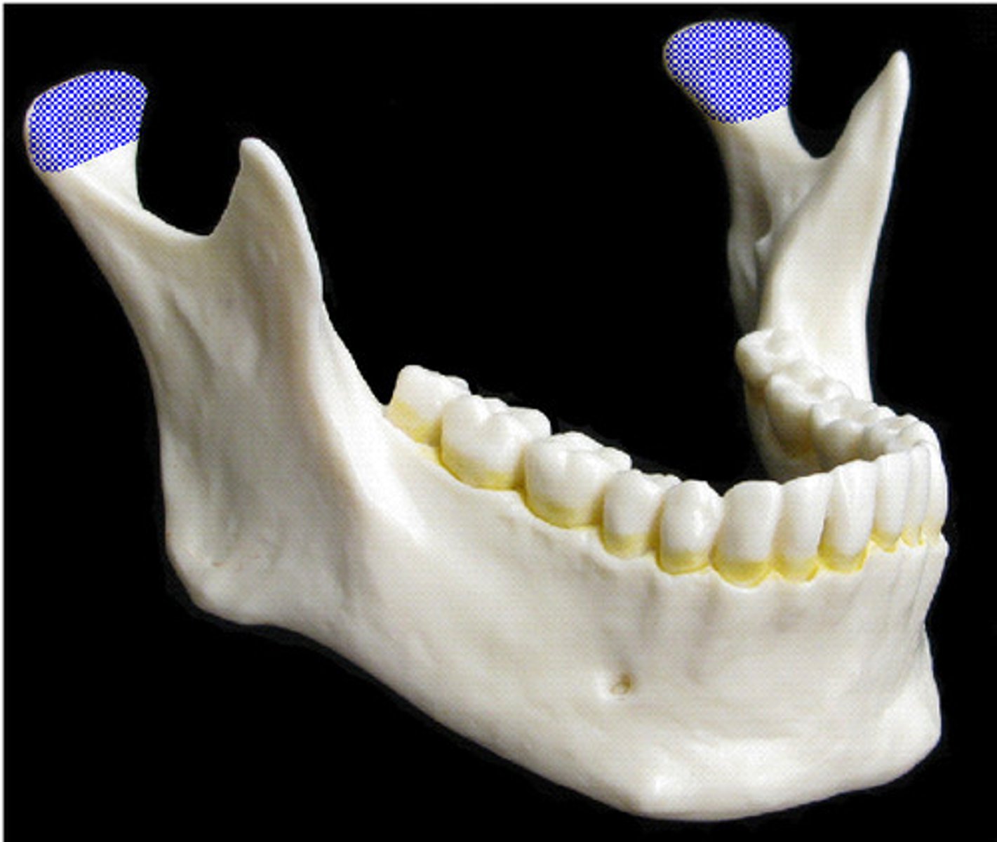
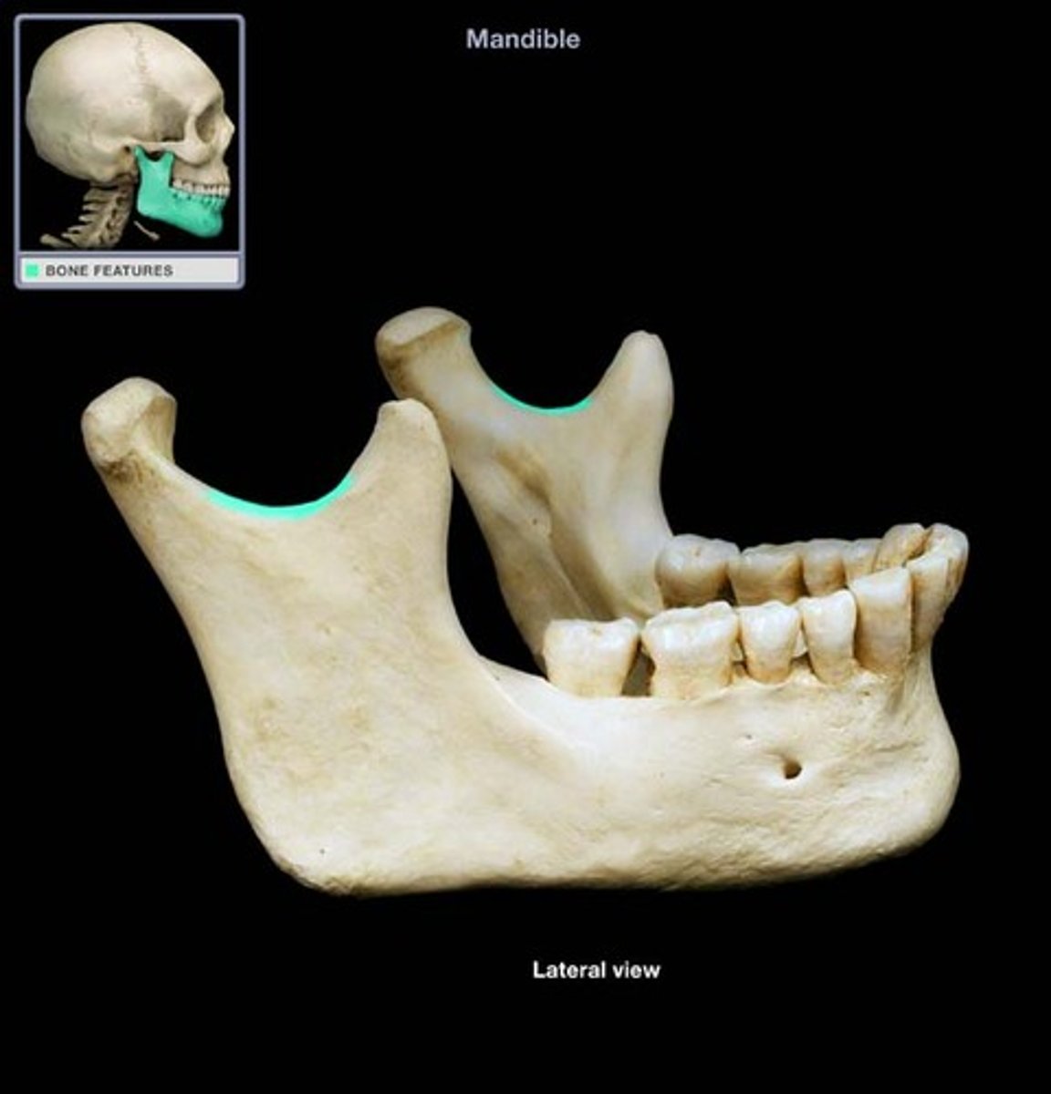
mandibular condyle
Articulation point of the mandible with the mandibular fossa of the temporal bone
condylar neck
slightly narrowed area just beneath the condyle
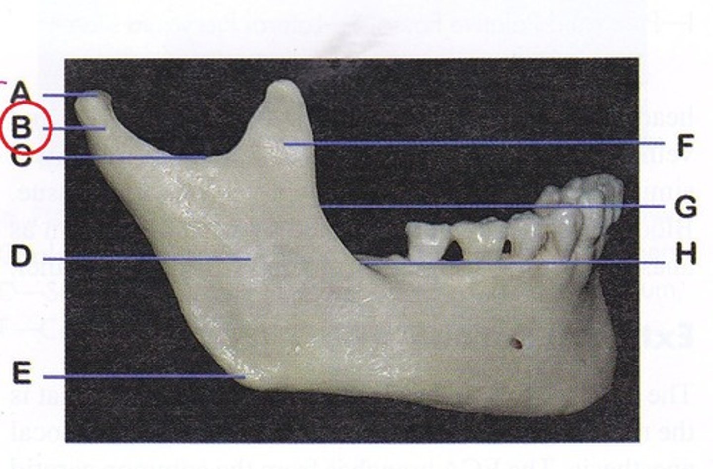
coronoid process
thin triangular extension of mandible bone
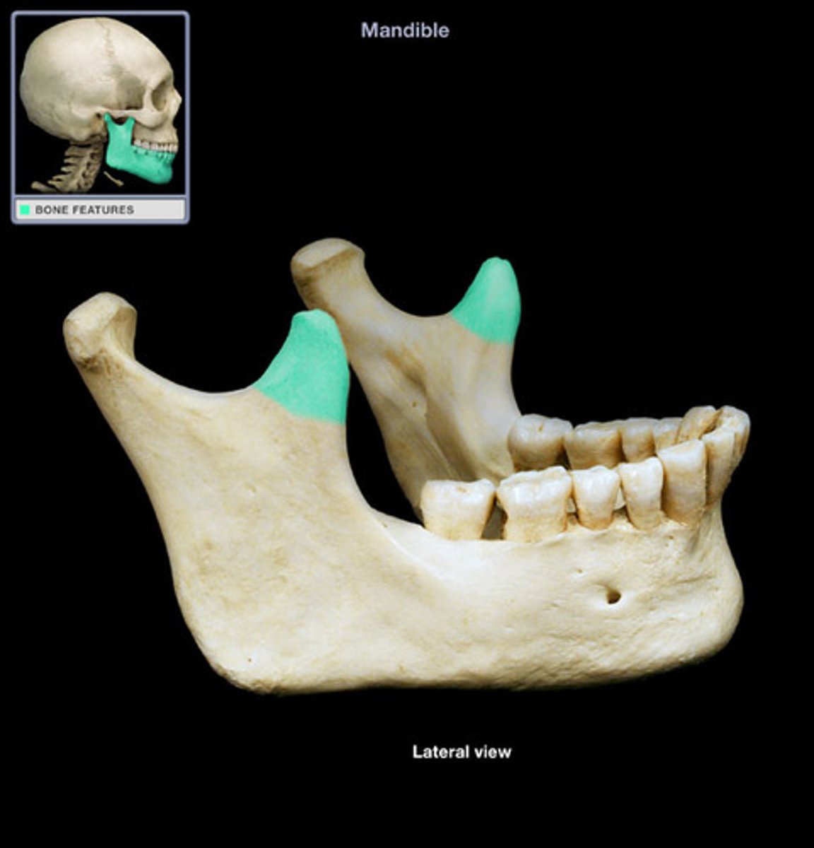
mandibular notch
ridge/depression from coronoid process
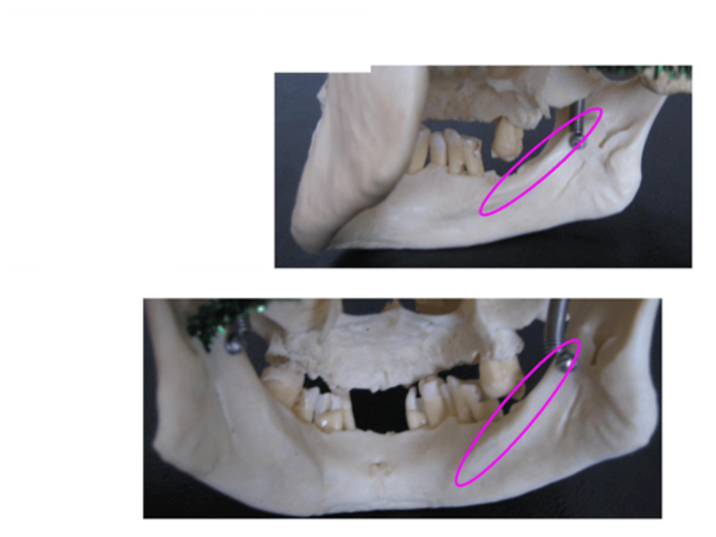
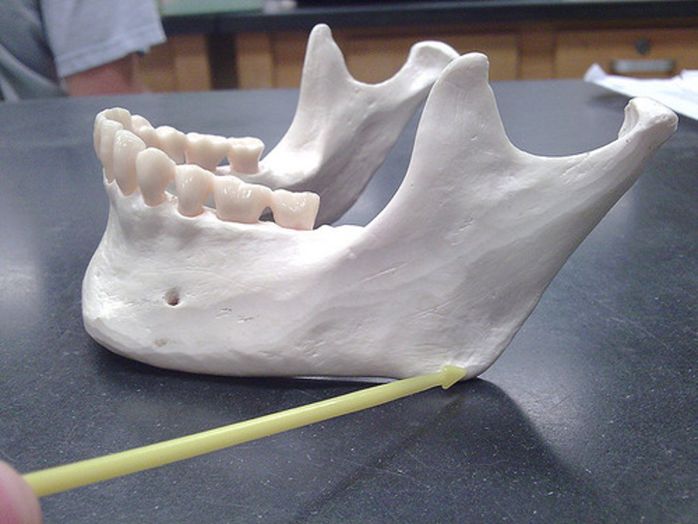
angle (gonial angle)
angle at the posterior border where the ramus and the body meet
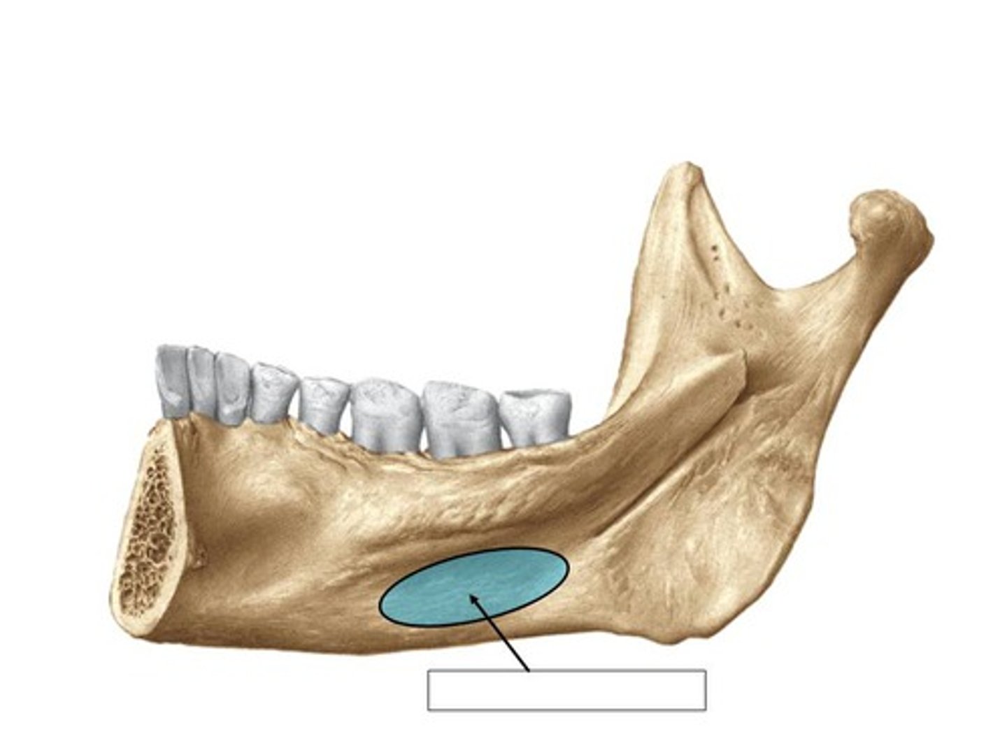
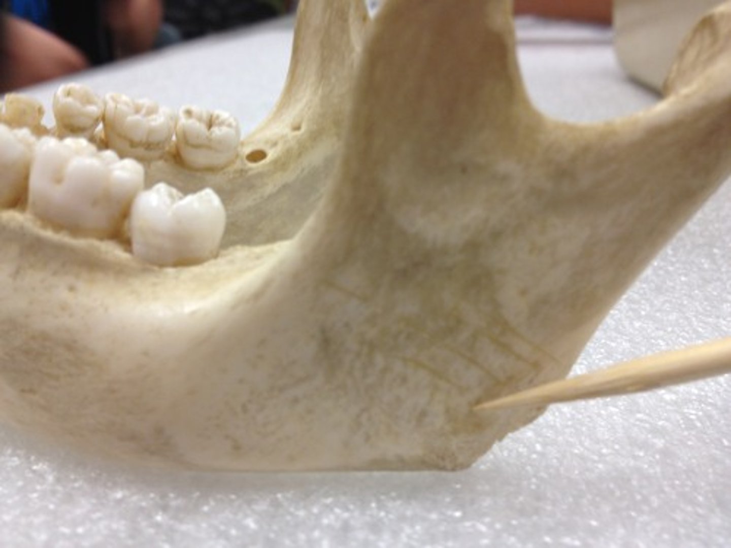
masseteric tuberosity/fossa
rough area & hollow on lateral surface of angle & ramus where masseter muscle attaches
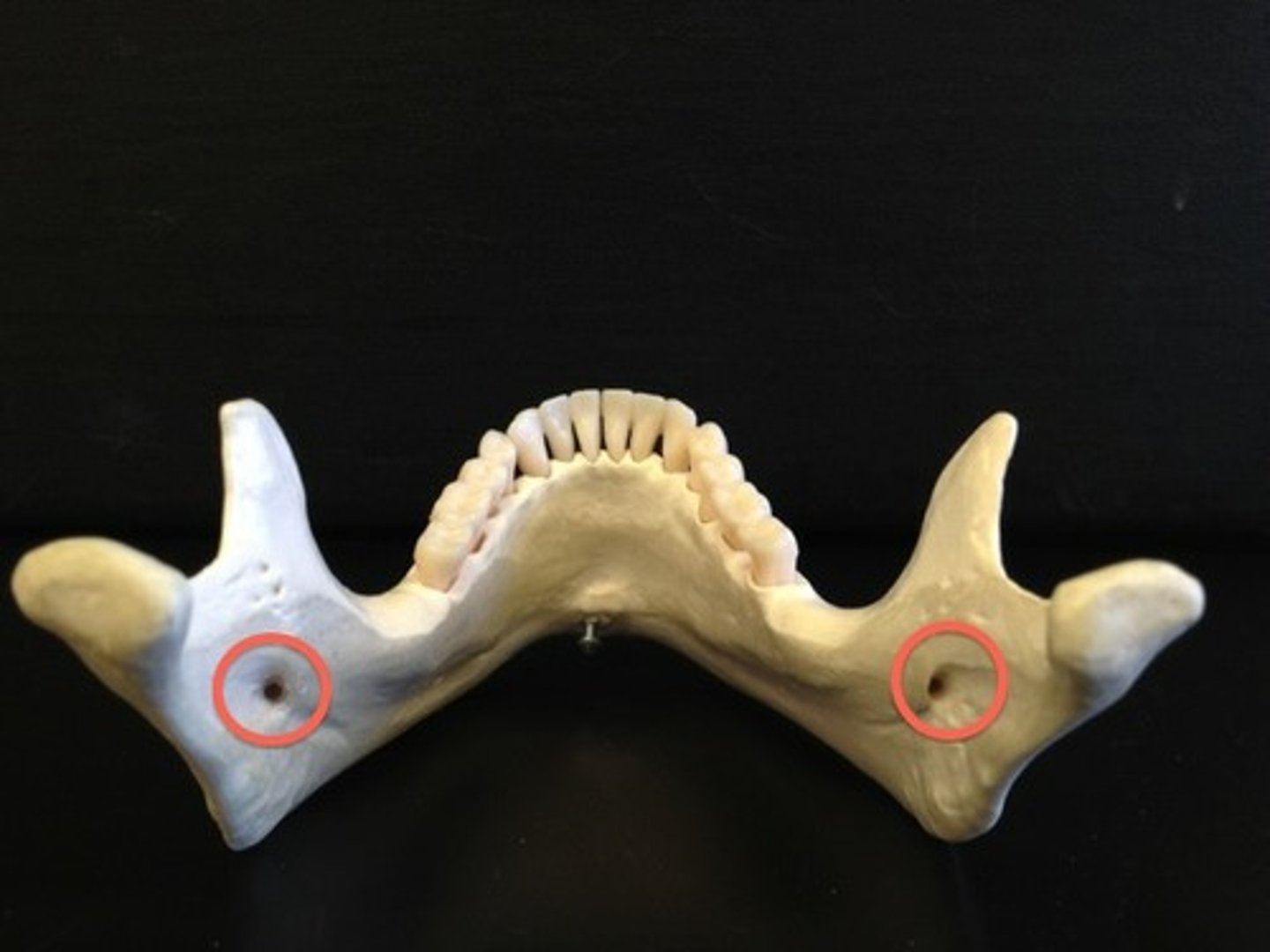
mandibular foramen
medial surface of ramus (exits via mental foramen)
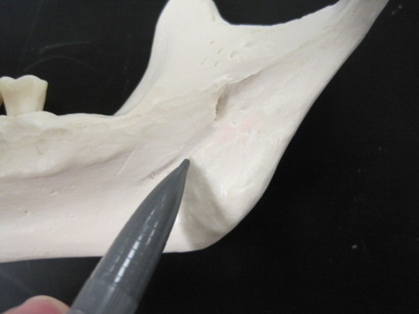
mylohyoid groove (sulcus)
Crosses the medial surface of the ramus, running anteroinferiorly from the edge of the mandibular foramen
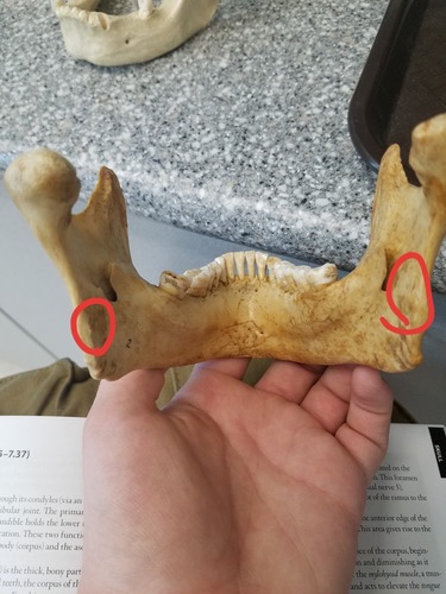
pterygoid tuberosities
Roughened patches on the medial surface of the angle for attachment of the Medial Pterygoid muscles
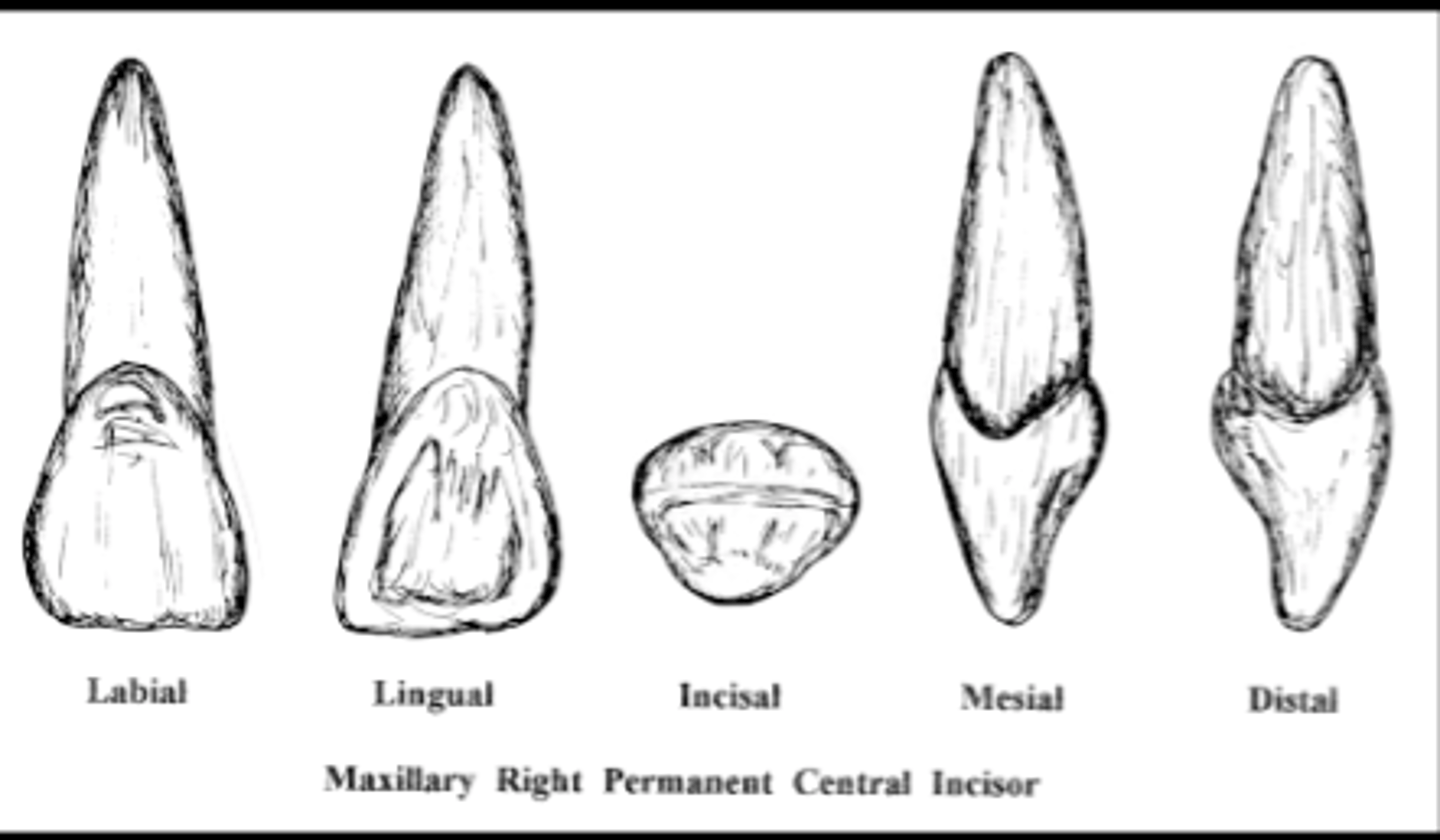
upper incisors
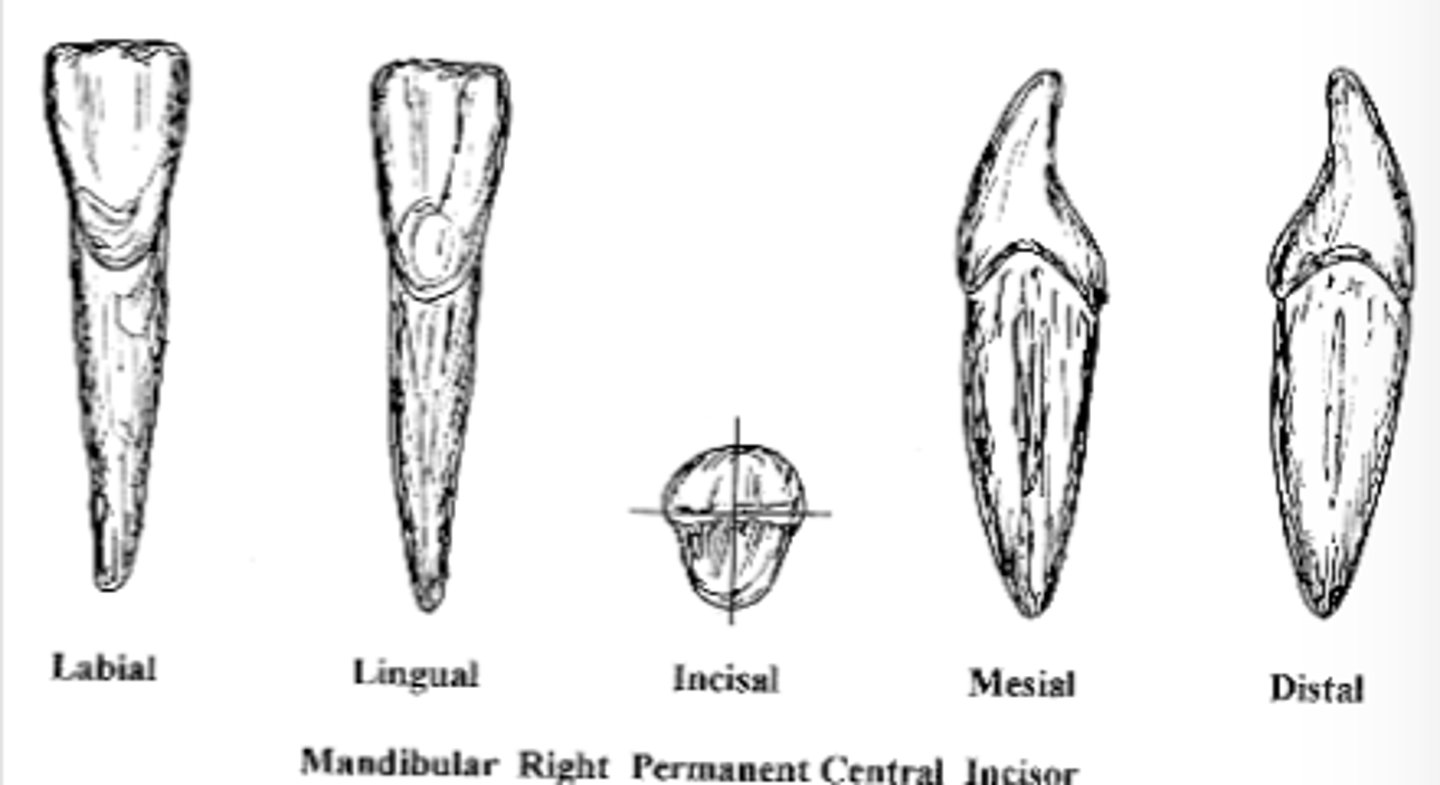
lower incisors
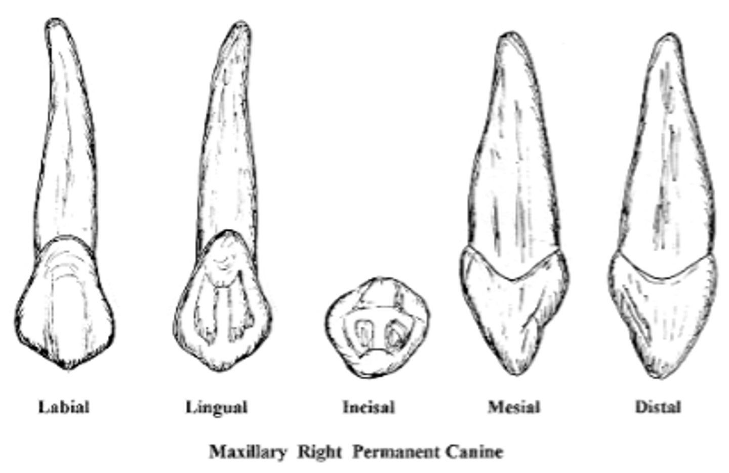
upper canines
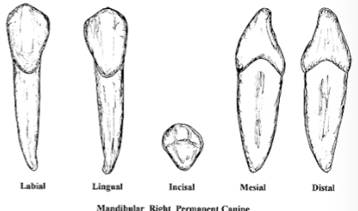
lower canines
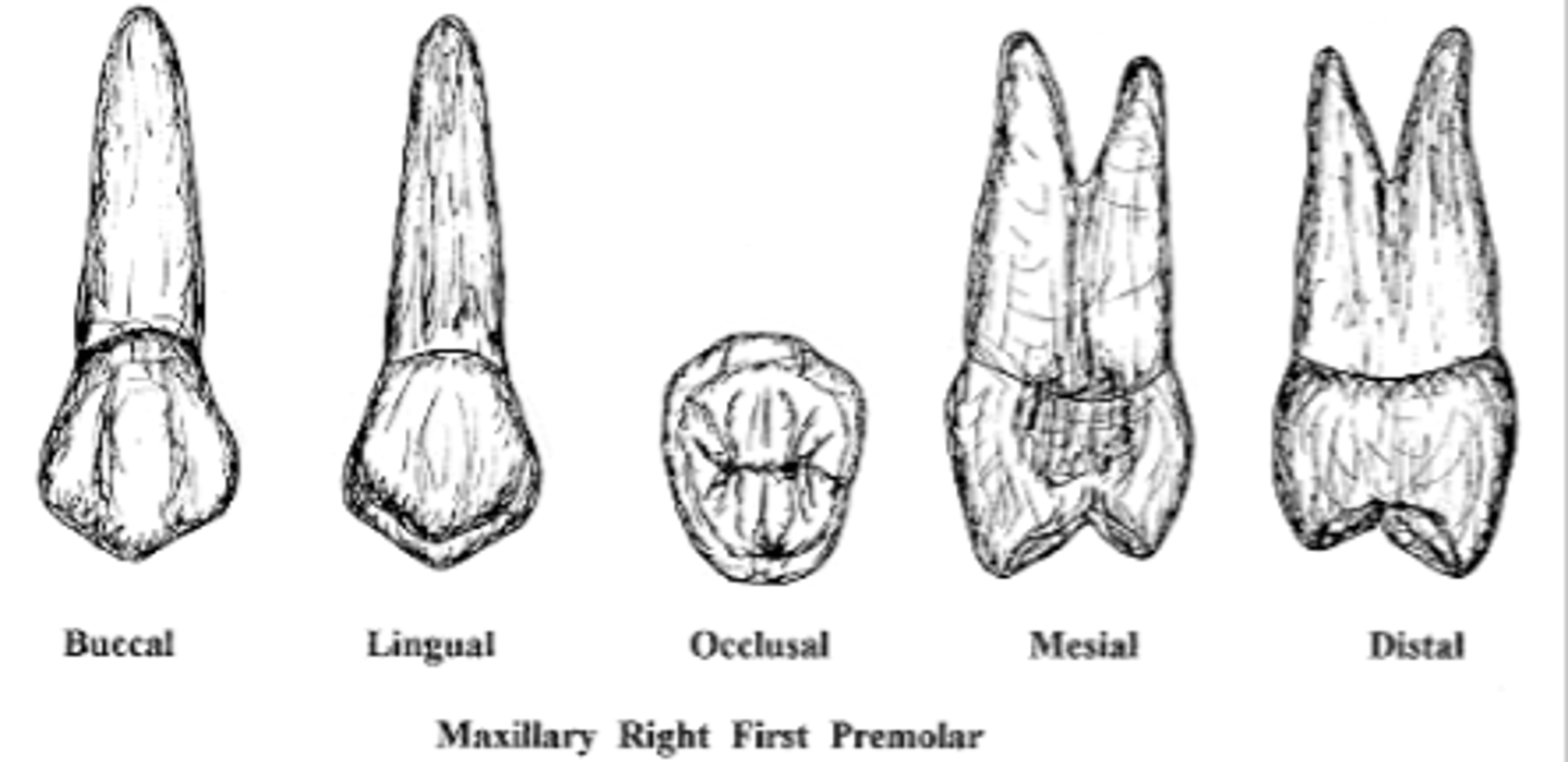
upper premolars
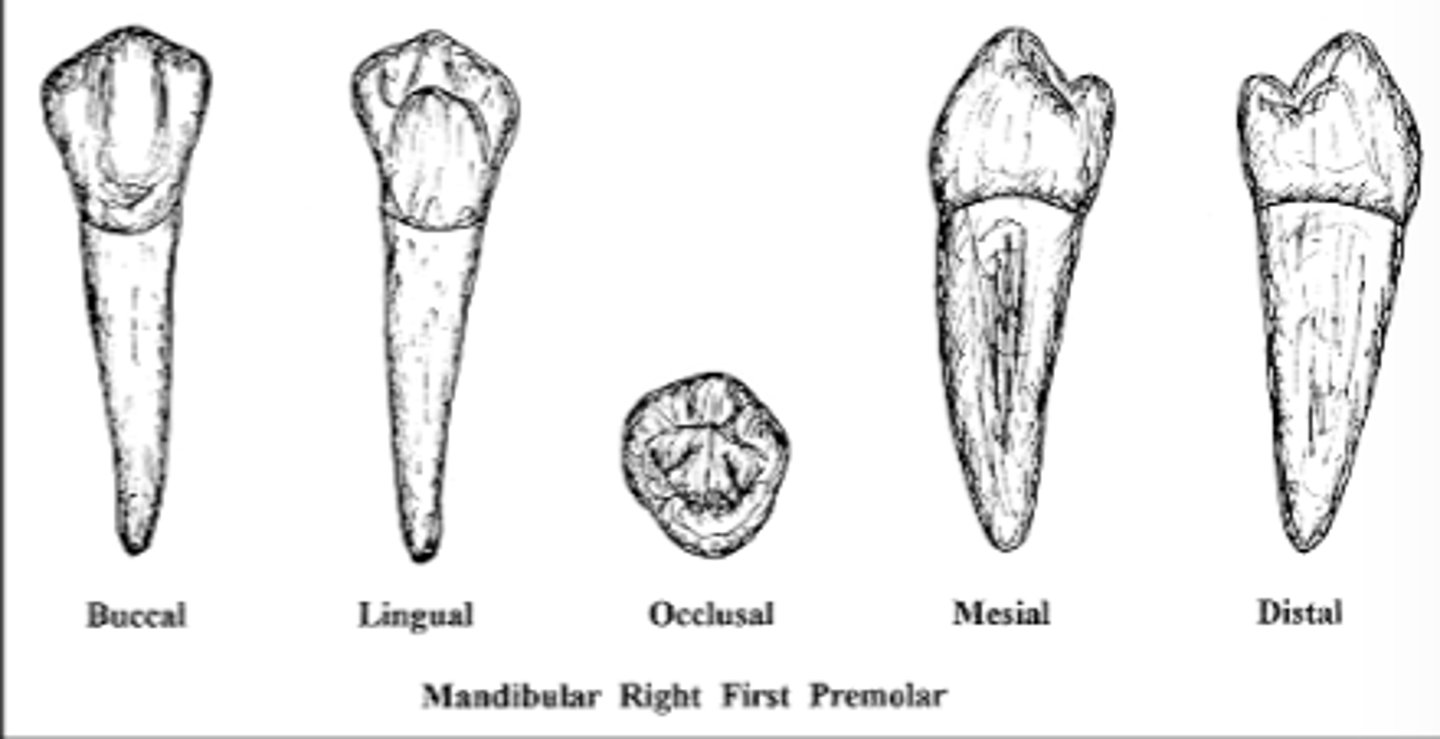
lower premolars
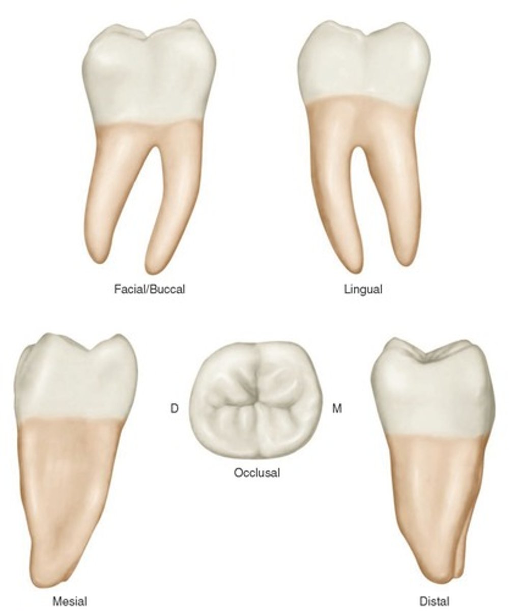
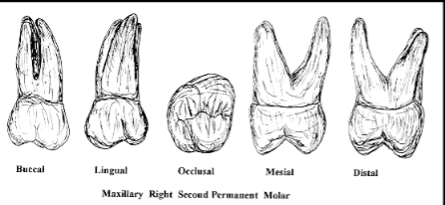
upper molars
lower molars