MIDTERM EXAM STUDY GUIDE - APHY 102 (Ivy Tech Online)
1/91
There's no tags or description
Looks like no tags are added yet.
Name | Mastery | Learn | Test | Matching | Spaced | Call with Kai |
|---|
No analytics yet
Send a link to your students to track their progress
92 Terms
What is a hormone and how does it act?
Hormones are chemical messengers that are responsible for regulation. They are secreted into body fluids, mainly blood and have specific actions on target tissues.
What is the role of negative feedback in control of hormone secretions.
a stimulus elicits the release of a substance; once the substance reaches a certain level, it sends a signal that stops further release of the substance.
Paracrine glands
hormones enter the interstitial fluid but affect only neighboring cells
Autocrine glands
affect only the secreting cell
endocrine glands
hormones are secreted from the interstitial fluid into the bloodstream and act on target cells
exocrine glands
secrete chemical substances into ducts that lead either to other organs or out of the body
Where can the different endocrine glands be found in the body?
Brain:
Pineal, pituitary and hypothalamus
Neck:
Thyroid and parathyroid
Between the lungs:
Thymus
Behind the stomach:
Pancreas
On top of the kidneys:
Adrenal glands
Abdomen:
Ovary
Scrotum:
Testes
Describe steroid hormones and their mechanism of action.
a steroid that acts as a hormone;diffuse through the cell membrane of the target cell.
Describe non-steroid hormones and their mechanism of action.
They are amino acid, peptides & protein hormones;produce their effect on target tissues by binding to the receptors.
How is the anterior pituitary gland different than the posterior pituitary gland?
the action of the anterior pituitary gland is regulated through vessels connected to the hypothalamus whereas the action of the posterior pituitary gland is regulated through nerves connected to the hypothalamus.
What regulates pituitary gland secretion?
The hypothalamus
Describe tropic hormones.
stimulate other endocrine glands to release hormones
Insulin vs Glucagon
Insulin activates facilitated diffusion of glucose through certain cell membranes, stimulates its storage. Lowers blood glucose
Glucagon stimulates the liver to produce glucose, increasing concentration of blood glucose. Also breaks down fat. Increases blood glucose
clinical conditions related to endocrine disorders
Hyperthyroidism-excess of PTH secretion usually caused by tumor
Goiter-reduced production of thyroid hormone caused by low iodine intake; neck edema
Ketoacidosis-elevated seton bodies in blood due to low production of insulin (glucose not then in by cells)
Cretinism-stunted mental and physical growth due to low production of thyroid hormone
Hirsutism-Excessive hair in women due to high androgen production
Hypoglycemia-Low blood glucose levels due to low production of glucagon
Polyuria and glycosuria-excessive urine production and glucose in the urine due to reduced insulin
Acromegaly-elevated growth hormone resulting in hyper growth all over the body
Grave's Disease-Over production of thyroid hormone caused by an autoimmune disorder
Diabetogenic effect-Polyuria due to low amounts of insulin
Diabetes insipidus-Excessive thirst, increase in urine due to low amounts of ADH (salt retaining hormone)
Insulin dependent diabetes mellitus (type 1)- Autoimmune destruction of insulin receptors
Non-insulin dependent diabetes mellitus- Insulin receptors become worn out
How does the stress response affect the body?
Stress causes hypothalamus to initiate response, so sympathetic branch of ANS is activated. Adrenal medulla releases adrenalin into bloodstream, increasing amount of oxygen in blood going to muscles-prepares body for 'fight or flight'.
How does aging affect the endocrine system?
As people get older, their endocrine glands decrease in size, muscular strength decreases as GH levels decrease, ADH levels increase due to slower breakdown in liver & kidneys. Calcitonin levels decrease, and insulin resistance may develop.
Describe the components of blood.
plasma, red blood cells, white blood cells, platelets
Compare the formed elements of the blood.
RBCs, WBCs, and platelets all act together to maintain life. RBCs transport oxygen to the body's tissues, WBCs fight infections in the body, and platelets clot wounds that occur.
What are normal levels and percentages of RBC?
4,600,000-6,200,000 in males.
4,200,000-5,400,000 in females.
4,500,000-5,100,000 in children.
RBCs are 45% of the blood.
How is the shape of a red blood cell important to its function?
enables oxygen and carbon dioxide to diffuse across the RBC's plasma membrane more readily.
What is hematocrit?
the ratio of the volume of red blood cells to the total volume of blood
Differentiate between the 5 different leukocytes (WBCs).
Neutrophils-- First Responder- Most abundant WBC that travels in the blood looking for infections.- 55%-70%
Lymphocytes-- B&T cells- release antibodies (B), Attack viruses (T)- 20%-40%
Monocytes- - Rebuild damaged tissue- Produce proteins & antigens- 2%-8%
Eosinophils-- Fight bacteria and parasites- 1%-4%
Basophils-- Responsible for allergic reactions- 0.5%-1% (smallest)
serum versus plasma
Plasma is the liquid portion of unclotted blood and still contains the clotting factors.
Serum is the liquid portion of blood that has been allowed to clot.
How are platelets involved in hemostasis?
enhancing vasoconstriction
platelet aggregation (cluster)
vessel repair
Describe the steps in clot formation.
Hemostasis - the stoppage of bleeding.
1. Blood vessel spasm - smooth muscle in blood vessel contracts
2. Platelet plug formation:
a. break in vessel wall
b. blood escapes through break
c. platelets adhere to each other, to end of broken vessel, and to exposed collagen
d. platelet plug helps control blood loss
3. Blood coagulation - clot forms (occurs extrinsically or intrinsically).
What happens if clots form within blood vessels?
A thrombus is made. If it breaks loose of the vessel wall and begins circulating through the body, it is then called an embolus, which can travel into tighter vessels and get trapped, causing death.
What is edema?
swelling
What antigens can be found on RBC?
Antigens A, B, AB, or none (O).
What antibodies can be found in the plasma?
Antibodies A, B, AB, or none (O).
How do these create different blood types?
Red blood cells on their membranes may contain two major antigens - antigen A and antigen B. These antigens are carbohydrates attached to glycolipids projecting from the surface of red blood cell. The ABO blood group is based on the presence or absence of these antigens and the antibodies circulating in the plasma. A person with only antigen A has type A blood; one with only antigen B has type B blood; a person with both antigens (A and B) has type AB blood; type O blood has a person with neither A nor B antigen. Antibodies that affect ABO blood groups are anti- A antibody and anti- B antibody. Anti- A antibody is produced when antigen A is absent on the red blood cells, and whenever antigen B is absent, anti- B antibody is produced.
What blood types can give/receive to/from other blood types?
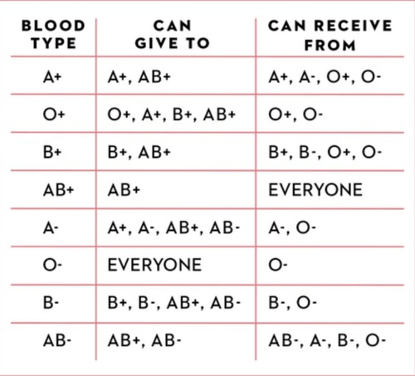
How does the Rh factor affect a developing fetus and its mother?
If woman is Rh negative and her baby is Rh positive (Rh incompatibilityRh incompatibility), woman's body might produce proteins called Rh antibodies after exposure to the baby's red blood cells. The antibodies produced aren't a problem during the first pregnancy. The concern is with next pregnancy. If woman's next baby is Rh positive, these Rh antibodies can cross the placenta and damage the baby's red blood cells. This could lead to life-threatening anemia, a condition in which red blood cells are destroyed faster than the baby's body can replace them.
What are the functions of the cardiovascular system?
The three main functions are to deliver materials throughout the body, to remove cellular wastes, and to fight against diseases.
Where is the heart found?
in the mediastinum of the thorax
Describe the layers of the heart and pericardium.
The outer layer of the heart is the fibrous pericardium (also the epicardium). The inner layer of the pericardium is the visceral pericardium (it lies against the heart). And the outer layer of the pericardium is the parietal pericardium which lies against the wall of the pericardial cavity.
The heart has three layers as well. The innermost layer is the endocardium, the middle layer is the myocardium, and the outermost layer is the epicardium (also the fibrous pericardium).
Describe the pathway of blood into, through, and out of the heart.
Blood flows first through the right atrium, then through the right ventricle. The blood stays in the right ventricle until the chamber is full, wherein the blood flows next to the lungs through the pulmonic valve and pulmonary artery. Then, the oxygen-rich blood from the lungs flows through the pulmonary vein into the left atrium. Next, it flows to the left ventricle and is temporarily stored here until the left ventricle is full. Finally, blood flows through the aortic valve into the aorta and is distributed to the rest of the body.
Describe the pathway of the cardiac conduction system.
Cardiac conduction system controls the heart rate. This system generates electrical impulses and conducts them throughout the muscle of the heart, stimulating the heart to contract and pump blood. Cardiac conduction system is composed of a group of specialized cardiac muscle cells in the walls of the heart. Crucial components of the cardiac conduction system are the sino-atrial (SA) node, the atrioventricular (AV) node, the bundle of His, the left and right bundle branches and the Purkinje fibers. Cardiac impulse is initiated in the sinoatrial node ( a small mass of tissue, situated in the wall of the right atrium) by spontaneous depolarization and sympathetic activity. Cells of the SA node are specialized to undergo spontaneous depolarization and generation of action potentials on their's own. From the SA node impulse spreads throughout the atria through internodal pathways to the AV node (located in the inferior part of the interatrial septum). Once the cardiac impulse reaches the AV node, it passes into a group of large conduction fibers that make up the atrioventricular bundle (bundle of His). The AV bundle enters the interventricular septum and divides into two atrioventricular bundle branches, commonly called the left and right bundle branches, which descend and reach the apex of the heart, where they connect with the Purkinje fibers. These larger fibers spread the impulse to the myocardial contractile cells in the ventricles, and extend throughout the myocardium from the apex of the heart toward the atrioventricular septum and the base of the heart. Contraction begins at the apex, travels toward the base of the heart, pushing the blood into the aorta and pulmonary trunk.
Describe an EKG
A recordable tracing of the electrical activity of the heart that the production and conduction of action potentials in the heart produces.
What is occurring within the heart during each part of the EKG?
At the P wave of the EKG, the atria are depolarizing. At the QRS complex, the ventricles are depolarizing and the atria are repolarizing. At the T wave, the ventricles are repolarizing and there is a brief refractory period between the T wave and the following P wave, which allows the heart a small rest.
Describe what is happening in the heart during atrial systole/ventricular diastole
Atrial systole/ventricular diastole - the atria are contracting and the ventricles are relaxed.Atrial diastole/ventricular systole - the atria are relaxed and the ventricles are contracting.
Describe what is happening in the heart during atrial systole/ventricular systole
How are the heart sounds made?
The first heart sound "lubb" occurs during ventricular systole as a result of the A-V valves closing.
The second heart sound "dupp" occurs during ventricular diastole as a result of the pulmonary and aortic semilunar valves closing.
What terms are used to describe abnormal heart rhythms?
Arrhythmia, atrial fibrillation, bradycardia, tachycardia, defibrillation, cardiac arrest, palpitations, Supraventricular tachycardia (SVT), Ventricular tachycardia, and ventricular fibrillation.
Compare and contrast pulmonary, coronary, and systemic circulation.
Pulmonary, coronary, and systemic circulation all work together to provide oxygen to the body and the heart and remove carbon dioxide waste from both. Pulmonary circulation is that which goes to and from the heart and lungs, oxygenating the blood and removing carbon dioxide from it. Coronary circulation is the blood supply provided to the heart and it oxygenates the heart.Systemic circulation is circulation from the heart to the whole body and back, dropping off oxygen to the body's tissues and picking up carbon dioxide to remove it from the body.
What factors can influence heart rate and/or blood pressure?
Cardiac output, blood volume, peripheral resistance, and blood viscosity.As blood volume, heart rate, stroke volume, blood viscosity, and peripheral resistance increase, BP increases.
How is cardiac output figured?
Stroke volume multiplied by the heart rate, expressed in bpm.
SV x HR = cardiac output
Where can pulse be found in the body?
Temple (temporal a.)
Neck (carotid a.)
Chin (facial a.)
Inner elbow (brachial a.)
Wrist (radial a.)
Groin (femoral a.)
Back of the knee (popliteal a.)
Front of the foot (dorsalis pedis a.)
Back of the ankle (posterior tibial a.)
At any given moment, where can blood be found in the body?
The veins.
Identify the major arteries/veins of the body and the body regions they supply/drain.
Major arteries: Ascending aorta->right and left coronary a. Brachiocephalic a., Left common carotid a., and the Left subclavian a.Descending aortaThoracic aorta->Bronchial artery, Pericardial a., Esophageal a., Mediastinal a., Posterior intercostal a.Abdominal aorta-> Celiac a., Phrenic a., Superior mesenteric a., Suprarenal a., Renal a., Gonadal a., Inferior mesenteric a., Lumbar a., Middle sacral a., Common iliac a.Major veins:Superior and Inferior vena cava, right and left external and internal jugular and subclavian v., right and left brachiocephalic v., hepatic v., splenic v., right and left common iliac v., external and internal iliac v., femoral v., great saphenous v., small saphenous v.
What is arteriosclerosis? How does it occur?
Chronic disease of arterial system.
Lipid and collagen fibers migrate into the vessel walls.
Thickening and hardening of vessel walls.
How does aging affect the cardiovascular system?
Cholesterol deposition in the blood vessels
Heart enlarges
Cardiac muscle cells die
Increase in fibrous connective tissue, adipose tissue and blood pressure
Decrease in resting heart rate.
What is the function of lymph?
filters the blood by removing toxins
Describe a lymphatic vessel.
Fine, thin-walled, transparent valved channels distributed through most tissues. They have 3 walls: intima, media, and adventitia.
Describe the pathway of lymph.
Lymph flows through the body into lymphatic capillaries which carry them to lymphatic vessels. From there, lymph flows into lymph nodes via the afferent lymphatic vessel and is cleaned before returning to circulation via the efferent lymphatic vessel. Lymph then flows into the lymphatic trunk and then the collecting duct before returning to normal body circulation.
Where can lymph nodes be found in the body
Cervical
Axillary
Supratrochlear
Inguinal regions and the pelvic, abdominal and thoracic cavities.
what is the structure of a lymph node?
Bean shaped and surrounded by a fibrous capsule.
What types of cells provide our immunity?
T cells and B cells
How does each cell type function?
T cells have the role of recognizing and distinguishing foreign substances from those belonging to the organism and destroying them.
B cells regulate the production of antibodies.
Compare an antigen to an antibody.
Antigens cause the disease and antibodies cure it.
Antigens are substances that provoke an immune response (they're the ultimate target for the immune system). Antibodies are simply proteins that are secreted as a result of the antigen provoked immune response.
Describe the thymus and its role in immunity.
The thymus is a soft, bi-lobed gland enclosed in a connective tissue capsule. It is the place where T cells mature.
Describe the different glands associated with the lymphatic system. What are their functions?
lymph-absorption of dietary fats. Also, filtered proteins are brought back into the bloodstream by the help of lymph. Lymph also takes part in carrying bacteria and viruses to lymph nodes for immune functions.
lymphocytes-absorption of dietary fats. Also, filtered proteins are brought back into the bloodstream by the help of lymph. Lymph also takes part in carrying bacteria and viruses to lymph nodes for immune functions.
lymphatic vessels-provide a conduit from the peripheral tissues to the venous system. Act as a one way valve-allows fluids, bacteria, viruses, fats to ender bot NOT return.
lymph nodes-filter possible substances that may harm the body
Tonsils-defense from the bacteria that can invade tissues in the area around the openings between the nasal and oral cavities.
Spleen-produces macrophages
Thymus-provides immunity by producing T lymphocytes
How does stress affect immunity?
With chronic stress, the immune system stays in low gear, leaving the body vulnerable to infection and disease. Basically, it suppresses it.
Compare and contrast primary versus secondary immune responses.
The primary immune response occurs after the first contact of B and T cells with a foreign antigen when there are still no previously produced antibodies. The time period in which antibodies are produced is long and can last up to several weeks or months.
The secondary immune response is the response of the immune body to the antigen, after prior exposure. This is helped by follicular dendritic cells, which due to memory begin to release antigens in the fight against infection
What is interferon? Complement?
Interferon is a protein produced by virus-infected cells. It helps other nearby cells to be better prepared to the virus attack and to heighten their defense against the virus.
Complement-is serum proteins ( 20 proteins ) synthesized in the liver ( and other body cells, macrophages, monocytes , intestinal epithelium ), and present in an inactive form in serum and tissue fluids of man , then interact with each other in a cascading fashion to protect against infectious agents.
What are macrophages and what is their role in immunity?
Effector cells of the innate immune system that phagocytose bacteria and secrete both pro-inflammatory and antimicrobial mediators. In addition, macrophages play an important role in eliminating diseased and damaged cells through their programmed cell death
Compare and contrast innate barriers versus adaptive immunity.
Innate immunity generates a non-specific immune response against the pathogen.
Adaptive immunity generates a specific immune response against a particular pathogen
Describe differences between T cells and B cells.
--B cells are most effective in fighting bacteria. Major function is to produce antibody.
--T cells are a type of white blood cell that is of key importance to the immune system and is at the core of adaptive immunity
How does an autoimmune disease work?
An autoimmune disease arises when the body's cells lose their ability to distinguish between self and non-self antigens. When this happens, the body mounts an immune response against what is really healthy tissue, (but they think is a foreign particle), and the body attacks itself causing tissue, organ, and joint damage as well as pain, fever, and swelling.
Passive immunity
the short-term immunity that results from the introduction of antibodies from another person or animal.
active immunity
A form of acquired immunity in which the body produces its own antibodies against disease-causing antigens.
Describe the roles of IgG, IgD, IgE, IgA, and IgM.
IgG- is in plasma and tissue fluids and is effective against bacteria, virsuses, and toxins and it activates complement proteins.
IgA- is in breast milk, tears, nasal fluid, gastric juice, intestinal juice, and urine and it is an exocrine gland secretion.
IgM- is a type of antibody produced in plasma in response to contact with certain antigens in foods or bacteria.
IgD- is on the surfaces of most B cells and acts as an antigen receptor and is important in activating B cells.
IgE- appears in exocrine secretions with IgA. It is associated with allergic reactions.
What is tissue rejection and how is it reduced through clinical practices/treatments?
Failure of transplantation that occurs when an organism recognizes transplanted tissue as foreign, and mounts an immune response.
Growth Hormone (GH)
Produced by: Anterior pituitary Released from: Hypothalamus
Target: All tissues
Controlled by: GH-releasing hormones and somatostatin from the hypothalamus
Body affect: Growth; carbohydrate, protein and fat metabolism
Prolactin (PRL)
Produced by: Anterior pituitary
Released from: Hypothalamus
Target: mammary gland
Controlled by: prolactin release-inhibiting hormone from the hypothalamus restrains secretion of prolactin
Body affects: Breast development & milk production
Thyroid Stimulating Hormone (TSH)
Produced by: Anterior pituitary
Released from: Hypothalamus
Target: Thyroid
Controlled by: the hypothalamus by secreting thyrotropin-releasing hormones
Body affects: controls Thyroxine secretion
Adrenocorticotropic hormone (ACTH)
Produced by: Anterior pituitary
Released from: Hypothalamus
Target: Adrenal cortex
Controlled by: the hypothalamus, by secreting corticotropin-releasing hormone
Body affects: regulates cortisol secretion
Follicle-stimulating hormone (FSH) and Luteinizing hormone (LH)
Produced by: Anterior pituitary
Released from: Hypothalamus
Target: Ovaries/testes
Controlled by: hypothalamus
Body affects: Ovarian follicle growth, estrogen secretion/ spermatogenesis
Antiduretic hormone (ADH)
Produced by: Posterior pituitary
Released from: Hypothalamus
Target: Kidneys/smooth muscle in arterioles
Controlled by:the hypothalamus
Body affects: causes the kidneys to excrete less water/blood pressure
Oxytocin (OT)
Produced by: Posterior pituitary
Released from: Hypothalamus
Target: Uterine smooth muscles, mammary gland
Controlled by:the hypothalamus
Body affects: uterine contractions, production and ejection of milk
Thyroxine and Triiodothyronine (T4 and T3) (thyroid hormones)
Produced by: Thyroid gland
Released from: Thyroid gland
Target: most tissues
Controlled by:the thyroid
Body affects: Metabolic rate; growth and development
Calcitonin (CT)
Produced by: Thyroid
Released from: Thyroid
Target: Bones
Controlled by: thyroid
Body affects: Plasma calcium and phosphate (lowers)
parathyroid hormone (PTH)
Produced by: Thyroid
Released from: Thyroid
Target: Bones, kidneys, intestine
Controlled by: the thyroid
Body affects: Plasma calcium and phosphate (elevates)
Thymopeotin
Produced by: Thymus
Released from: Thymus
Target: T-lymphocyte cells in blood
Controlled by: Thymus
Body affects: Immune response
Insulin
Produced by: Pancreas
Released from: Pancreas islets
Target: most tissues, notably muscle and liver
Controlled by: Pancreas
Body affects: Glucose utilization; lowers blood glucose
Glucagon
Produced by: Pancreas
Released from: Pancreatic islets
Target: Liver
Controlled by: The pancreas
Body affects: elevates blood glucose
Melatonin
Produced by: Pineal gland
Released from: pineal/thalamus
Target: various tissues
Controlled by:the pineal gland
Body affects: Circadian rhythm, reproduction
Intestinal mucosa hormones
Gastrin->stomach->acid secretion
Secretin->pancreas->digestive secretions
Cholecystokinin->Gallbladder->release of bile
Somatostatin->intestine-.inhibits acid and intestinal secretions
adrenal medulla hormones
Adrenaline/ noradrenaline->all tissues-> Metabolism; heart rate and output; response to stress and exercise
adrenal cortex hormones
Cortisol/ corticosterone-> all tissues-> Metabolism; response to stress and exercise
Kidney hormones
Renin (converts to angiotensin-II)-> blood vessel smooth muscle, adrenal cortex-> Blood pressure, aldosterone secretion
Ovary hormones
Oestrogens-> reproductive organs-> reproductive development; also has effects on oestrus behavior
Progesterone-> Uterus-> uterine condition
Testes hormones
Testosterone-> reproductive organs-> reproductive development; also has effects on behavior