L16 - Protein Delivery
1/15
There's no tags or description
Looks like no tags are added yet.
Name | Mastery | Learn | Test | Matching | Spaced | Call with Kai |
|---|
No analytics yet
Send a link to your students to track their progress
16 Terms
overview
fwd transport pathway using transport vesicles is the default pathway
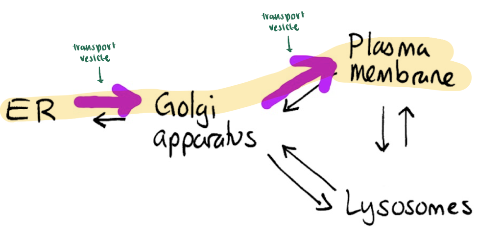
Protein Synthesis at ER (fwd pathway)
in nucleus: ribosome reads mRNA → get signal peptide to go to ER
ribosome attaches to ER protein complex on ER membrane
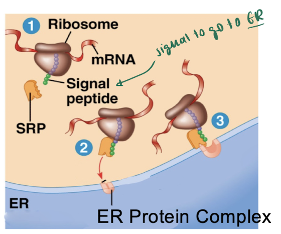
signal seq enters complex & stays, while the growing polypeptide emerges from ribosome + enters the ER lumen
mature & soluble proteins stays in ER lumen & the signal peptide is cleaved at membrane
uses chaperone proteins = small proteins that help other proteins achieve + maintain proper shape
small & soluble & within ER lumen
these ER lumen proteins may be packaged into transport vesicles leaving the ER bc they’re small & soluble
ex. help maintain a proteins shape at higher temps (heat shock proteins/Hsp)
→ discovered by fruit flies exposed to heat
ex. BiP = loading control & chaperone protein that work sin the ER lumen

Will any ER membrane proteins be packaged into transport vesicles?
No, they are too large to fit
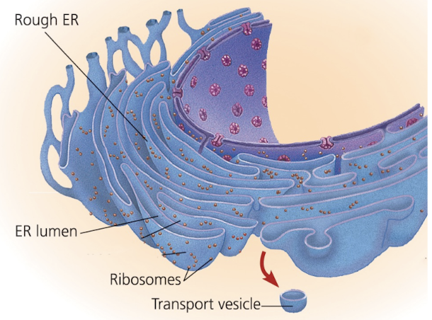
Will any ER lumen proteins be packaged into transport vesicles?
ex. chaperon proteins like BiP
Yes, they are small & soluble
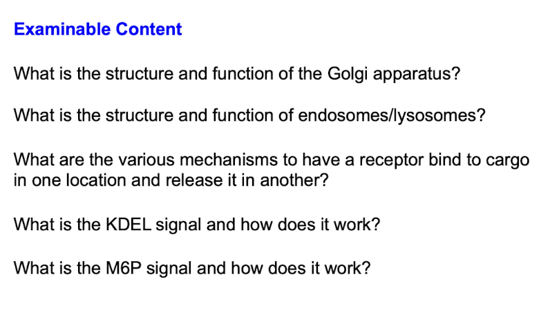
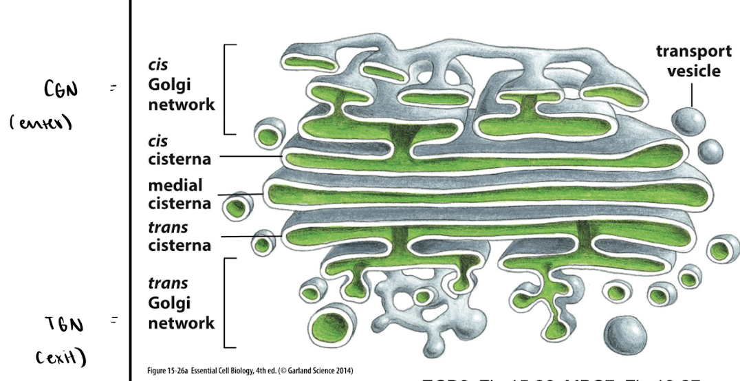
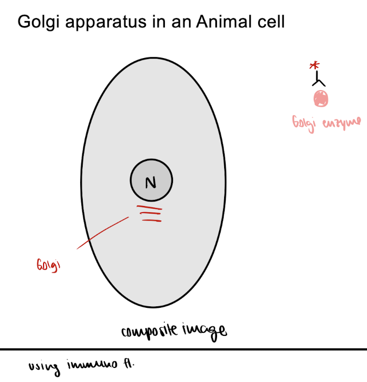
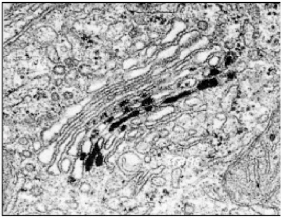
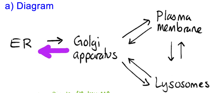
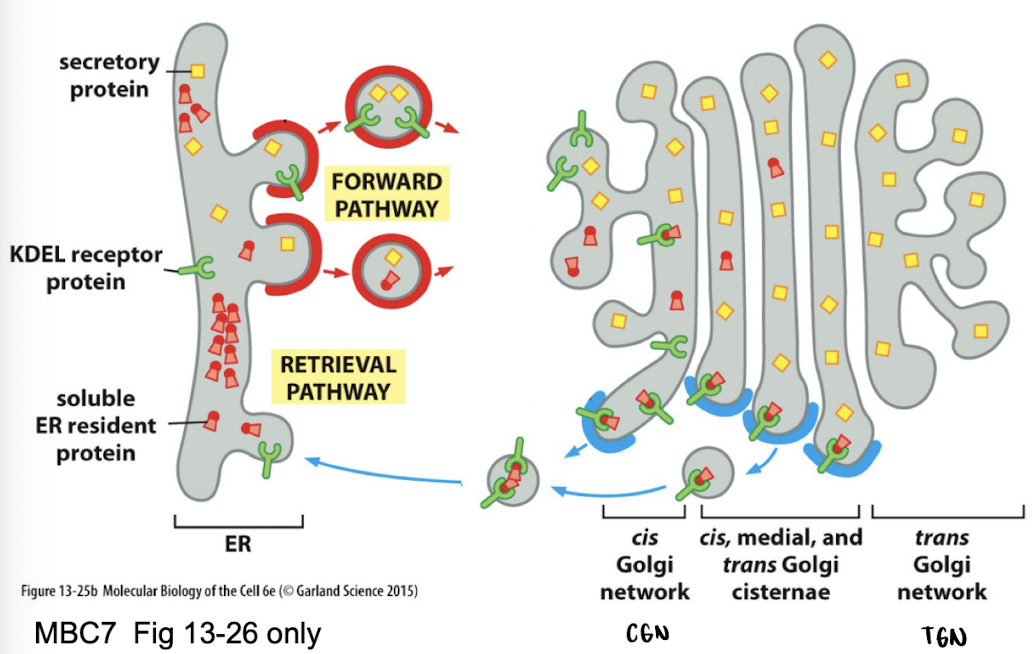
Golgi apparatus
= modify & distribute proteins made in ER
each compartment has diff enzymes
→ to view these Golgi, u can use techniques to fluoresce/mark the enzyme within the Golgi
Structure
cis face = receiving side from ER
trans face = shipping side to plasma membrane
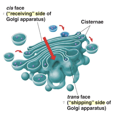
has diff levels of flattened vacuoles
→ EGN = enter (cis Golgi network)
→ TEN = exit (trans Golgi network)
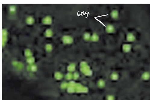
Animal cells : have ONE Golgi apparatus
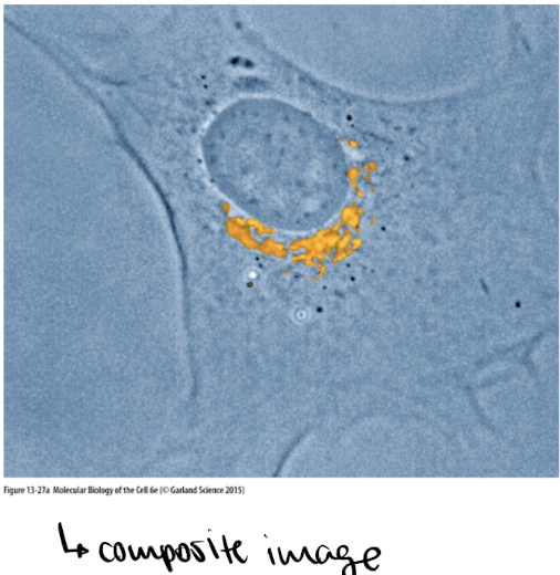
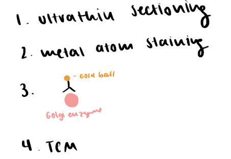
ex. looking at a fibroblast (skin cell) using DIC + wide field fluorescence to see Golgi w antibodies against the Golgi enzyme
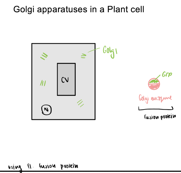
Plant cells: have MANY little Golgi
ex. looking at a plant cell using a fluorescent microscope with a fusion protein (Golgi enzyme : GFP)
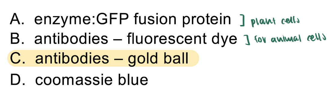
How was this enzyme labelled?
a). enzyme: GFP fusion protein
b). antibodies - fluorescent dye
c). antibodies - gold ball
d). coomassie blue
looking at an enzyme within the Golgi apparatus
c). antibodies - gold ball
Procedure:
why the others dont work:
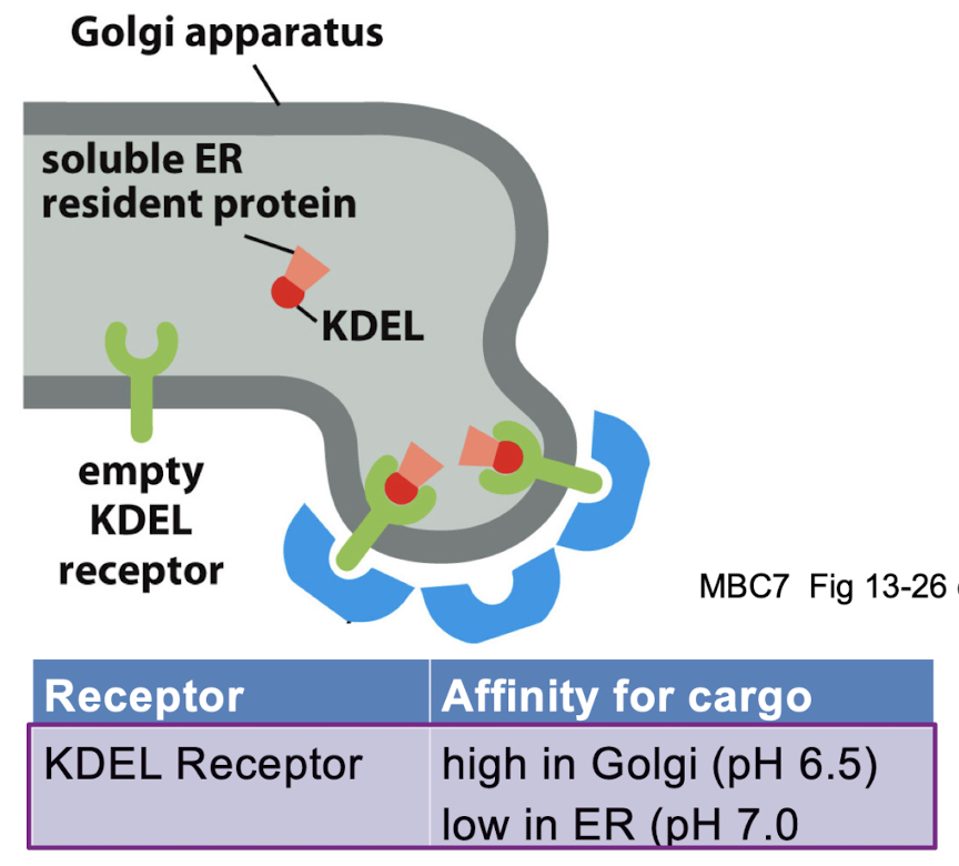
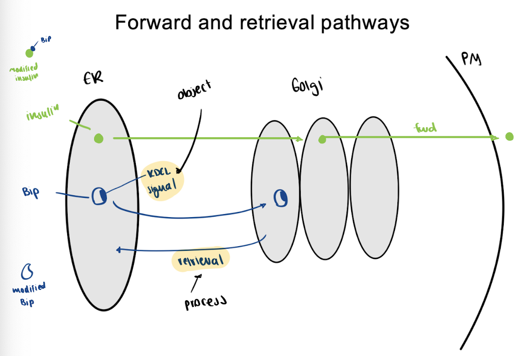
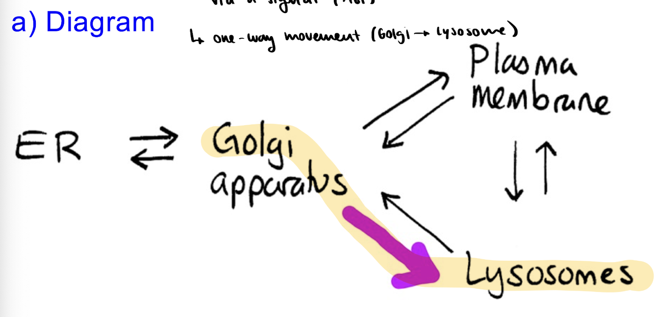
Protein Transport: The Retrieval Pathway
Retrieval Pathway = return things from Golgi BACK to ER
ex. returning BiP (a small chaperone protein) to the ER lumen - via KDEL signal + KDEL receptors
VIA:
KDEL signal = return to ER lumen
→ ONE directional
ex. BiP have KDEL signal
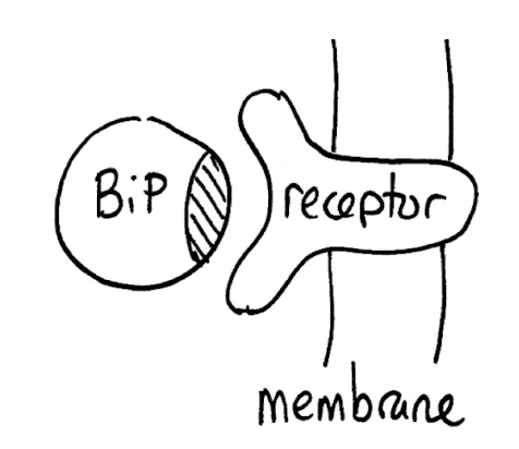
KDEL receptor = high affinity for cargo in the Golgi (bc of lower pH) & low affinity in ER (bc of higher pH)
Protein Transport: Forward vs. Retrieval Pathway Summary
Protein Delivery: Experiments
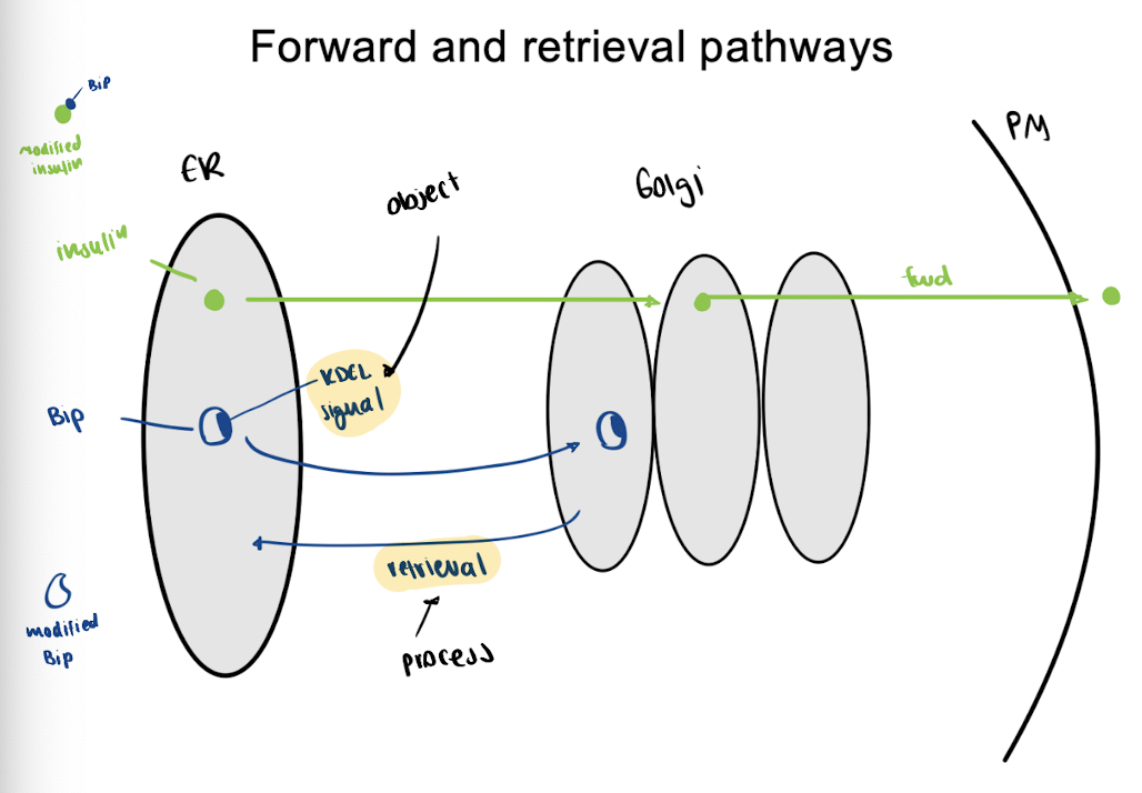
Experiment 1: Remove KDEL sequence from BiPs
result : modified BiPs exported from the cell (thus retrieval DIDNT occur)
conclusion: KDEL sequence is NECESSARY for retrieval
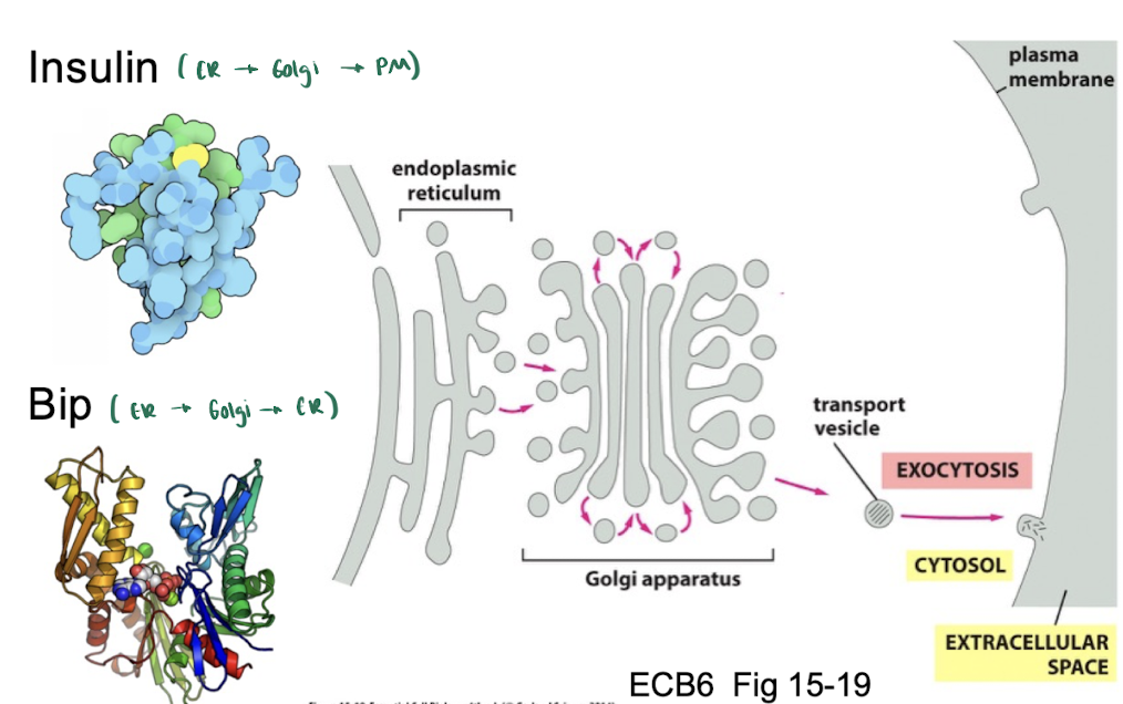
Experiment 2: KDEL sequence is added to insulin
result : modified insulins cycle b/w Golgi and ER (thus added BiP with KDEL results in retrieval)
conclusion: KDEL sequence is SUFFICIENT for retrieval
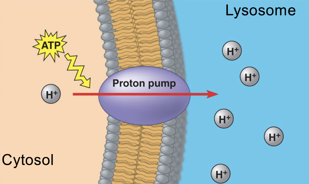
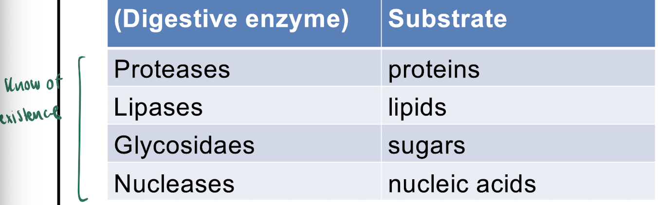
Lysosomes/endosomes
= contain digestive enzymes that can breakdown ANYTHING
acidic - bc of ATP-powered proton pumps
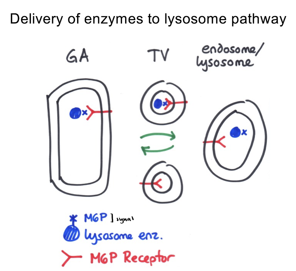
Delivery of Enzymes to Lysosomes
via: M6P signal
ONE-way movement (Golgi→ lysosome)
M6P receptor = high affinity for cargo in Golgi (high pH), low in lysosome
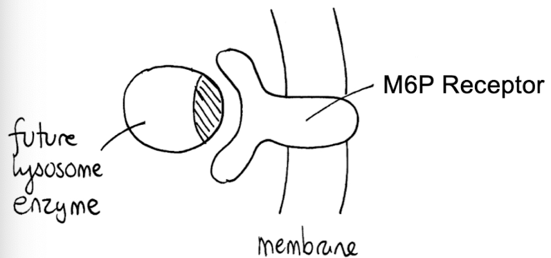
Mechanism:
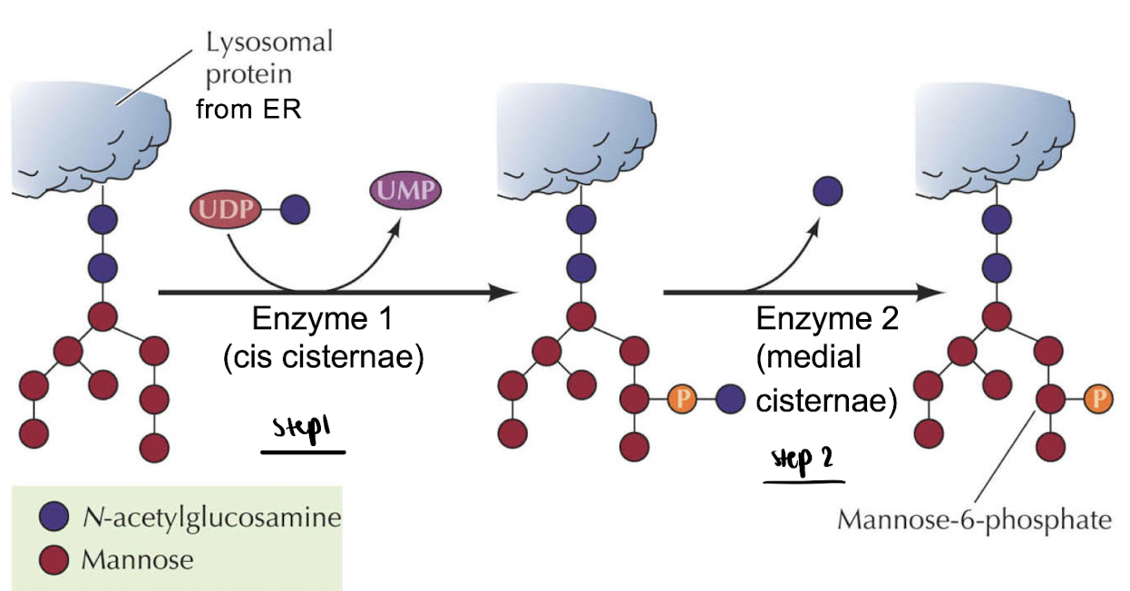
enzymes going to lysosome are given M6P signal in Golgi
2 steps:
→ allowed by diff. levels of the golgi
cis cisterna : UDP → UMP
medial cisterna : M6P attached
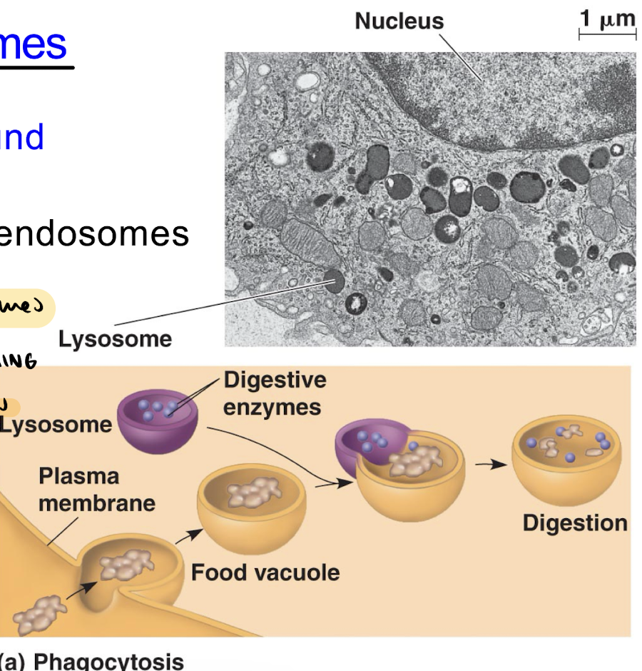
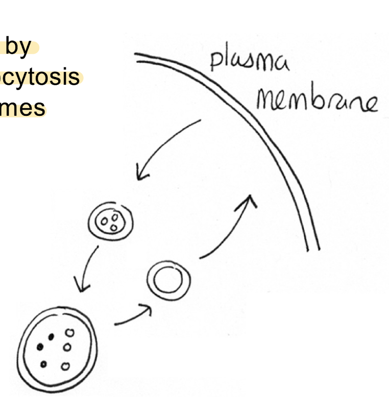
Delivery of Substrates to Lysosomes
substrates from plasma membrane → lysosomes
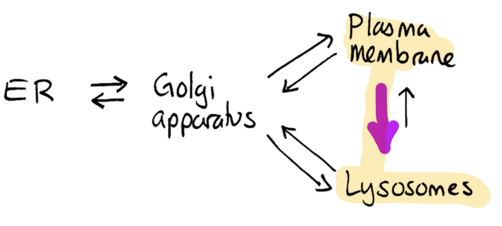
vesicles formed by RME & phagocytosis at the plasma membrane fuse with lysosomes
ex. trasferrin (iron utilization) : imported into future RBCs with RME
Mechanism:
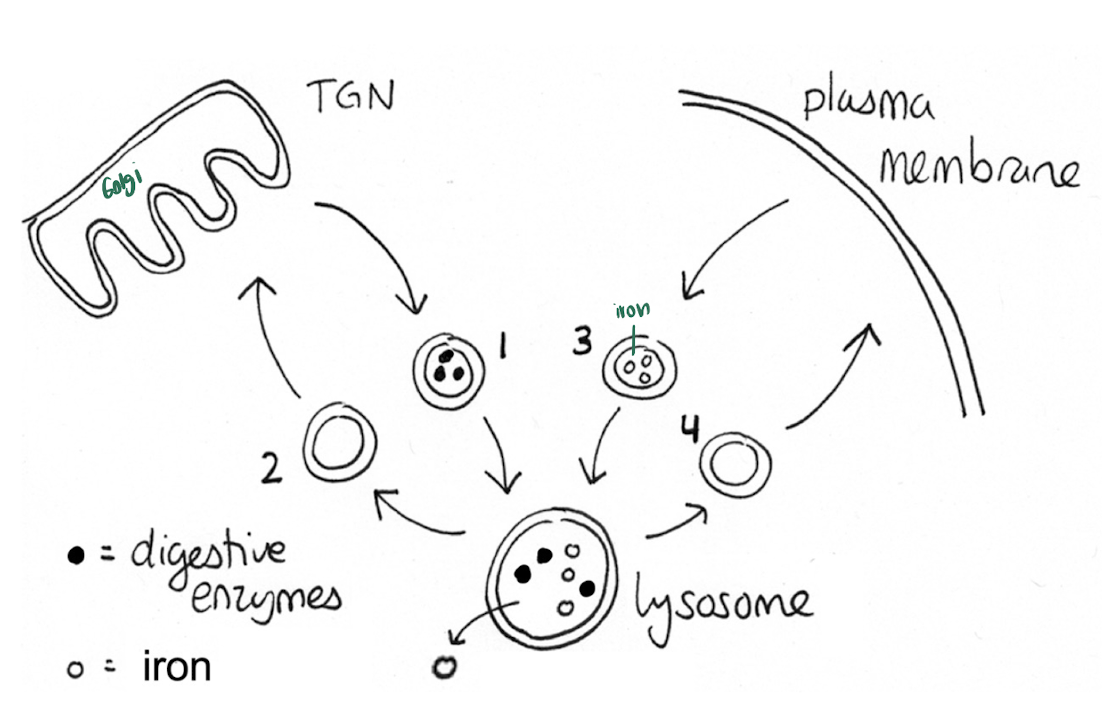
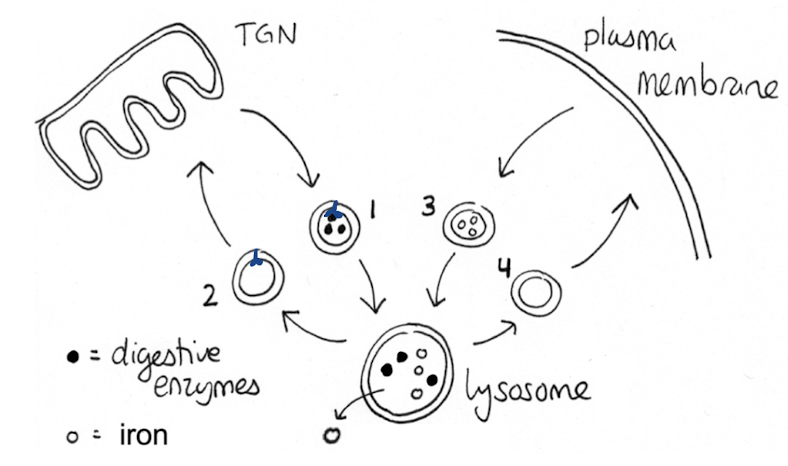
Which of these vesicle contains M6P receptors?
1 & 2
bc: M6P receptors are for digestive enzymes
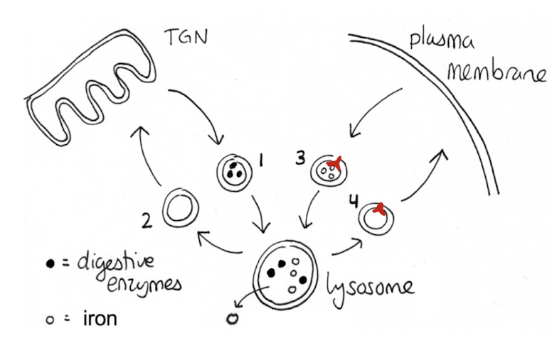
Which of these vesicle contains Transferrin receptors?
3 & 4
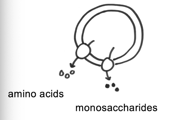
Transport of digested materials into cytosol
done w membrane transport proteins
ex. DMT1 transports iron

Examinable Content