4.Nematoda
 Call Kai
 Learn
Learn Practice Test
 Spaced Repetition
 Match
Match Flashcards
 Knowt Play
1/26

Earn XP
Name | Mastery | Learn | Test | Matching | Spaced | Call with Kai |
|---|
No analytics yet
Send a link to your students to track their progress
27 Terms
1.NEMATODA, general morphology, classification, life cycles, characteristics of orders
Nematodes = roundworms, found everywhere, many are parasites of animals/plants.
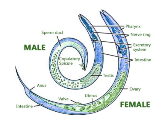
Morphology: Body is long, cylindrical, worm-like.
Body cover:
Transparent cuticle - protective, resistant to host digestive enzymes, shed during moulting. has circular striations, semi-permeable.
Layers: epicuticle, cortex, basal layer. Hypodermis under. Glycocalyx covers cuticle.
Nutrition - absorbed by body surface
Body cavity: pseudocoel - fluid-filled, high pressure, helps with movement.
Movement: only longitudinal muscles. Whip-like movement due to muscle contraction, the fluid pressure, and elasticicity of cuticle.
Systems:
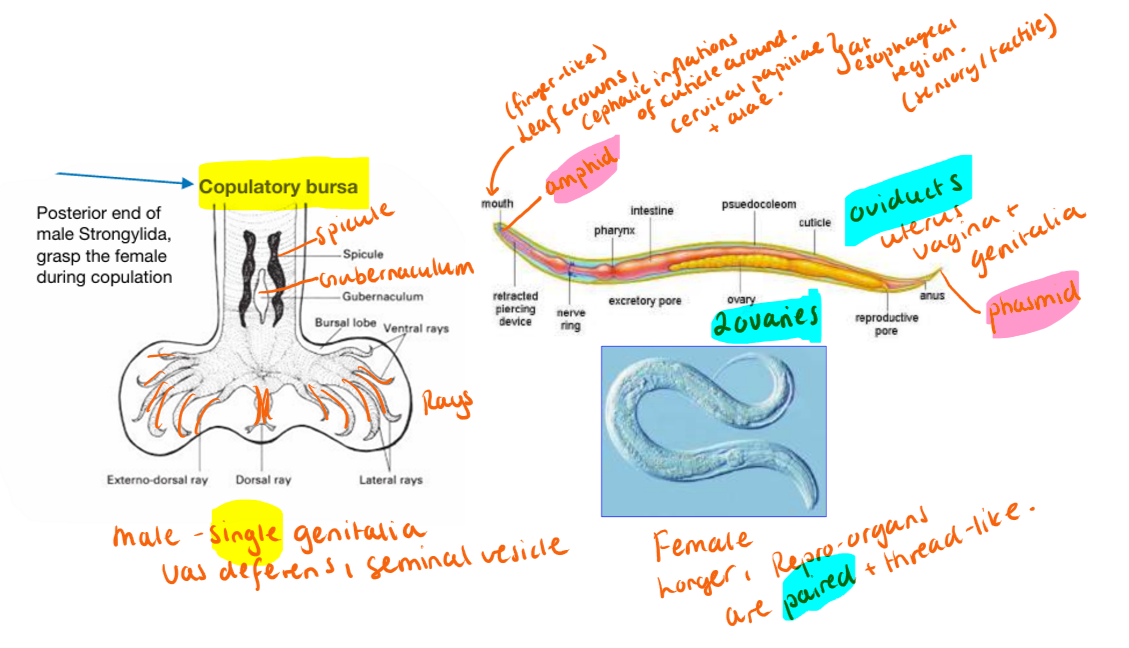
Digestive system: Complete tubular tract - mouth → buccal cavity → esophagus → intestine → anus/cloaca. Mouthparts vary, teeth can be present. Leaf-crown.
esophagus shape types:
Rhabditiform → free living larvae
filariform → infective larvae
bulb-shaped/ascaroid, muscular, glandular, trichuroid
Nervous system: nerve ring around pharynx. sensory organs - amphids, phasmids, papillae.
Respiratory/circulatory = ABSENT
Excretory - poor, lateral canals in body wall, glandular/tubular.
Reproductive system: most have separate sexes
Male: smaller, curved tail, can have spicules, gubernaculum, copulatory bursa (strongylida)
Female: larger, ovaries → oviduct → uterus → vagina. Vulva position varies. End of oviduct - spermatheca.
All reproduce sexually, except strongyloides.
Life cycle:
All nematodes develop through: Egg → L1 → L2 → L3 → L4 → (L5 - juvenile form) Adult.
Types of egg production: oviparous (unembryo), ovovivi (embryonated eggs with larva), viviparous (live larvae)
Types of LC:
Direct: No IH, development occur in environment
Indirect: needs IH, larvae develop to infective stage inside host/vector
Infection routes: Oral ingestion of eggs/larvae, skin, transplacental - hypobiotic larva via uterus/transmammary, vectors.
Classification:
1) Class secermentea (phasmidia)
(has sensory structures around esophagus)
Orders:
Rhabditida - strongyloides (small, rhabditiform esophagus, direct LC)
Strongylida - strongyloidea, ancylostomatoidea, trichostrongyloidea, metastrongyloidea (large buccal capsule, often teeth/cutting plates, male have Copulatory bursa, direct LC)
Ascaridida - ascaridoidea, heterakoidea (large thick bodies, 3 lips, direct LC)
Oxyurida (small, pinworms, posterior bulb of esophagus, long pointed tail)
spirurida (indirect LC, mouth poorly developed)
2) Class Adenophorea (Aphasmidia)
Order: Enoplida, include: trichinella, trichuris, capillaria (no phasmids)
Typical - infective stage = L1 larva!
2.Swine Ascariosis
Order: Ascaridida
Fam: Ascarididae
Genus: Ascaris, Parascaris, toxascaris
Species: Ascaris suum
Morphology: Large, white roundworm, spindle-shaped, has 3 lips around the mouth. No pharynx/buccal capsule.
Males are smaller with a curved tail. Has 2 spicules with small alae sometimes. NO bursa copulatrix. The spicules (penial setae) - what they use for sperm transfer into female!
Anterior end of many genera have cervical alae (T.cati has biggest).
Male - single reprod. female - paired reprod. organs.
Location: SI. Worldwide.
FH: Pig, human (Zoonotic)
IH: earthworms, dung beetles (can be paratenic host)
Epidem: Common parasite of pigs. resistant + survive up to 5 years. Pigs infected → poor growth - need more feed → economic loss. Sunlight + exposure to drying = destroys them.
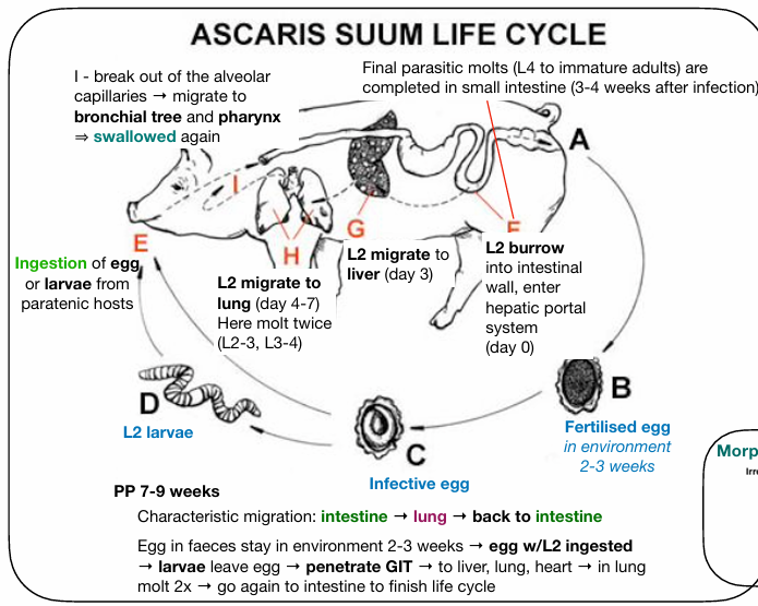
LC: Direct/indirect, has hepato-tracheal migration (liver/lung)
Eggs passed in feces → embryonates in environment (L1→L3) → pig eats eggs → hatch in intestine → migration (liver → heart → lungs) → L4 → goes up trachea, swallowed → back to SI (L5) → adults, eggs.
If paratenic host - larva stays inside (L2) - then eaten by pig
Patho/CS: usually mild/asymptomatic
Liver lesions: liver damage/intralobular destruction, white fibrotic spots = “milk spots”, sign of recent infection - if infected many times → fibrotic.
Lung lesions: Hemorrhage, edema, eosinophilic inflammation → pneumonia, cough, nasal discharge.
Intestine: atrophy of villi, poor growth, red.feed intake
In humans: migrating larva can affect liver + lungs → get resp. signs (fever, cough, wheezing, pain) or no signs. Cross infections of humans with A.suum + A.lumbricoides. Can get larva migrans visceralis (but more common in toxocara!?)
Can also affect other animals (lambs - milk spots liver, ru - pneumonia)
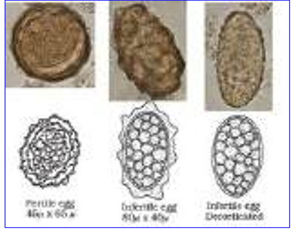
Diagnosis: fecal examination - eggs in feces (fertilized/unfertilized, decorticated eggs) - in older pigs. In young - CS (resp. signs)
Eggs: typical irregular shape, 3 layers, thickwalled, oval/round, yellow/brown.
Treatment: Levamisole, benzimidazoles (fenbendazole, albendazole), macrocyclic lactones (ivermectin)
Control: regular deworming, treat piglets after weaning, treat sows before farrowing.
Prevent - hygiene, remove feces, wash farrowing area.
3.Equine Parascariosis
Order: Ascaridida
Fam: Ascarididae
Genus: Parascaris
Species: P.equorum
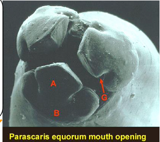
Morphology: Large - females up to 50cm, males 20cm, yellow-white, mouth with 3 lips
Location: SI
Hosts: Horse, donkey. Mainly affects foals (<1y)
Adults usually immune
Epidem: worldwide. Common in nursing and weaned foals. High exposure due to contaminated environment.
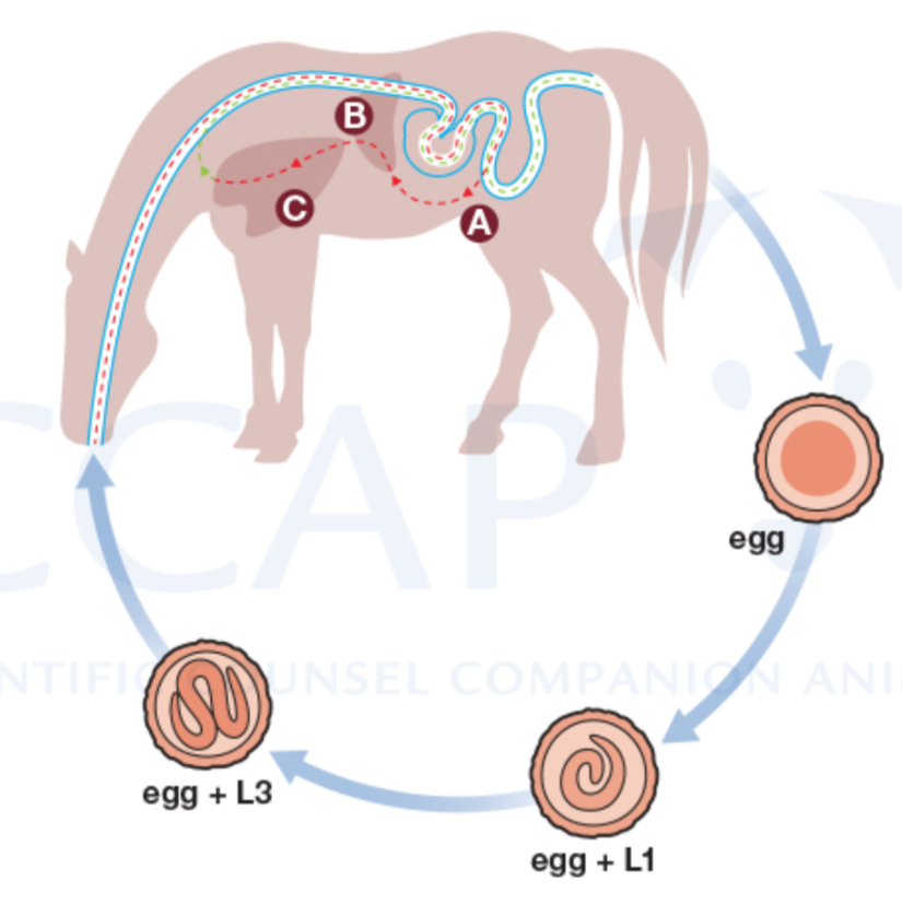
LC: Direct. With hepato-tracheal Migration.
Eggs in feces → environment, L1 → L3 (infective) → horse eats egg → larvae hatch in SI → migrates, L4 (intestine → liver → heart → lungs) → travels up trachea then swallowed → return to SI → becomes adult, eggs.
Patho/CS:
lung damage: petechial hemorrhage, inflammation
Cough, nasal discharge (grey/white), frothy mucus in trachea - in foals up to 6m age.
Chronic/heavy infection: poor growth, colic, diarrhea, lethargy, intestinal blockage/rupture (fatal)
Liver and lung lesions: scarring, nodules under pleura (raised, grey-green) with dead larvae + immune cells - more seen in older foals with multiple reinfections.
Diagnosis: CS (foals with cough + poor growth). Fecal flotation.
Treatment: Benzimidazoles, pyrantel, macrocyclic lactones.
Possible ivermectin resistance
Prevent: regular cleaning, weekly removal of feces from pasture, deworming programs in foals (foals - first time at 8w of age, then deworming following fecal examination every 3 m until 1year)
4.Roundworms of carnivores and Human Toxocarosis (LMV, LMO)
Order: Ascaridida, Fam: Ascarididae (Toxocaridae-bok for Canis/cati), Genus: Toxocara, Toxascaris
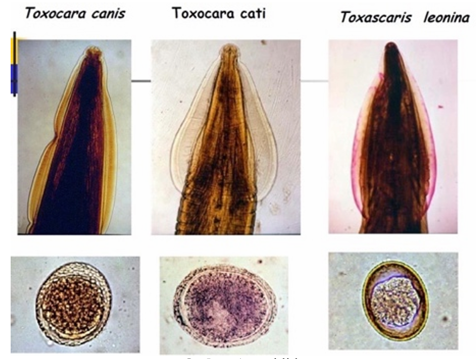
1) Main species: Toxocara canis, T.cati, Toxascaris Leonina
General Morphology: T.canis + cati are similar, except for the cervical alae.
Alae of T.canis = long, anrrow
Alae of T.cati = Short, broad.
Adults = alike ascaris, but a lot smaller. 3 lips, but has prominent cervcial alae in both sexes. Male with 2 spicules, NO bursa. Tail of male is constricted behind anus. While in toxascaris it tapers gradually.
Eggs: brown, spherical, superficial pits.
Location: SI. All - worldwide.
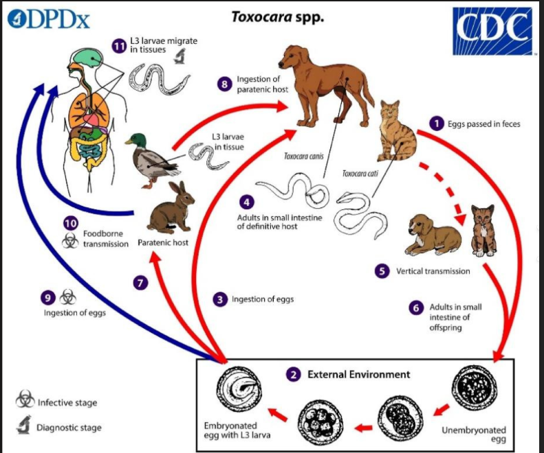
1) T.CANIS
FH: dog, man. IH: reptile, rodents can be paratenic host.
Epidem: Most young puppies and 20% adult dogs are actively infected with toxocara canis. Puppies bw. 3w and 3m of age → alot of eggs excreted → hazard to environment.
Life cycle: Unembryonated eggs shed in feces → after 4w, becomes embryonated with larvae - L3 (infective).
Has Hepato-tracheal migration. (intestine, liver, lungs) + somatic
Inside FH:
Tracheal: larvae hatch in intestine → wall → migrates into liver, lungs -bronchioles - L4 → trachea, swallowed → SI again, adults develop.
Somatic: dogs over 3 months - Migrate into tissues incl. liver, lung, kidney, brain, heart, muscle + GIT walls - development stops and it can remain dormant (hypobiotic stage)
Prenatal/transplacental infection: dormant larvae reactivate during pregnancy → puppies born infected
Transmammary infection: larvae passed in milk
Paratenic host infection: Dog eats infected rodent/bird → larvae develop directly in intestine into adult, no migration.
Patho/CS: Can be mild (asymptomatic) in dogs
Heavy infections in puppies: vomit, diarrhea, mucous defects, retarded development. Tissue damage, intestinal infection if severe. Obstruction.
Paratenic host: hemorrhagic lesions in lungs, migration cause inflammation of lungs & liver.
Diagnosis: fecal examination of eggs, sometimes larvae/adult. USG.
Treatment: Febantel, fenbendazole. Puppies should be treated every month to 6 months of age, and 4 times a year from 1-2 years old. Firstly treated 2 weeks after birth. Pregnant females can be treated during late gestation and after weaning to reduce transmission.
2) T.CATI
Only transmammary infection, eating eggs + paratenic host. NO Transplacental.
Host: Cats, wild felids. Paratenic host: earthworm, coackroaches, chicken
CS: Kittens get same as dogs - Pot belly, vomit, diarrhea, cough, poor coat, weight loss (resp. issues/GIT). Often asymptomatic in adults. Common in older cats also.
Treatment: Benzimidazoles. Deworm kittens beginning at 3w. monthly from 6m. 2x a year for adults.
3) TOXASCARIS LEONINA
LC: animal eats infective egg/paratenic host → larvae stays in intestine → develops in intestinal wall → return to lumen → adults. NO liver-lung migration!
No migration in FH. Ascaridioid type.
Host: dog/cat. Humans can be accidental host.
Patho/CS: low, but in heavy infections → enteritis
Dg: fecal examination. Treat with benzimidazoles.
4) HUMAN TOXOCAROSIS
By mainly T.canis/Cati
Human eats infective eggs from such as soil, dirty hands, veggies, cotnact with pets.
Humans = accidental hosts. Larvae do NOT become adults!
Types of disease:
Visceral larva Migrans (VLM) - larvae migrates through organs, liver, lungs, heart → fever, eosinophilia, enlarged liver/spleen, myocarditis. Common in children - esp. history of eating dirt!
Ocular larva Migrans (OLM) - larvae goes to eye → retinal inflammation, vision loss, red painful, strabismus. Often one eye affected.
Dg: ELISA, PCR, Western blot
Treatment: Alben/Mebendazole, anti-inflammatory drugs
5.Calf Toxocarosis
Order: Ascaridida, fam: Ascarididae (Toxocaridae - in book).
Genus: Toxocara
Species: Toxocara vitulorum
Hosts: Mainly calves of cattle and buffaloes, especially water buffaloes.
Morphology: Same as other toxocara, 3 lips, cervical alae - long & narrow. Body flexes ventrally. Tail of male - spike-like appendage.
Location: Small intestine.
geo: Common in tropical regions; Rare in Central Europe.
Epidem: Infects - all calves in endemic areas with bad control measures.
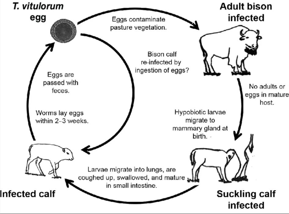
infected through contaminated food/water. Or through transmammary (from mother, after larvae develop directly in calf intestine (NO migration in this case).
Larval migration through tissues - migration via blood → organs/tissues (Somatic route)
Life cycle: Toxocaroid type (same as t.canis, t.cati). Direct.
Larvae undergo hepato-tracheal migration in calves younger than 6months.
Calves older, can be infected by eating eggs, but this infection rarely results in becoming detectable (meaning that they DO NOT develop into adults). In these older calves → somatic migration → larvae dormant in tissues, mainly liver, lungs. → infection then after in calves, when they get pregnant.
Complete EHP (intestine, liver, lung) migration of larvae in host.
Patho/CS:
Main effects are caused by adult worms in intestines of calf under 6m age.
Severe infection: cough, catarrhal enteritis, diarrhea, colic
Intestinal obstruction + perforation (peritonitis + death)
Migrating larvae - can damage many organs in adult cattle - esp. lungs → infection by sec. bacteria, pneumonia
Characteristic acetone smell is present in breath/urine + Meat of calves
Diagnosis: Fecal examination - eggs
but pregnant calves without eggs can still be infected with dormant larvae in tissues → passes to offspring. In some countries → ELISA. or detecting the acetone odor!
Treatment & Control: check pastures. Anthelminthics - (benzimidazoles) albendazole, levamisole, ivermectin. Disinfection, manure removal.
Untreated heavy infections can lead to high mortality in calves.
6.Ascaridiosis and Heterakidosis of Birds
1) Ascaridiosis
Fam: Ascaridiidae, by superfam: Heterokoidea
Genus: Ascaridia
Main species: A.galli (chicken mainly), A.columbae (pigeons), A.dissimilis (turkey)
Morphology: largest nematode of GIT in poultry, up to 12cm long, yellow-whitish color, transparent. Females are longer. Males have preanal sucker + 2 spicules (equal). Narrow caudal alae.
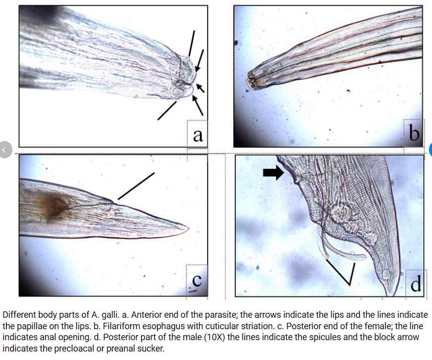
Eggs: Oval symmetrical, 3 thick and smooth shells, non-embryo - one germ cell, dark gray.
Location: SI (esp. in chicken), may reach cloaca and oviduct
FH: chicken, turkey, pigeon, peacock, pheasant
Earthworms can be paratenic host.
LC: Direct. Eggs in feces → embryonates in environment (L1→L3) → birds get infected by eating the eggs or eating infected eartworms → larvae develop in intestinal wall (encyst in SI) → invade mucosa, L4 → return to lumen → L5, adult. PP: until infection - 30-50d.
Ascaridioid type. No migration! (stuck to mucosa, then reenters into lumen → adult).
Patho/CS: typ. intestinal issues!
Destruction of lieberkuhn glands (epithelial, tissue damage)
Inflammation (enteritis) + obstruction
Young - more affected. Reduced food intake, not gaining weight, anemia.
Immature + adult worms → may go up to stomach, claoca, oviduct, liver..
When infection is over, poultry is immune!
Diagnosis: Fecal examination - inspection at necropsy
Treatment: Piperazine, levamizole. Keep litter dry, keep changing litter, never feed directly from the litter.
2) Heterakidiosis
Fam: heterakidae, Genus: heterakis
species: H.gallinarum, H.dispar (water poultry)
Morphology: Caudal alae + precloacal sucker in males (sometimes described as having small-bursa like structure), 2 unequally long spicules. Adult heterakis are small, very thin, whitish. Esophagus with large posterior bulb (in all pinworms).
eggs: ovoid, thick, smooth shell. Similar to A.galli, but smaller.
Location: Cecum
FH: chicken. Paratenic host = Earthworms
Geo: Worldwide. Seen mostly as poultry parasites.
LC: same as ascarida. Direct. Vectors of h.meleagridis (protozoan).
Patho/CS: digestive disorder
inflammation of cecum, hemorrhage, nodules in mucosa
not eating, diarrhea, weak, not growing, reduced egg prod.
Dg: eggs in feces, necropsy
Therapy: Flubendazole, levamizole
7.Anisakidosis
Fam: Anisakidae, Genus: Anisakis (Of order ascaridida)
Main species: Anisakis simplex
Morphology: General ascaridoidea - wormlike, pseudocoel, complete dg. system, cuticle, longitudinal muscles. 3 Lips with labial paillae. Males - simple spicules to hold female.
Hosts: Marine mammals are the definitive hosts; humans are accidental hosts (zoonosis)
IH: 1st: Crustaceans (ex. lobster, shrimp). Paratenic host: marine fish (salmon/tuna), squid (no development in this host)
Location: stomach, SI
Geo: Worldwide, especially where raw or undercooked fish is commonly eaten.
Epidem: The occurence of the parasite is increasing due to more people eating sushi, other raw seafood dishes. More common in cheaper marine fish species.
Life Cycle: Anisakoid type of migration (GIT). Indirect.
Unembryonated eggs released in ocean from feces → L3 hatch and becomes free-living larvae → Larvae eaten by IH host/paratenic host → Humans can become accidental infected by eating fish (paratenic) with L3.
FH - like dolphins, whales → becomes infected when eating infective IH. → L3 settles in the stomach/intestine and matures - L4-L5-adult.
(rare) - Human-consumed live larvae can penetrate into stomach wall, then into internal organs. But mostly it stays in stomach, no development.
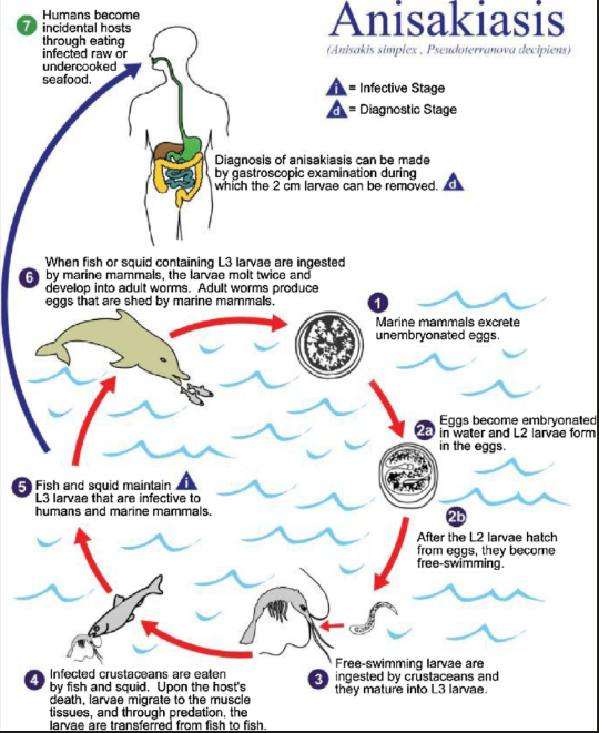
Patho/CS: Many are asymptomatic, but larvae that gets to stomach or intesitnal wall can cause: Abd. pain, nausea, vomiting, if wandering → mimic other diseases.
allergic reactions - gastroenteritis, skin issues (allergy → can be severe - humans due to larvae)
Can be gastric (pain, fever, nausea), intestinal (colicky pain), extraGIT (LN, spleen, pancreas)
Diagnosis: Endoscopy (often directly visualizes larvae), Serology, PM
Transillumination of fillets - or screening.
Examine immediately or store in fridge, clean tissue, place in transparent plastic bag, compression, thickness of tissue after should be 1-2mm.
Diagnosis in humans is difficult because symptoms are nonspecific.
Treatment: Removing larvae by endoscopy/bopsy forceps, usually no effective drug treatment - albendazole can be tried. Avoid raw/undercooked fish. COOK/FREEZE.
8.Oxyurosis of horses
Order: Oxyurida
Family: Oxyuridae
Species: Oxyuris equi (pinworm)
Morphology:
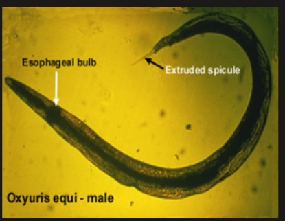
Females: slender with a sharp-pointed posterior end; 5–15 cm long
Males: smaller, about 1 cm; have a single pin-shaped spicule
Large esophageal bulb; anterior cuticle is swollen. The 3 lips are small/not easily visible in many.
Eggs: oval, with a mucoid cap (single operculum)
Location: Large intestine
FH: Horses and donkeys
Geo: Worldwide
Life Cycle: Direct.
Larvae L3 hatch in SI → LI → enters mucosal crypts of cecum and colon→ molts to L4 → after fertilization, gravid females goes to anus (adult) → emerges head-first and lays eggs in clumps, in a yellow-grey gelatinous material on perineal skin. Develops to L3 while eggs are attached to skin!
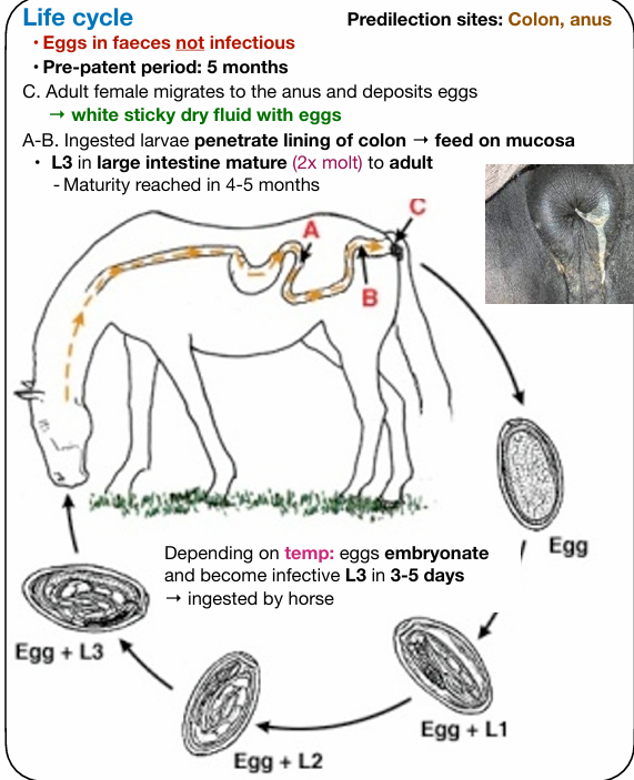
Patho/CS:
Larval stage (L4): Causes mild mucosal erosions while feeding on intestinal mucosa; typically little clinical effect.
Adult stage - lives freely in lumen, feeds on gut contents.
Main patho effect from egg-laying behavior of females.
Females through anus → perineal irritiation, itching (pruritus).
horses may show tail rubbing, broken tail hairs, “rat tail”.
Diagnosis: CS (tail rubbing, anal irritation). Perianal swab + microscopic exam for eggs.
little or no immunity develops - reinfection is common
Treatment: Anthelmintics - ivermectin, moxidectin, febantel.
Hygiene - wash perineal area with soap and water to remove eggs, regular cleaning of stalls and bedding to prevent reinfection.
9.Other oxyuroids of mammals (Passalurus, Skrjabinema, Enterobius, Aspiculuris, Syphacia)
Order: Oxyurida
General features:
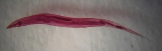
Pinworms, small nematodes in mainly LI. Females = long pointed tail.
Direct LC, infection occur by eating eggs - the eggs often become infective quickly.
in some - females lay eggs around perianal region → irritation
In enterobius vermicularis, syphacia obvelata, skrjabinema ovis. While in aspiculuris tetraptera & passalurus ambiguus = eggs passed in feces is more common.
Reinfection + environmental contamination = common
Diagnosis: detecting eggs in feces/adults in LI (PM).
Treatment by single dose of piperazine + benzimidazoles (passulurus). Also piperazine for lab rodent pinworms.
Typical LC:
Adults live in LI → female migrate to anus/perineum → eggs layed around → eggs embryonate → L3 develops → falls down, host eats eggs → larvae develop in intestine → adults.
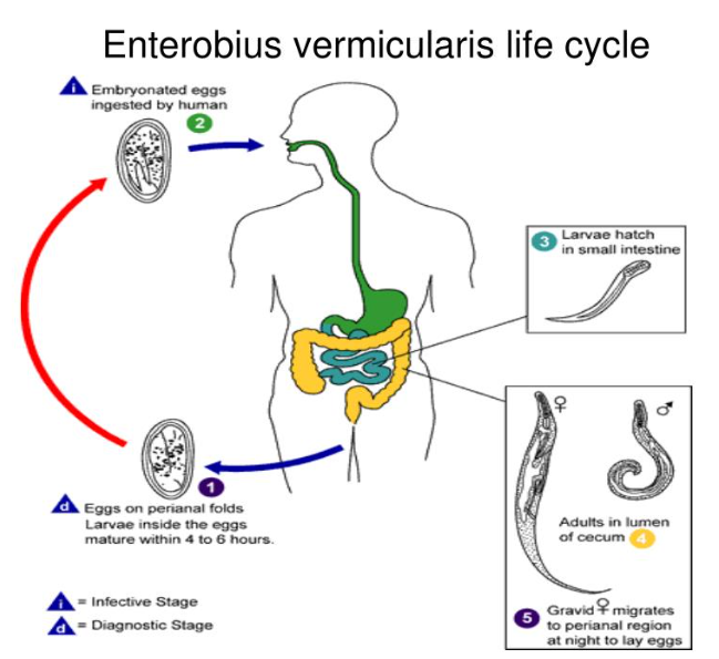
Main patho: Usually not intestinal damage, but more of irritation caused by the eggs, itching, self-trauma, contamination of environment.
Heavy infection: enteritis, weight loss, mucosal inflammation
Differences:
1) Passalurus - P.ambiguus:
Males has unpaired spike, tail with whip-like protrusion. Female tail tapers into a point. Cuticle has circular grooves. Esophagus has prebulbus + strong bulbus. Eggs - M/L, asymmetrical, pig + non-embryonated.
FH: rabbits. Cause usually mild infection. heavy - appendicitis, LN inflammation. Larval stages can cause necrotic inflammation, diarrhea, weight loss.
2) Skrjabinema:
S.Ovis (sheep) & S.caprae (goat)
both of minimal importance, cause mild irritation. Small eggs.
3) Enterobius - E.vermicularis:
Humans, very common in children (eating snow). Worldwide.
More a mental condition than a medical one - eggs can be found everywhere (bedding, clothes, stuffed animals) → sleep issues, decr. appetite, weight loss, irritation, vaginal discomfort. Cause strong perianal itching.
dg: Scotch tape technique - early morning around anus → microscope with acetone. Or by flashlight on anus. Treat whole family - mebendazole. Everything washed!
4) Pinworm of lab rodents:
Aspiculuris tetraptera (mice)
Syphacia obvelata (mice) & S.muris (rodents)
Cause slowed growth (moderate infection), or cattharal enteritis (massive).
10.RHABDITIDA, Strongyloides: Strongyloidosis of mammals.
Order: Rhabditida of class Secernentea (Phasmida)
Family: Strongyloididae (superfam: Rhabditoidea)
Genus: Strongyloides (strongyloidosis)
Species | Main Host |
|---|---|
Strongyloides ransomi | Pigs |
Strongyloides papillosus | Ruminants, rabbits |
Strongyloides westeri | Horses, donkeys |
Strongyloides stercoralis | Dogs, humans, cats, monkeys |
Strongyloides ratti | Rats |
Morphology: Very small, thin nematodes, small lips. Male spicules of equal length. Characteristics Rhabditoid esophagus with prebulbus, and bulb with valvuar apparatus.
Location: SI
Geo: Worldwide, esp. tropical/subtrop. climates
Epizoo/Transmission: S.stercolaris = Zoonotic.
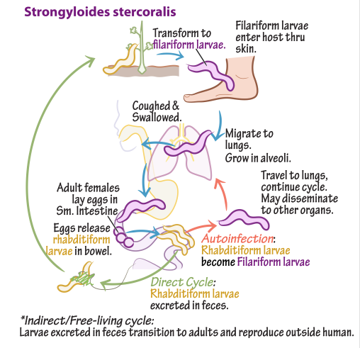
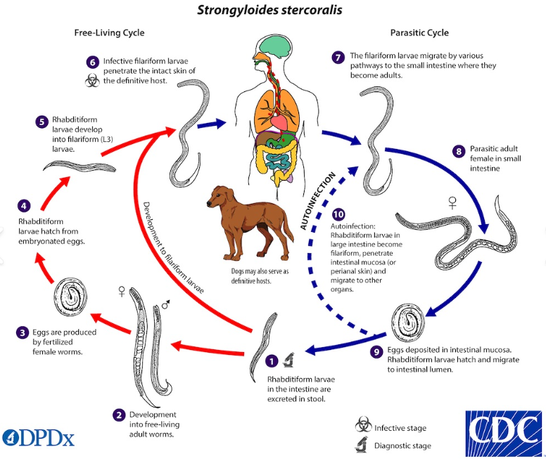
By skin penetration - infective L3 → skin, enters blood → migrate to lungs → trachea → swallowed → SI. This is the “Tracheal-Migration route”. Can also go to muscle in older host → dormant (somatic cycle).
Oral infection - ingestion of larvae via food/water/pasture/milk (lactogenic)
Lactogenic/colostral - dormant larvae in mother activates → goes to mammary gl.
Autoinfection - mainly with S.stercoralis. L3 inside intestine, then goes to mucosa, perianal skin → cause self-infection.
Infection often occurs in young. Thrives in warm, moist and dirty environment.
Life cycle: Direct. Only females are parasitic. Alternate bw. parasitic and free-living cycle. Asexual reprod. in FH → free L1 or L1 inside egg.
Homogonic cycle: Eggs/larvae develop directly into infective L3 (filariform) → reinfect through skin/eating → migrate through bv, lung, trachea, SI.
Heterogonic cycle: Larvae become free-living males and females in environment before producing infective larvae
Patho/CS:
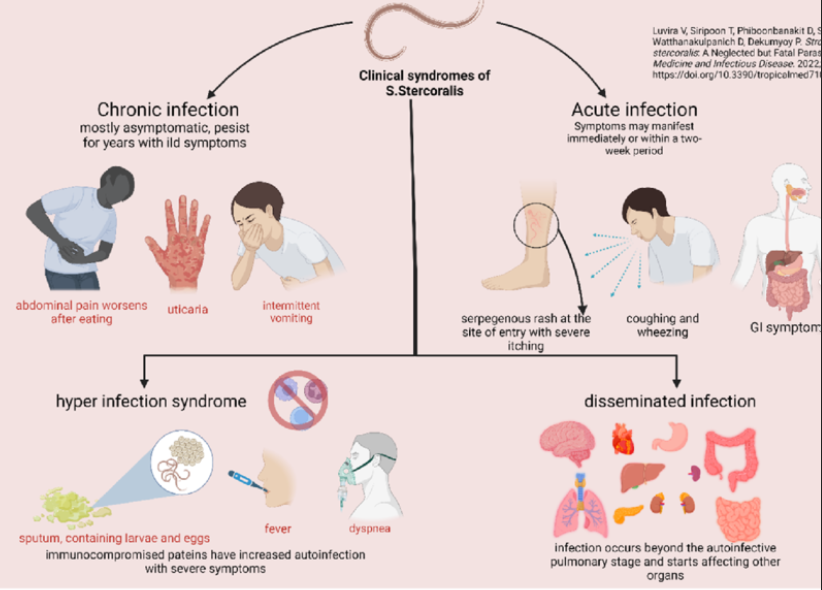
Many infection are mild, asymptomatic.
disease is more severe in young, immunosuppressed.
Humans (S.Stercolaris) - GI signs, diarrhea, abd.pain. Pulmonary signs - during larval lung migration → cough, resp. irritation. Skin signs - urticarial rash, perianal irritation.
Severe: septicemia, shock, neurologic/pulmonary complication - can be fatal.
Eosinophilia - common finding
Pigs (S.ransomi) - severe in piglets, bloody diarrhea, weight loss, anemia, dehydration. high mortality in heavy infection.
Dogs: usually mild - diarrhea, poor appetite. Puppies - severe dehydration, weak, fatal.
Cats: usually asymptomatic. but can get chronic diarrhea, white nodules in colon.
Diagnosis: fecal exam - eggs/larvae, larvoscopic method - fresh fecal smear may reveal motile larvae - important as strongyloides often pass larvae instead of typical eggs.
Treatment: thiabendazole-most common, fenbendazole, ivermectin - off label.
Control - larvae are sensitive to cold, dryness. Prevent - clean, dry housing, fecal hygiene, treat young during nursing.
11.Equine Strongylosis (Strongylus, Cyathostoma)
Order: Strongylida, under class secernentea (phasmidia)
This order - bursate nematodes. Incl. some of most common pathogenic GI or resp. parasite. Has copulatory bursa in adult males, general shape + eggs - very similar.
Superfam: Strongyloidea
Main families:
Strongylidae - Subfam: Strongylinae (large), cyathostominae (small)
Chabertiidae
syngamidae
1) Strongylidae
General morphology: Well developed buccal capsule, median thickening in capsule - dorsal gutter. Has external + internal leaf crown or “corona radiata”. + teeth in buccal cavity. Named bloodworms - eats blood → red colored worms in feces. All infect via direct LC (pasture ingestion of L3).
Genus: Strongylus
Main species:
S.vulgaris (smallest, most pathogenic, intestinal wall → arteries → intestine)
S.edentatus (Intestine → liver → intestine)
S.equinus (intestine → liver (less defined wandering) → intestine)
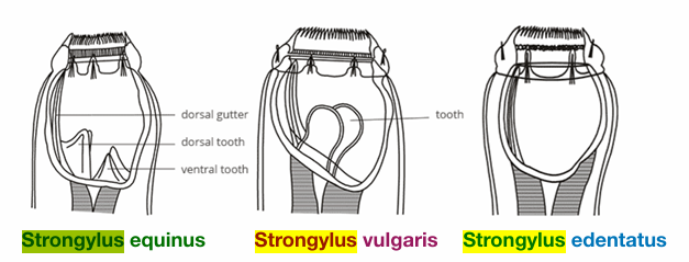
These together with triodontophorus = large strongyles.
Location: LI - cecum & colon. Worldwide.
Host: horse, donkeys
Life cycle:
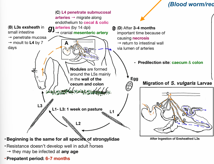
Patho/CS:larval migration → damages blood vessels → inflammation, obstruction → intestine affected → colic, diarrhea, poor condition.
Diagnosis: fecal flotation & necropsy
Treatment: moxidectin - best.
Control - clean, not mixing young foals with older horses, rotation on pastures, regular treatment programs, dewormer types changes.
2) Genus: Triodontophorus
Colon, does NOT migrate. Contribute to overall worm issue in mixed strongyle infection, cause mild/moderate intestinal damage.
3) Small strongyles (Cyathostomes), under subfam: cyathostominae
genus: Cyathostomum (insigne), cylicocyclus, coronocyclus
Life cycle: L3 eaten from pasture → into intestinal wall → encyst in mucosa of cecum, colon, forms nodules → L4 inside nodules → into large intestine - L5 early → adult. Only migrates locally! does not go through systemic.
Patho/CS: range from poor performance, to severe enteritis, diarrhea, weight loss
12. Parasites of large intestine of ruminants and pigs (Chabertia, Oesophagostomum)
Subfam: Cyathostominae (small strongyles)
Fam: Chabertiidae (under order: strongylida)
Subfam: Chabertinae, oesophagostominae
GI worms of ru & pigs.
Both treated by pyrantel (ru), benzimidazoles, ivermectin. Diagnose by fecal flotation, PM.
1) Chabertia ovina (Chabertiosis)
Morphology: End is ventrally curved, large buccal capsule, oral aperture - surrounded by double row of small cuticular elements = Leaf cowns. There is a shallow ventral groove + cephalic vesicle. Male bursa is well developed with gubernaculum.
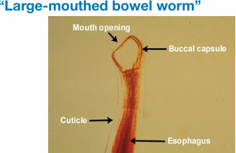
Location: Colon/LI for adults, larval in SI + cecum, host: sheep
Life cycle: eggs in feces → Free-living L1→L3 → eated by FH → SI/cecum → L5 + adult in LI → draw intestinal mucosa into oral capsule.
Patho/CS: catarrhal nodular enteritis colitis - column wall is edematous, bloodied, thickened with hemorrhage.
diarrhea with blood, anemia, weight loss
2) Oesophagostomosis
Main species: oesophagostomum columbianum (green diarrhea), O.venulosum, O.radiatum, O.dentatum (pig)
Morphology: Cylindrical buccal capsule, cervical groove, cephalical vesicle + papillae. General called as nodular worms.
Location: Adults in LI, colon. Larvae from pyloric - rectum.
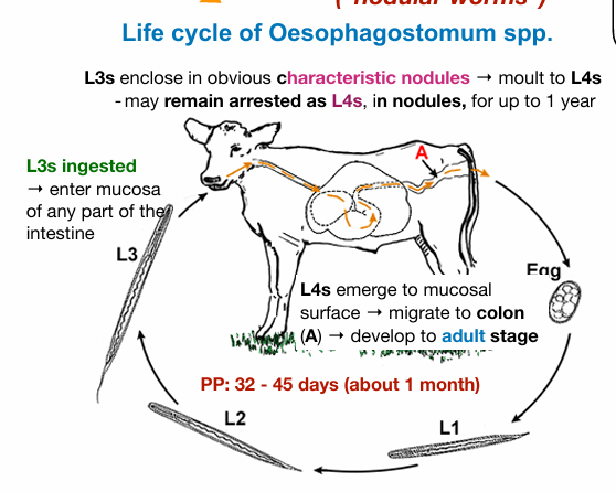
Patho/CS: Severe enteritis, local inflammation (encapsulation), stops normal intestinal motility + absorption, thickening of wall, mucus production. (all have nodules, except venulosum). Pot belly.
PP - can be a bit longer for pigs (43-55d)
LC: Direct, eggs by FH → environment, embryonates → L1 rhabditiform → L3 (filariform) → FH infected → enters mucosa of small/large intestine - cyst → L4 → migrate back to LI → adults, forms nodules in cecum.
13.Amidostomosis and Trichostrongylosis of poultry
Superfam: Trichostrongyloidea, fam: Amidostomatidae & Trichostrongylidae
The superfamily = biggest among bursate nematodes. Has copulatory bursa, lacks corona radiata. Also lacks the buccal cavity, lips reduced.
Life cycle - direct. Egg in feces → L3 eaten.
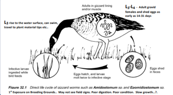
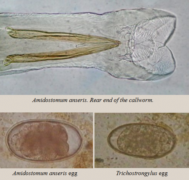
Amidostomosis - Amidostomum anseris
Morphology: adult worm is slim and red. Male spicules are the same. Large eggs.
Location: Stomach of aquatic birds. Under the keratinin layer, in proventricule or in esophagus.
Patho/CS: loss of appetite, exhaustion, weak, anemia, diarrhea. (general intestine)
Worms are very pathogenic to young, while in older - becomes carriers.
severe inflammation, hemorrhage, necrosis. Extreme blood loss can occur in heavily infected birds.
Dg: fecal examination of eggs, necropsy
Treatment: benzimidazoles, ivermectin
Bird Tricostrongylosis - Trichostrongylus Tenuis
Location: SI of galliformes & aquatic birds
Patho/CS: similar
14.Ancylostomatidosis of carnivores and ruminants
Superfam: Ancylostomatidae
subfam: ancylosomatinae (ancylostoma, globocephalus, uncinaria)
General: in small intestine, have teeth or cutting plates, attach to mucosa → blood loss/tissue feeding. Infection via skin or oral ingestion.
LC: Direct, eats L3/skin → lungs → trachea → swallowed → SI
Diagnosis: Fecal exam, History, CS
+PCR - uncinaria
1) Genus: Ancylostoma - in dogs/cats
Species: Ancylostoma caninum, A.braziliense, A.tubaeforme (cat)
morphology: medium sized, head is bent, large buccal capsule with teeth. Bursa copulatrix with spicules, gubernaculum.
Paratenic host: Rodents. Accidental host: Humans (zoonotic)
Epizoo/Transmission: per skin, oral, lactogenic, paratenic host.
Migration - trachea, somatic, transmammary, transplacental (but not really proven acc. to alicia)
Life cycle:
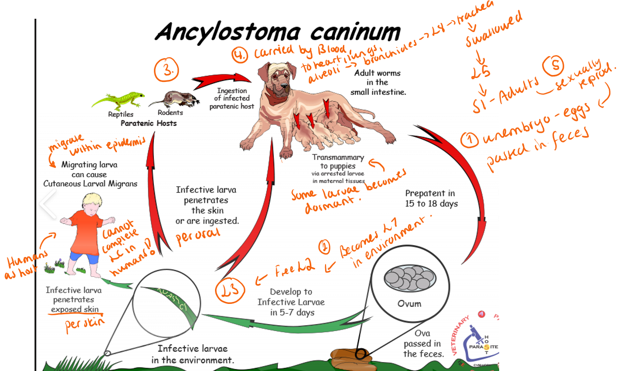
Patho/CS: suck microvilli of SI, eats mucosa - blood suckers. → use the blood for O2, does not feed on it.
Larva migrans cutaneous = humans.
Caused by both A.caninum & brasiliensis. Wanders in subcutaneous tissue → inflammation, painful swelling.
Most CS are due to adults + L4 taking blood from intestines.
In young puppies = blood loss, anemia, black tarry diarrhea
Anorexia + depression from blood loss/low O2
Treatment: ivermectin, milbemycin oxime (toxic for cats).
Supportive + transfusion if anemic.
2) Genus: Uncinaria
Main species: U.stenocephala
Large-funnel-shaped oral capsule with 2 cutting plates.
In SI, dogs. North america.
Small mammals can be paratenic host.
Patho/CS: mild, can cause enteropathy, diarrhea, poor growth.
Treatment: Benzimidazoles, macrocyclic lactones
3) Ancylostomatidosis of RU (Necator, Brunostomum)
subfam: bunostominae (bunostomum, necator)
A) Genus - bunostomum.
B.trigoncephalum (lamb), B.phlebotomum (calves)
large funnel-shaped oral capsule. Large buccal cavities with teeth/cutting plate. Most are blood suckers.
Larva - likes moist places, grass.
Patho/CS: anemia, hemorrhage, enteritis, intermandibular edema, bloody diarrhea. Not blood feeders! Older - asymptomatic.
Treatment: benzimidazoles & macrocylic lactones.
B) Necator Americanus
Small, bent head, mouth with teeth/plates. FH = humans!
Skin penetration + sec. bacterial infection → “ground itch”.
Pulmonary phase = asymptomatic.
blood feeders, anemia, iron deficiency, malnutrition, weak, fatigue.
Treatment by mebendazole.
15.Trichostrongylosis of Ruminants and Lagomorphs
Superfam: Trichostrongylidea - largest, has copulatory bursa, buccal cavity is reduced/absent, generally small, slender, delicate in looks. L3 = free-living.
Fam: trichostrongylidae
General: Very small, thin intestinal worms, live in abomasum or SI. Direct LC.
Eggs → hatch in environment → L1→L3 on grass. Infection by eating L3. NO systemic tissue migration in host. Local movement can occur. Some undergo hypobiosis (lays dormant) - esp. ostertagia, haemonchus, nematodirus.
Mainly cause: Gatroenteritis (Diarrhea, weight loss)
Diagnosis: combo of CS, necropsy, eggs in feces, fecal cultures to identify L3. Trichostrongylus eggs are not distinguishable from other in its family.
Genus - trichostrongylus, Haemonchus, nematodirus, cooperia, ostertagia
1) Abomasal strongyles
a) Haemonchus - blood suckers, red color in feces. Uses lancet in their buccal. Female has white ovaries around red blood filled intestine. Cop. bursa is species - has lobe with Y-shaped dorsal ray.
Haemonchus contortus (sheep/goat)
Haemonchus placei (cattle)
b) Ostertagia ostertagi (small ru) - small, reddish brown. In warm areas.
Both cause severe disease, death.
Haemonchus = blood sucking → severe anemia, ascites, bottle jaw
Ostertagia → form nodules in mucosa, gastric glands damage → diarrhea, weight loss, not eating. 2 types - summer + winter (higher lethargy)
Treatment: mebendazole, resistance in small ru (haemonchus), benzemidazoles & macrocyclic lactones (ostertagia)
2) Small intestinal strongyles
Trichostrongylus - T.axei (also abomasum), T.columbriformis
Cooperia - C.curticei (small ru), C.punctata (cattle)
Nematodirus - N.helvetianus (cattle), N.spathiger (small ru) - in necropsy, a lot is seen as cotton wool. Typ. inflated cuticle at one end.
Lives in Small intestine - mostly cause mild to moderate enteritis.
Trichostrongylus - role is to usually help in gastroenteritis, where ostertagia or haemonchus are the primary pathogens in ru. T.Axei - can cross infect bw. horse & ru. General inflammation, but severe - erosion of epithelium. Diarrhea = black scours - watery/darkgreen-black in severe.
Nematodirus - also role in gastroenteritis, as an additive effect in mixed infections! → dehydration, diarrhea.
Cooperia - mild, contribute secondary effect to primary.
Treatment: benzemidazoles (albendazole), macrocyclic lactones (ivermectin)
3) Rabbits & Hares
Trichostrongylus retortaeformis
Graphidium strigosum
Obeliscoices cuniculi (hares)
Mostly affects stomach or SI. Can cause hemorrhagic gastritis, anemia, diarrhea, weight loss. Often mild unless heavy infection. Feeds on blood (sometimes also T.retortaeformis).
Treat by fenbendazole, ivermectin in rabbits.
16.Dictyocauluosis of sheep, cattle and protostrongylidosis of small ruminants
General for all: Adults are in lungs (trachea, bronchi), similar patho - bronchitis, inflammation of airways, resp. issues/cough, Similar Dg - baermann/larvoscopic method, similar treatment - benzimidazole (albendazole), macrocyclic lactones (ivermectin).
Order:Strongylida
1) Dictyocauluosis
Superfam: trichostrongyloidea
Fam: dictyocaulidae
Genus: Dictyocaulus
Species: Dictyocaulus Filaria (sheep/goat)
Host: Sheep/goat. Mainly lambs. Adults usually mildly affected.
Morphology: Similar to Dictyocaulus viviparus of cattle. Small cuticular knob at end, tail blunt, larvae contain dark brown/black food granules except head and tail.
Location: Bronchi, trachea
Usually less pathogenic than cattle lungworm.
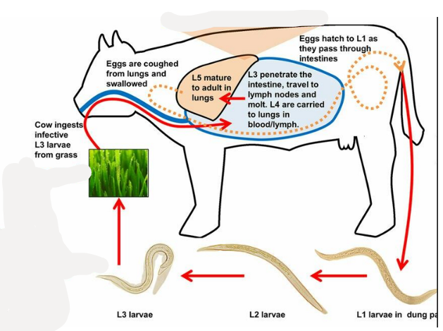
LC - direct!
2) Protostrongylidosis of small Ru
Superfam: metastrongyloidea (Mostly lungworms, Indirect LC)
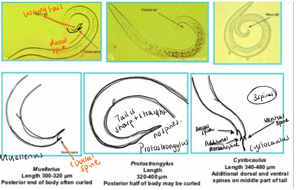
Fam: Protostrongylidae - genus protostrongylus, cystocaulus, muellerius
Species: Protostrongylus rufescens (sheep/goat), P.brevispiculum (sheep), Muellerius capillaris, Cystocaulus ocreatus
Morphology of superfam: rudimentary buccal cavity, 6 lips, thin spicules, reduced/absent copulatory bursa.
Host: Small ru, IH: snails
Location: Small bronchi and bronchioles, lung parenchyma & pleura (cystocaulus)
Life cycle: Similar to dictyocaulus, Larvae - L4 in LN, but with IH (indirect).
Patho/CS: Affects lungs
Inflammation of bronchioles & lung tissue
nodules, lobular pneumonia, pleuritis
sometimes asymptomatic
M.capillaris → 6m of age, cause interstitial pneumonia, small nodules, calcification of tissue, hyperemia.
signs: poor condition, nonspecific resp. signs, sec. infections
Diagnosis: Larvoscopic methods (L1) or PM
Treatment: Benzimidazoles, macrocyclic lactones
3) Lung strongylidosis of cattle (Dictyocaulus viviparus)
Superfam: trichostrongyloidea
Fam: Dictyocaulidae
Host: Cattle, Paratenic host: Pilobus fungi. Location: trachea & bronchi
Morphology: Gray color, larvae contain fat/protein granules, rounded head, oval blunt tail.
Life cycle: same as dictyocaulus filaria.
L3 can migrate onto pilobolus fungus (paratenic host) → fungus explodes and spread larvae on pasture.
Patho/CS:
Bronchial epithelial damage, bronchitis, inflammation - even with only a small number of worms
signs: resp. signs - cough, dyspnea/intensive breathing, growth retardation, poor performance
Diagnosis: Baermann larvoscopic method (L1)
Treatment: Macrocyclic lactones, levamisole, benzimidazoles
Prevention: use clean pasture, avoid overcrowding, move calves to clean pasture after 12w, pasture previously mown is preferred.
17.Metastrongylosis and stephanurosis of Suis
1) Metastrongylosis
Superfam: Metastrongyloidea
fam: metastrongylidae
Genus: metastrongylus
Main species: M.pudendotectus, M.apri (most prevalent), M.salmi, M.confusus
Morphology: Thin, thread-like, white, 6 lips, rudimentary buccal cavity, thin spicules, reduced/absent cop. bursa.
FH: pigs, IH: Earthworm. Location: Lungs (bronchi/bronchioles)
Life cycle: Indirect. Similar to other metastrongylid lungworms.
Eggs in feces → earthworm eat larvae → develops to L3 → pig eats earthworm → larvae migrate - intestine → lymphatics/blood → lungs → adults in bronchi/bronchioles.
Patho/CS: migrating larvae cause mechanical damage in intestinal mucosa, LN, bv, pulmonary alveoli, bronchial walls, bronchioles
Bronchitis, cough, severe vesicular breathing (resp.)
non-specific - not eating, weight loss, anemia, poor immunity, delayed hair change.
Severe - accelerated resp. syndrome
Dg: fecal flotation, PM
Treatment: Benzimidazoles (albendazole ex.)
2) Stephanurosis
Fam: Syngamidae
Species: Stephanurus dentatus
Morphology: 25mm male, 40mm female
FH: pigs, paratenic host: earthworms
Location: around kindeys, in cysts near kidneys/ureters
Epizoo: common in warm, moist tropical areas. Infection by skin, ingestion and paratenic host.
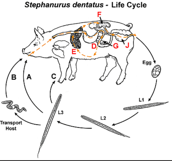
LC: unembryo eggs passed in urine → L1→ L3 → can go by skin, eaten or through earthworm → then FH → intestine → liver via blood → across peritoneum to kidneys → adults develop in renal/perirenal cysts → eggs enter ureter → urine.
patho/CS:
Larval migration cause: Liver abscess, liver cirrhosis, inflammation of abdominal/thoracic cavities
Percutaneous infection → skin nodules, swelling, enlarged regional LN.
CS: relates to liver damage, chronic inflammation and poor condition.
Dg: eggs in urine or necropsy
Treatment: Levamisole, ivermectin, benzimidazoles
18.Lung strongylidosis of carnivorous animals (Filaroides, Aelulostrongylus, Angiostrongylus, Crenosoma)
General - belongs to (mostly) Metastrongyloidea (superfam) and are lung-adapted nematodes.
All of them live in or affect the respiratory system - either lungs (aeululostrongylus), bronchi (crenosoma), pulmonary arteries/heart (angiostrongylus) and trachea (filaroides)
Infective stage: L3. Usually indirect LC, can involve snails as IH.
Larval development and migration is similar.
L3 enters host (eats/skin) → migration, intestine → blood/lymph → final settlement in resp. organs → adults make eggs/larvae → L1 in feces.
ALL = fenbendazole! (avoid ivermectin in cat - toxic)
Diagnosis: Baermann/larval detection.
1) Angiostrongylus Vasorum (Dog)
Fam: angiostrongylidae
Morphology: slim, L1 = wavy tail with dorsal thorn. Rudimentary buccal cavity, 6 lips, small/slender, thin spicula. Bursa copulatrix is reduced/absent.
Location: Pulmonary arteries/right heart🫀
FH: dogs/other canids. IH: snails
Geo: endemic in western Europe, Canada. Have been reported in east-Europe.
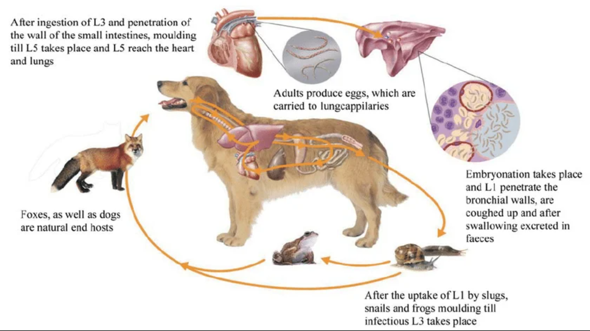
LC: L1 in feces → snail (develops to L3) → dog eats snail or paratenic host like frog → L3 into intestine → heart, lungs (L5) → eggs carried to lung capillaries → embryonates → L1 hatch → alveoli - pharynx, swallowed → feces.
Patho/CS: Usually chronic course, months - years. Presence of adults - congestion - circulatory failure - heart failure.
In weak infection - stay latent
In more severe - resp. issues of tachycardia/tachypnea, cough - can be with blood/nasal discharge.
Chronic - anemia, fatigue, anorexia, weightloss
Severe - heart failure, death
dg: baermann test (L1), sputum examination
2) Aelulostrongylus abstrusus (Cat lungworm)
Morphology: Thin, very fine, small bursa and short & stout spicules. Larvae has a dorsal spine on tial.
Location: pulmonary parenchyma & peribronchial tissues.
FH: cat
LC -similar to angio, snail IH, paratenic host: birds, rodent, amphibians
Patho/CS: greyish Nodules + granulomas in lungs, mild-moderate pneumonia.
often mild/subclinical signs, cough. Severe - resp. distress
Dg: baermann test, fecal flotation, x-ray - for nodules
3) Crenosoma Vulpis (Fox lungworm)
fam: crenosomatidae
Cuticle of anterior end of this is thrown into crenated folds.
Location: trachea, bronchi, bronchioles
Host: fox, wolf, dog, IH: snail
LC: indirect. Similar to angio.
Patho/CS: bronchitis, bronchopneumonia. Can be asymptomatic in small infection.
signs - massive infection - non-specific. chronic cough, sneezing, nasal discharge. death in severe.
Dg: baermann, sputum exam.
4) Filaroides osleri (Tracheal worm of dogs)
fam: filaroididae - larvae has s-shape tail.
Location: trachea, bronchi
Host: dogs + other car
LC - NO IH. Direct. Eggs (L1) in trachea → swallowed → feces. Also transmission from female to puppies - cleaning/licking.
path/CS: fibrous nodules in trachea. Signs of dry cough, often mild/subclinical.
Dg: fecal baermann = unreliable. Best is bronchoscopy + nodule biopsy.
filaroides hirthi..
19.Syngamosis and cyathostomosis in poultry and waterfowl
General:
Order: strongylida
Fam: Syngamidae
Genera: Syngamus & cyathostoma
General similarities - respiratory nematodes of birds, mainly affecting trachea, bronchi and larynx.
Cause resp. distress & asphyxiation
infect mainly young birds, Infective -L3. often involve earthworms as paratenic/transport host.
Diagnosed by eggs in feces. Treated by benzimizoles (ex. fenbendazole)
1) Syngamosis (“gapeworm”)
by syngamus trachea
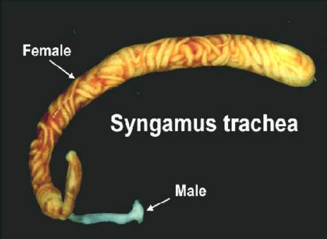
Morphology: red worms, male permanently attached to female, Typical Y-shape.
Buccal capsule - cup-shaped, teeth at base. Leaf crown absent.
Eggs: ellipsoidal, thick shelled, operculated at both poles, contain segmented embryo.
Host: chicken, turkey, guinea fowl
Location: Trachea.
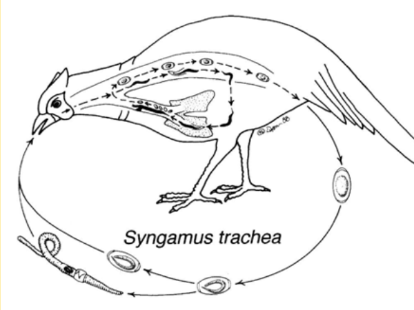
LC: L3 can be in embryonated egg, free in environment or in earthworm → bird eats L3 → larvae migrate - intestine, blood, lungs → L4/L5 in bronchioles → migrates up trachea → adults attach and makes eggs.
Patho/CS: adults/larvae cause tracheitis, hemorrhage, mucosal inflammation, nodules/knots. bronchopneumonia, pulmonary edema.
Signs - mostly in young: gasping (gapes), open-mouth breathing, cough, dyspnea, weak, possible death by asphyxiation).
2) Cyathostomosis
By cyathostoma bronchiale
Host: aquatic poultry/waterfowl
Morphology: similar to syngamus. BUT male and female live separately!
Location: larynx, trachea, bronchi
Patho/CS: VERY pathogenic, even 2-3 worms can cause death by asphyxiation!
signs similar to syngasmus - resp. distress, dyspnea, cough, suffocation/asphyxiation.
20.Spirocercosis in dogs
Order: Spirurida
General: small to medium, usually indirect LC, insect IH, adults found in organs communicating with the exterior - stomach, eyes, skin, esophagus.
2 lips around mouth, esophagus - anterior muscular & post. glandular part, males - 2 spicules (equal or unequal), females are ovoviviparous (embryonated eggs) or viviapara (larvae inside eggs).
Spirocercosis
superfam: spiruroidea
fam: thelaziidae - riktig ifølge bok / (Fam: spirocercidae ifølge pres. )
Species: Spirocerca lupi
Morphology: pink-red worms, males have spirally coiled posterior end, caudal wings, two unequal spicules.
Eggs - oval, thin shelled, embryonated, transparent.
Host: dogs, sometimes cats. IH: dung beetle.
Paratenic host - reptile, birds, small rodents, chickens can also.
Location: adults/nodules in distal esophagus, stomach wall, intestinal wall & thoracic aorta.
Geo: mainly tropics, subtropics
LC: embryonated eggs in feces → dung beetle eats → L3 developed → dog eats beetle/paratenic host → L3 into stomach wall → arteries → thoracic aorta → stays abit, then to esophagus → form nodules in wall → adults.
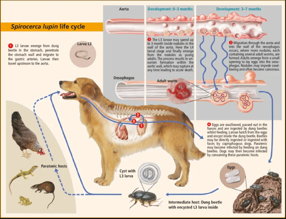
Patho/CS:
Aortic damage - migrating larvae cause hemorrhage, aneurysms, stenosis + possible aortic rupture
esophageal damage → submucosal granulomas + in CT, obstruction, possible rupture
Signs: can be asymptomatic, but can be general like vomit, regurgitation, dysphagia, weight loss, resp. difficulties.
Dg: imaging (endoscopy, radiography, sonography), eggs in feces, PM of nodules
Treatment: doramectin, milbemycin oxime
Prognosis - poor often, prevention difficult due to many paratenic hosts, dogs may eat infected chickens/reptiles, cooking meat helps prevent infection.
21.Thelasiosis of ruminants and carnivores
Superfam: Spiruroidea
Fam: Thelaziidae - genus: Thelazia (Eye worms)
small nematodes (roundworms) that live in the eyes & tear ducts of animals.
Main species:
Th.callipaeda (dog/cat, man - zoonotic - Eu, Asia)
Th.californiensis (dog/cat, sheep, man, deer, other mammals in western Us)
Th.rhodesi, Th.gulosa (cattle)
Th.lacrimalis (eq)
Morphology: Very small, white-ish, slender tubular form, Has a striated cuticle (fine transverse lines).
2 lips, buccal capsule well developed, males can have caudal alae (“wing like” expansions of tail). Male - spicules for holding female.
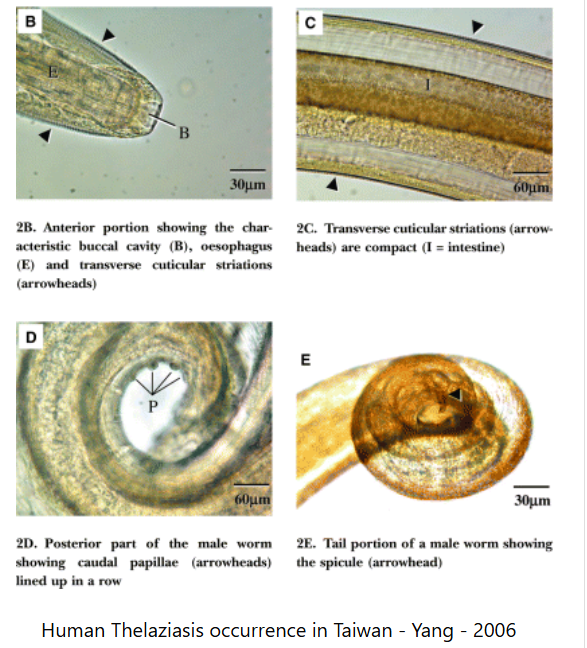
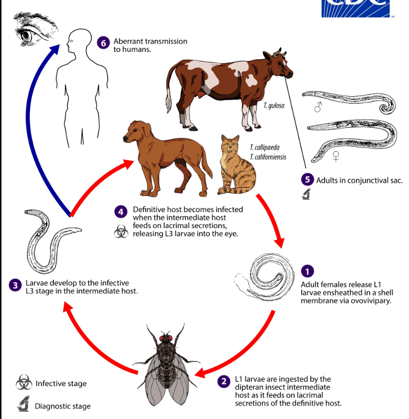
Location: adults in conjunctival sac, lacrimal/nasolacrimal ducts, sometimes under 3rd eyelid.
feed on tears, ocular secretions
FH: mostly mammals (Can be zoonotic!), IH: face flies that feed on tears.
exact fly species depend on region/species - drosophilid flies, muscid face flies.
Life cycle: adult worms live in conjunctival sac → eggs with larva (eggs hatch internally in female) (ovoviviparous) → free L1 in tears/secretions → face fly feeds on tears, get L1 → L1 migrate into fly tissues → L3 → L3 leave when fly feeds again on tears → enters sac → adult.
Patho/CS:
worms irritate the eye - damage occur as worms move across conjunctiva/cornea, inflammation, sec. bacterial infection
10-15 worms → conjunctivitis, excessive tearing (lacrimation), light sensitive, swollen.
Severe - keratitis, cloudy/opaque cornea, ulcers/perforation of cornea, weight loss
In dogs: Eye irritation, conjunctivitis, itching, swelling, light sensitivity. sec. infection.
Dg: visual examination of eye + tissues, or in sediment of centrifuged wash obtained from eye or lacrimal duct rinsing. They are active + move quickly.
Treatment: Levamisole, ivermectin, doramectin, fenbendazole. May need surgical removal.
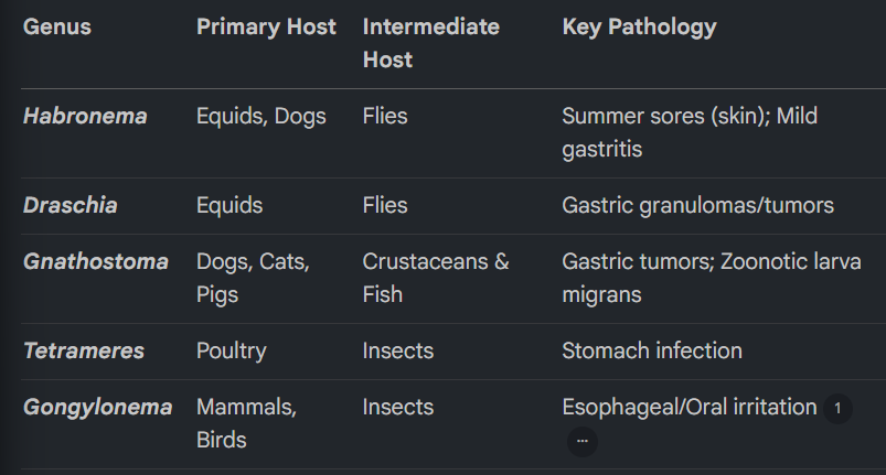
22.Other spiruroids: tetrameres, Habronema, Draschia, Gnathostoma, Gongylonema
superfam: spiruroidea
fam: spiruridae - Genera: Habronema, Drachia
fam: Thelaziidae - genera: Gongylonema
fam: tetrameridae - tetrameres
Superfam: Physalopteroidea
Fam: gnathostomidae - gnathostoma
Location: stomach, gongylonea - esophagus/oralcavity
1) Habronema & Draschia
Habronemiasis - “summer sores”, granular dermatitis, in donkey, eq, mules, zebra + dogs. Most common in temperature, subtropical/tropical regions.
Species: H.majus & H.muscae, D.megastoma
Morphology: Habronema - medium sized, females are about twice as long as males.
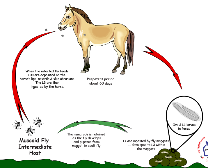
FH: equids & dog, IH: house flies, stable & face flies
LC: Embryonated eggs in feces → L1 eaten by fly maggots → L3 → adult flies carry the L3 to lips (around face), genitalia & wounds
normal route (gastric) → horse swallows larvae → goes to stomach → adult in 2m.
Aberrant route (cutaneous/ocular habronemosis) → larvae stay in skin/eye tissue instead of reaching stomach → cannot mature, but triggers inflammation & hypersensitivity.
Patho/CS: signs with local sensitivtiy, non-healing lesions, ulcers of moist area, intense itching for cutaneous/conjunctival.
Cutaneous: granulomatous lesions.
conjunctival (mainly Habronema): Conjunctivitis, tearing, irritation
Gastric: Draschia megastoma cause granulomatous nodules/tumors in stomach wall. Usually clinically silent, unless perforation.
H.majus & muscae - mild gastritis (NO tumors)
Dg: fecal flotation, visual detection in wound/lesions
Treatment: Many different used, with poor results. Insect repellents to prevent.
Ivermectin or moxidectin - broad spectrum against adult - can be effective. surgical removal/cauterization of excesssive granulation tissue can be needed.
2) Gnathostoma
G.spinigerum (dog/cat, man as accidental)
G.hispidum (pig)
Morphology: adults - thick + short, head is with bulb armed with hooks. Body has cuticular spines, mouth with tri-lobed lateral lips.
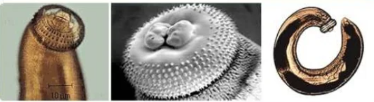
IH: 1st: water crustaceans, 2nd: fish. Birds = paratenic.
Most common asia, thailand, japan. Infection occur from undercooked fish, chicken, duck/bird. Can cause cutaneous larva migrans.
LC: unembryo eggs in feces → hatch, L1 → eaten by first IH → L2 → humans infected by eating water with L2 (crustaceans) or by 2nd IM host (L3) → FH eats fish or through paratenic host → liver, muscle, CT → back to stomach.
in G.spinigerum - forms tumors in gastric wall, embed its spiny head into mucosa
Patho/CS: migration - 3months → clinical signs.
Gastritis, perforation of stomach, fatal peritonitis.
Painful, pruritic swellings (cautaneous larva migrans) - in subcutan.tissue
Migrating to other tissue - visceral larva migrans → cough, hematuria, ocular issues - meningitis, myeloencephalitis. High eosinophilia.
Dg: fecal exam, eggs operculated
Treatment: unknown.
Tetrameres spp. - only infect poultry - stomach.
Gongylonema pulchurum - mammals/birds. Inhabit upper resp. tract. irritate esophageal/oral.
23.Dirofilariosis of carnivores
Superfam: Filarioidea
Fam. Onchocercidae, subfam: dirofilariinae
Genus: Dirofilaria
Species:
D.immitis (Heart (right ventricle) & Lungs - Heartworm disease)
D.repens (subcutaneous tissue - subcutan. nodules)
Morphology: long, thin, whitish, thread-like “hairy” worms. Females are bit bigger. D.repens is smaller. Mouth is very small, No lips.
post. end of male is spirally coiled.
Females - ovoviviparous - gives birth to live L1 (microfilaria)
FH: dog (main), cats, humans - accidental. IH: mosquito
Geo: worldwide, esp. warm/tropical regions (many mosquito)
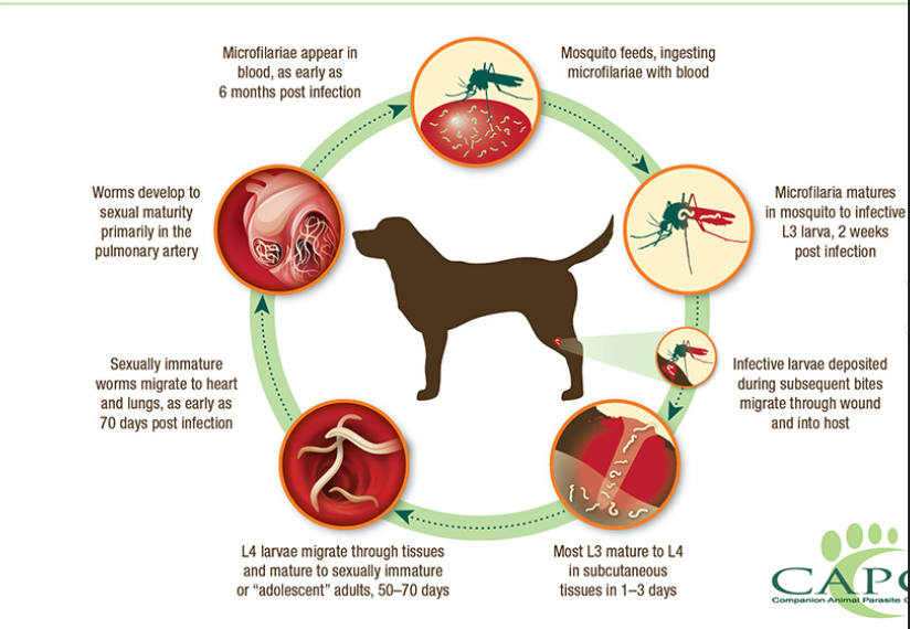
LC: mosquito → bites host, L3 enters → stays in subcutan. tissue → L4 → migrates to heart (1month) → L5 → Adults in pulmonary arteries, right side of heart sometimes → microfilariae produced - circulates in blood → mosquito take blood meal with the microfilariae.
Patho/CS:
Inflammation of bv, thickening of blood vessel walls, thrombosis
Lungs develop inflammation, alveoli filled with MF + eosinophils
Signs - often chronic, progressive
In dogs: chronic cough, resp. distress, fatigue, weight loss vomiting. Death from cardiopulmonary failure.
In humans: usually worms do not fully mature. often mild/asymptomatic but can cause cough, chest pain, fever, blood with cough, sometimes pulmonary nodules on x-ray are found accidentally.
D.repens - adults stay under skin → local swelling, itching subcutan.nodules.
Dg: blood tests - ELISA, Ab tests. Detection of microfilariae in blood. Radiographs, CS.
Treatment: difficult. Killing adult worms cna be dangerous, as dead worms may block pulmonary vessels → worsen inflammation. Early cases can be cured.
Drugs - ivermectin during mosquito season for prevention.
D.repens - removal of worm under skin.
24.Bovine and Equine Onchocercosis (Onchocerca, Parafilaria, Stephanofilaria, Setaria)
Filarial nematodes transmitted by biting insects. Mainly affecting eq, cattle, ru. Most cause skin disease, nodules, dermatits, hypersensitivity.
1) Onchocerca - genus
O.cervicalis (eq, in nuchal lig.)
O.gutturosa (cattle, CT)
cattle: O.lienalis (gastrospl.lig), gibsoni & ochengi (subcutan.nodules), O.armillata (aortic wall)
Morphology: Sharply pointed tail, long, slender. Typical filarial worms.
Location: usually in ligaments, tendons, CT, tissues.
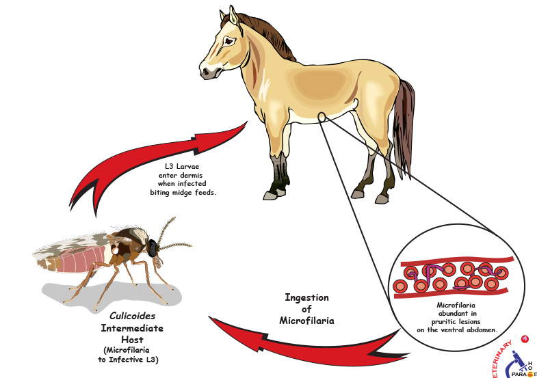
IH: biting midge (culicoides)
Worldwide.
LC: larvae (microfilaria) released in blood → eaten by biting midges while feeding on the horse → L3 → given back to FH → migrate through tissues, mature into adults.
Patho/CS: Most horses are asymptomatic. Supposed hypersentivity reaction to microfilaria mostly → swelling, skin irritation.
if going to eye → blindness.
Nodules under skin + dermatitis, lesions
Dg: ELISA, PCR, histology
Treatment: Endectocids, kills only microfilariae. Ivermectin & moxidectin.
2) Parafilaria
P.multifilaria (“summer bleeding, bloody sweats)
P.multipapillosa
P.bovicola (cattle)
Small and thin white worms. In subcutan. CT.
IH: blood sucking flies. Worldwide.
LC: Flies with microfilariae → L3 → gets into FH → into dermis, form nodules → female lays eggs in the nodules → rupture, bleeding on skin → eggs + microfilaria exit through the bleeding nodules.
Patho/CS:
In horse: bleeding skin spots, edema, inflammation & painful nodules.
chronic: lesions can become gelatinous, green-yellow, metallic smell.
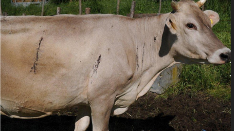
Dg: CS, eggs or microfilaria in exudate. ELISA.
Treatment: Difficult, due to long prepatent period (5-7months).
macrocyclic lactones, nitroxinil
use of insecticide impregnates ear tags - good for vetor control
bleeding spots still occur in treated animals due to long PP.
Stephanofilaria stilesi - form ventral midline dermatitis. Similar to others.
ventral abdomen - dermatitis, itching, alopecia, thickening
Setaria tundra - in reindeer, Setaria cervi - ru
live in peritoneal vacity, often relatively harmless. can cause neurologic disease
25.Filariosis of man (Onchocerca, Brugia, wuchereria, Loa, Dracunculus)
These are filarial worms transimitted by biting insects or contaminated water. The main diseases are: lymphatic filariasis, river blindness, loiasis, guinea worm disease. Same ish LC. Same treatment except for guinea worm.
Superfam: Filarioidea, Fam: Onchocercidae
1) Lymphatic Filariasis (Wuchereria, brugia)
Main species: Wuchereria Bancrofti, brugia malayi, brugia timori
Location: Vessels (lymph vessels) + LN
Transmission: by mosquitoes (Anopheles, mansonia)
Geo: Millions infected in over 80countries in Africa, Asia, america. Can live -ish 15 years. Transplacental transmission.
LC: mosquito injects L3 → worms mature in lymphatics → microfilariae enter blood → mosquito eats microfilariae.
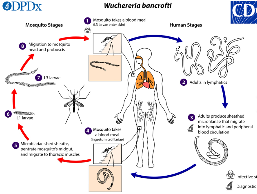
Patho/CS: Damage to lymphatic system/drainage.
Early: fever, LN inflammation, lymphangitis
Chronic: lymphedema, hydrocele, elephantiasis (massive swelling)
Usually effects legs, arms
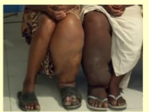
Dg: Blood tests, ELISA, microfilariae in blood. CS
Treatment: Ivermectin.
2) Onchocerca Volvulus - “River Blindness”
Males smaller, sharply pointed tail, long slender
Location: subcutaneous tissues.
Vector: Blackflies (Simulium)
Mostly in africa.
Patho/CS: Main damage by microfilariae in skin & eyes.
skin: itchy nodules, lesions in skin/subcutan.tissue, eosinophilia
Eyes: photophobia (light sensitive), tearing, corneal damage, BLINDNESS
Dg: microfilariae in skin biopsies, eye exam
Treatment: Ivermectin
3) Loa Loa - “African Eye Worm”
By filarial parasite Loa Loa
Location: Subcutaneous tissues
Vector: Deer flies/horse flies
Patho/CS: adult worm migrates under skin. Sometimes visible crossing conjunctiva.
transinet swelling (Calabar swellings), itching, redness. Usually not very dangerous.
Dg: eosinophilia, microfilariae in daytime blood (as they move to lungs at night)
Treatment: Ivermectin
4) Dracunculus Medinensis - “Guinea worm”
Fam: dracunculidae (under same superfam)
Tramsmits by drinking water with Daphnia (“water fleas”)
LC: drinking contaminate water → larvae gets into intestine → adult female migrates under skin → blister forms → worm releases larvae into water
Patho/CS: About 1y later → painful skin blister forms, usually on legs/feet. Worm slowly emerges. Patients often place leg in water because of burning pain → in which blister rupture, releasing larvae.
burning pain, blister, ulcer, sec. bacterial infection, difficulty walking
Dg: clinical signs
Treatment: No reliable drug cure. Main treatment is to slowly take out adult worms by rolling it a few cm a day, or by surgical incision. Metronidazole can be okay in killing the worm, ATB for bacterial infection.
Prevention - clean drinking water, filter water, prevent infected people entering water sources.
26.Trichinellosis in animals and humans
Order: ENOPLIDA - all nematodes in this order is characterized by filariform anteriorly, No lips. Extremely long, thin + non-muscular pharynx called Stichosome with stichocytes. Most are oviparous. Males have a single spicule or none.
ALL have L1 = Infective stage (Except fam: dictophymatidae - with L3).
fam: Trichinellidae
Genus: Trichinella
Main species:
High pathogenicity for humans: T.spiralis, T.nativa, T.pseudospiralis & T.murrelli
Low patho: T.britovi & Nelsoni
T.T6, T.papuae, T8, T9 and Zimbawanesis
Morphology: Very small worms. Long esophagus, no spicules in male but it has a pair of flaps on sides of cloaca opening + 2 pairs of papillae. Female is viviparous (release larvae, not eggs).
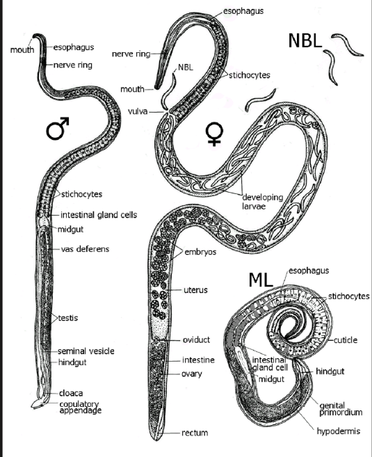
Location: Adults in SI, Larvae in striated muscles.
Esp. diaphragm, tongue, limb muscles
FH: mammals, sea-mammals, birds, rodents, fish etc..
IH can be either -
Geo: one of most widespread parasites - except in deserts. In Europe - T.spiralis, britovi, nativa.
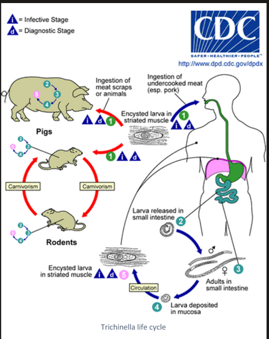
Transmission/Epidem: Infection by eating raw/undercooked meat with encysted L1 larvae (pork, wild boar, bear/horse meat, sausage).
main reservoir: rats & rodents. Maintained by cannibalism and scavenging.
2 cycles - domestic cycle (Mainly pigs, rats - mainly by T.spiralis) & sylvatic cycle (wildlife carnivores)
Life cycle: Host eats infected meat → Larvae leave cysts and enters intestine → molt into adults in intestinal mucosa → produce L1 larvae, releasing into lymph/blood → migrate to muscles and form nurse cells/cysts - L1 coil inside these cysts.
Patho/CS: Most domestic animals are asymptomatic. Disease in humans depends on number of larvae ingested. Signs:
Intestinal phase: first few days - diarrhea, abd.pain, inflammation
muscle phase (1-2w after): myositis, muscle pain, fever, eosinophilia. Periorbital edema, conjunctivitis, splinter hemorrhages.
Severe: myocarditis, encephalitis, resp. problems → death.
Dg: trichinoscopy (compression of muscle sample) OR digestion method
In living animals/humans: ELISA, Serology, muscle biopsy, PCR
Treatment:
humans: mebendazole, albendazole
Control/prevent: inspect meat, cook, freeze, rodent control, avoid feeding raw scraps to pigs
27.Trichurosis and capillariosis of animals.
Fam: trichuridae (under order Enoplida - class Adenophoea (Aphasmida))
1) Trichurosis
Subfam: trichurinae, genus: trichuris
Main species: T.ovis, T.discolor, T.globulosa (Ru)
T.suis (pig), T.vulpis (dog)
T.campunula, T.serrata (cats)
T.trichiura (human)
Morphology: “whipworms” - thin anterior, thick posterior end. Looks like a whip, whitish/yellowish color. Male has only one spicule.
Location: adults in LI (cecum/colon)
LC: direct.
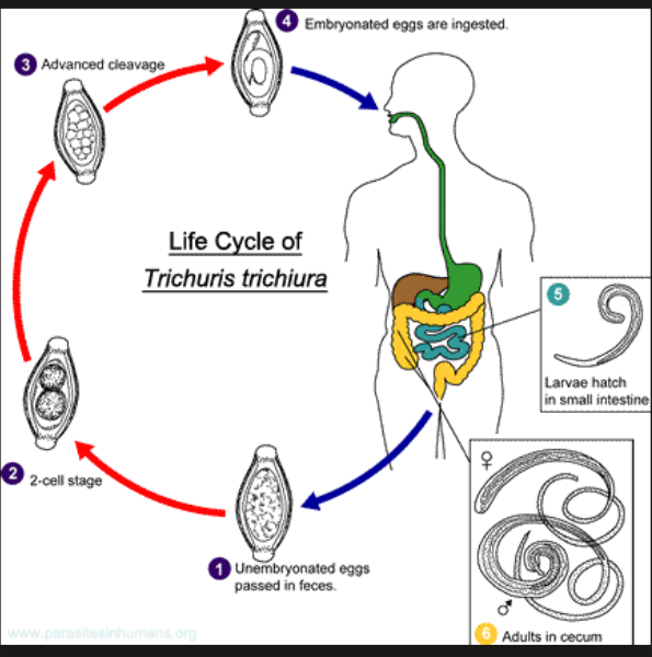
Eggs in feces → embryonate in environment → infective eggs eaten (L1) → hatch in LI → adults in intestinal mucosa
Adults embed thin ant.end into mucosa and feed on blood
Patho/CS: Damage caused by blood feeding & tissue penetration
adults tunnel into mucosa and damage bv.
Light infection: often asymptomatic
heavy: diarrhea, mucus/blood in feces, dehydration, weight loss, anemia and colotis
severe - electrolyte imbalanace (hyponatremia/hyperkalemia), seizures
Dg: coprology - eggs in feces
Treatment: fenbendazole
2) Capillariosis (Capillaria) - Subfam: capillariinae
Species:
Birds: C.caudinflata, C.contorta, obsignata
Mammals: C.plica (urinary bladder), aerophila (lungs), hepatica (liver), felis, philippinensis (human)
Some species have earthworm as IH - C.plica, caudinflata
Morphology: Very thin and slender, Posterior end is NOT thickened.
Location: depends on species. SI.
LC: depends on species
Direct: eggs in feces → embryonate → host eats → adults
Indirect: some have earthworms as IH
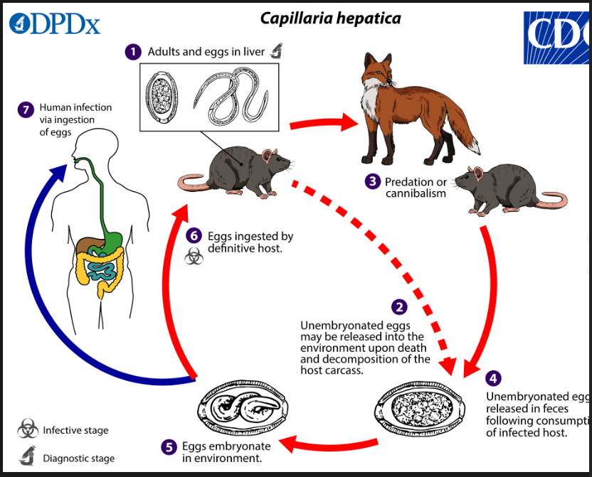
C.hepatica - in liver, adults lay eggs inside liver tissue. Eggs do not leave in feces directly. Eggs are released ONLY when host dies or predator eats infected liver. Then eggs goes to environment and embryonates. Can have paratenic host.
C.plica - in urinary bladder. Uses earthworm IH. Eggs shed in urine.
C.aerophila - in resp. tract/Lungs - eggs are coughed up, swallowed and then passed in feces.
Patho/CS: depends on organ affected.
Intestinal: enteritis, diarrhea, weight loss
Bird infections: inflammation of crop, hemorrhagic enteritis
lung species: coughing, resp. irritation
urinary species: cystitis, urinary irritation
Dg: finding eggs in feces, urine or resp. secretions depending on species.
Treatment: Fenbendazole