Choroidal Rupture, Commotio Retinae, Pathological Myopia, Angioid Streaks, Central Serous, Solar Retinopathy, AMN, Krill Disease - Posterior Segment & Ocular Disease Spring 2026
1/113
There's no tags or description
Looks like no tags are added yet.
Name | Mastery | Learn | Test | Matching | Spaced | Call with Kai |
|---|
No analytics yet
Send a link to your students to track their progress
114 Terms
BLUNT TRAUMA to the eye
What is choroidal rupture a common complication of?
usually crescent shape w/ concavity toward the disc
What is the usual shape of a choroidal rupture?

-typically in the posterior pole
-can be more in the periphery (posterior to the equator)
Where is a choroidal rupture usually located?

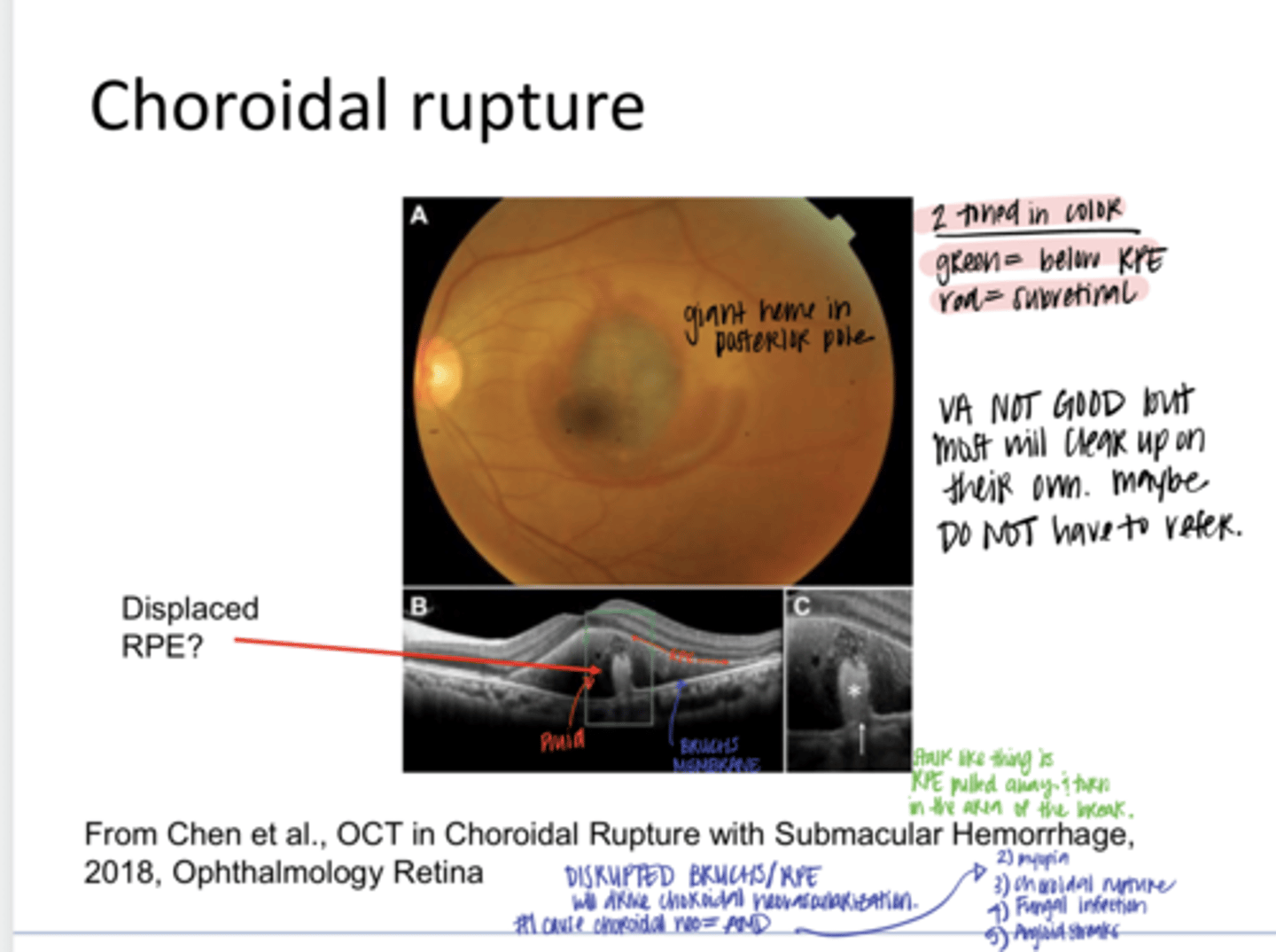
-Green = subRPE
-Red = subretinal
Why is a choroidal rupture 2-toned?
hemorrhaging into the choroid, RPE, subretinal space, intraretinal space, or vitreous
What can be associated with a choroidal rupture?

Yes
Can hemes obscure the choroidal rupture at first?

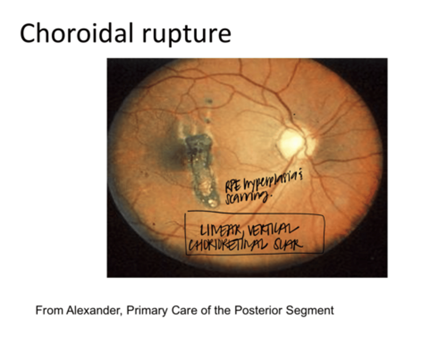
scarring, RPE hyperplasia
When the blood resorbs after a choroidal rupture, what can be seen?

Yes
Can the overlaying retina be OK after a choroidal rupture?

Choroidal rupture (Pic)
Choroidal rupture (Pic)

RPE Displaced and pulled away/town in the area of the break
What is the stalk like thing?

Possibly choroidal neovascularization
What happens ANYTIME Bruch's membrane breaks or an RPE break is present?

AMD
REVIEW: What is the #1 cause of choroidal neovasc?

Choroidal Rupture -- Scarring w/ RPE Hyperplasia (Pic)
Choroidal Rupture -- Scarring w/ RPE Hyperplasia (Pic)
**Linear, Vertical Chorioretinal Scar
1) No treatment in the acute phases for the choroidal rupture but must ensure that the globe is intact; may require imaging of the globe (orbital CT, Seidel's sign)
2) Can get neovasc up to 5 years later
3) Monitor at home with an Amsler grid, esp if near the macula
4) Examine yearly
What is the management of choroidal rupture?
Haze d/t traumatic disruption of outer segments of photoreceptors
What is commotio retinae?
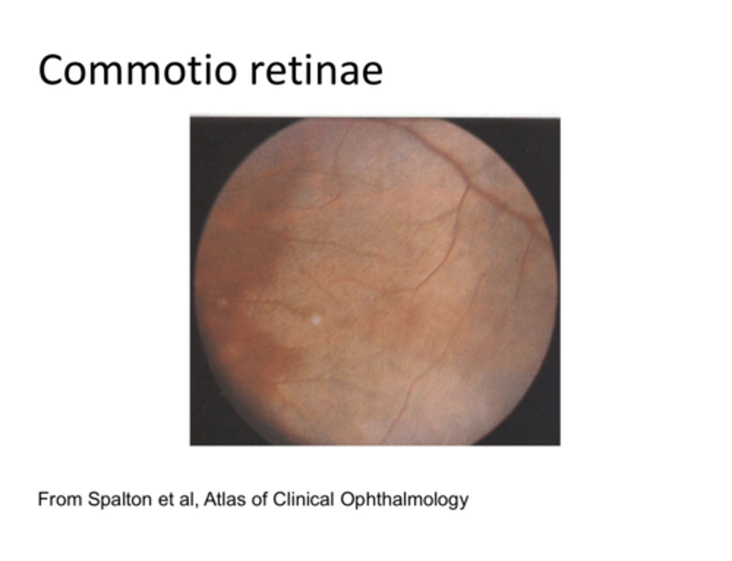
Edema may appear d/t damage to RPE with/without subretinal hemorrhage; not enough to rupture the choroidal BVs
What is the characteristic of commotio retinae?

Berlin's edema
If commotio retinae is found in the macula, what is it called?

Pigmentary changes, including RPE dropout and RPE hyperplasia
What can commotion retinae lead to long term?

Commotio retinae (Pic)
Commotio retinae (Pic)

3
About ____% of the world's population has degenerative or pathological myopia
No
Is degenerative myopia the same thing as refractive myopia?
No
Does refractive myopia commonly exceed 6-9D?
myopic maculopathy
Pathological myopia now primarily refers to the presence of what?
1) Patient has A LOT of myopia w/ pathological changes
2) Patient's myopia keeps getting worse
What are the common differences between pathological myopia and refractive myopia?
1) Primary change is elongation of the eyeball posteriorly d/t the progressive thinning of the sclera, RPE, and choroid
2) Vascular insufficiency of choroid and probably the retina
3) Early vitreous liquefaction
What are the changes that are associated with degenerative (pathological) myopia?
1) peripheral retinal degenerations (lattice)
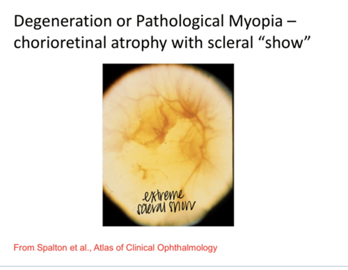
2) Patches of chorioretinal atrophy
3) Scleral cresent, vessel straightening
4) Posterior staphyloma
5) Disc tilting (supertraction)
What are the CLINICAL FINDINGS associated with pathological myopia?
1) Retinal breaks and detachment
2) Choroidal neovasc and possibly an associated Fuch's spots
3) Lacquer cracks
4) Regional optic nerve head damage
What are the DEGENERATIVE/DISEASE FINDINGS associated with pathological myopia?
Lacquer Cracks w/ Scleral Cresent (Pic)
Lacquer Cracks w/ Scleral Cresent (Pic)

breaks in Bruch's membrane that are NOT as extensive or wide as angioid streaks. D/t vascular insufficiency
What are lacquer cracks?

Choroidal neovasc from breaks in Bruch's membrane
Lacquer cracks are correlated to a high risk of what?
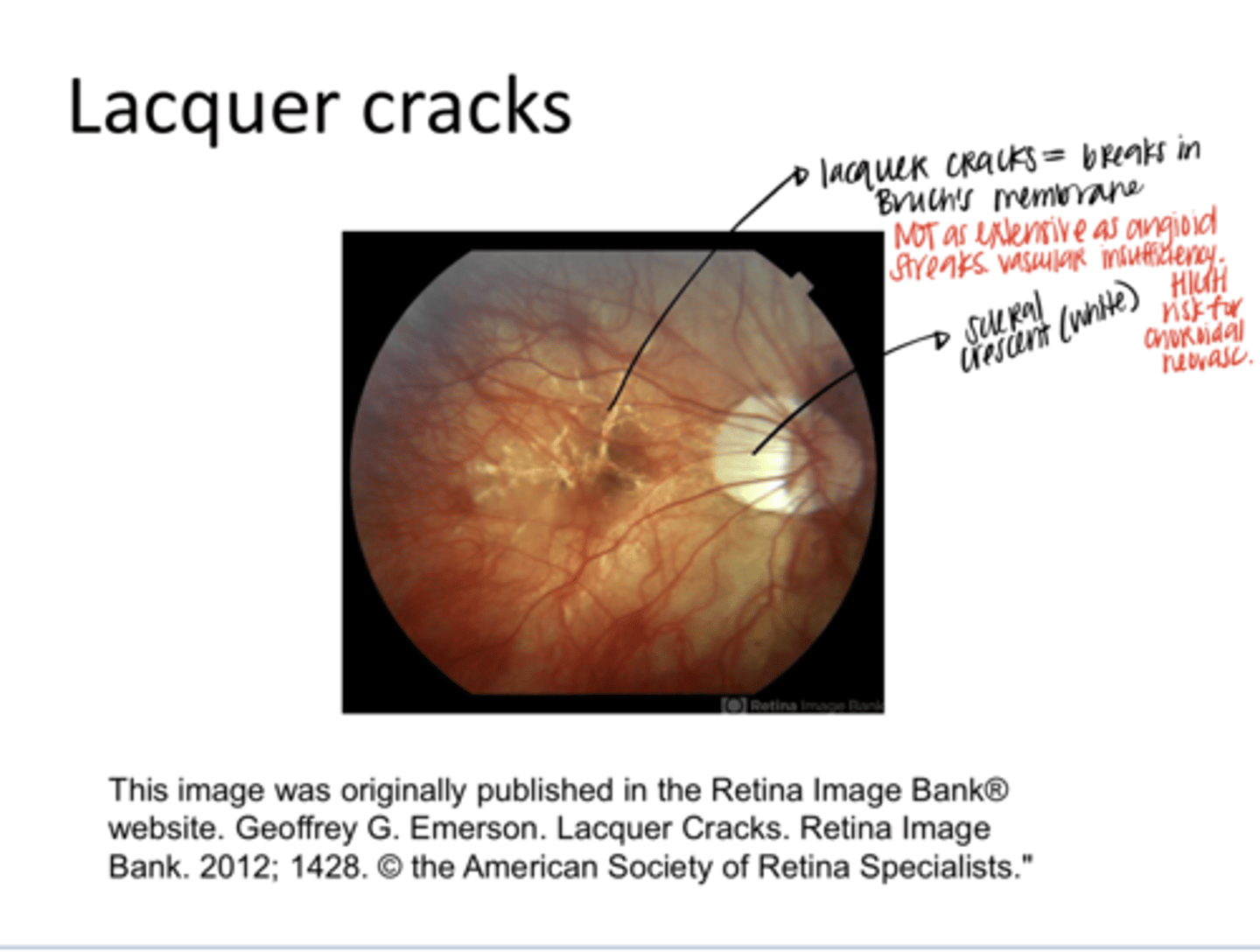
SCLERAL crescent
What is the white around the optic nerve?

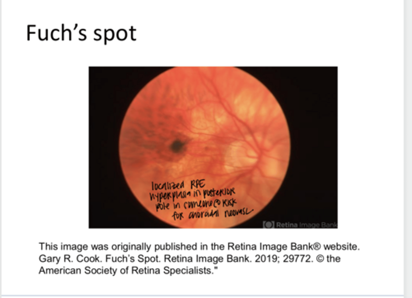
localized RPE hyperplasia in a patient who is at risk for choroidal neovasc
What is a Fuch's spot?
Degeneration or Pathological Myopia -- Chorioretinal Atrophy with Scleral Show (Pic)
Degeneration or Pathological Myopia -- Chorioretinal Atrophy with Scleral Show (Pic)
Posterior Staphyloma (Pic)
Posterior Staphyloma (Pic)
**Tissue is so thin that the retina jams into the tissue and causes a posterior POP OUT?
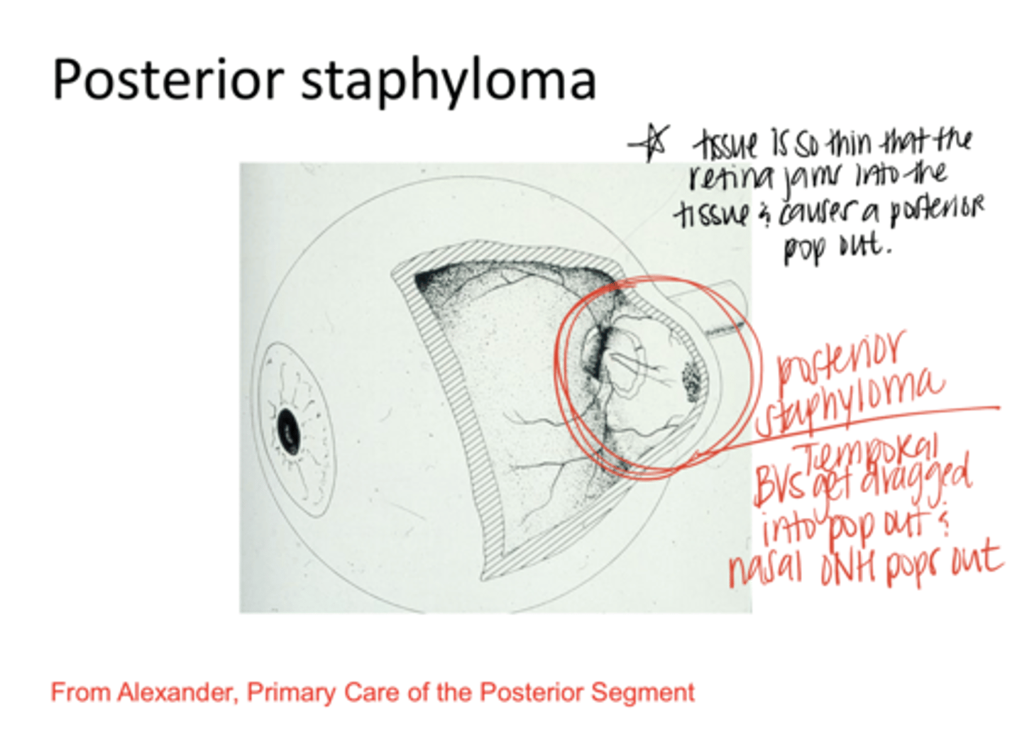
They get dragged into the pop out
What happens to the temporal optic nerve BVs with a posterior staphyloma?

Foveoschisis (myopia maculopathy)
What is an eventual consequence of a posterior staphyloma?

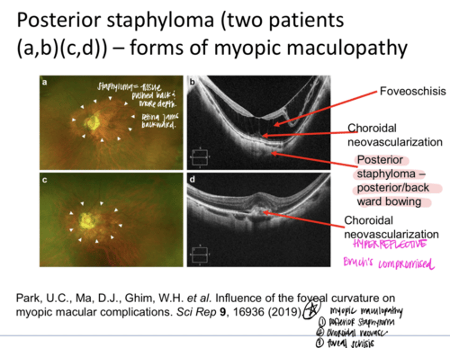
Posterior Staphyloma on Fundus Photo and OCT (Pic)
Posterior Staphyloma on Fundus Photo and OCT (Pic)
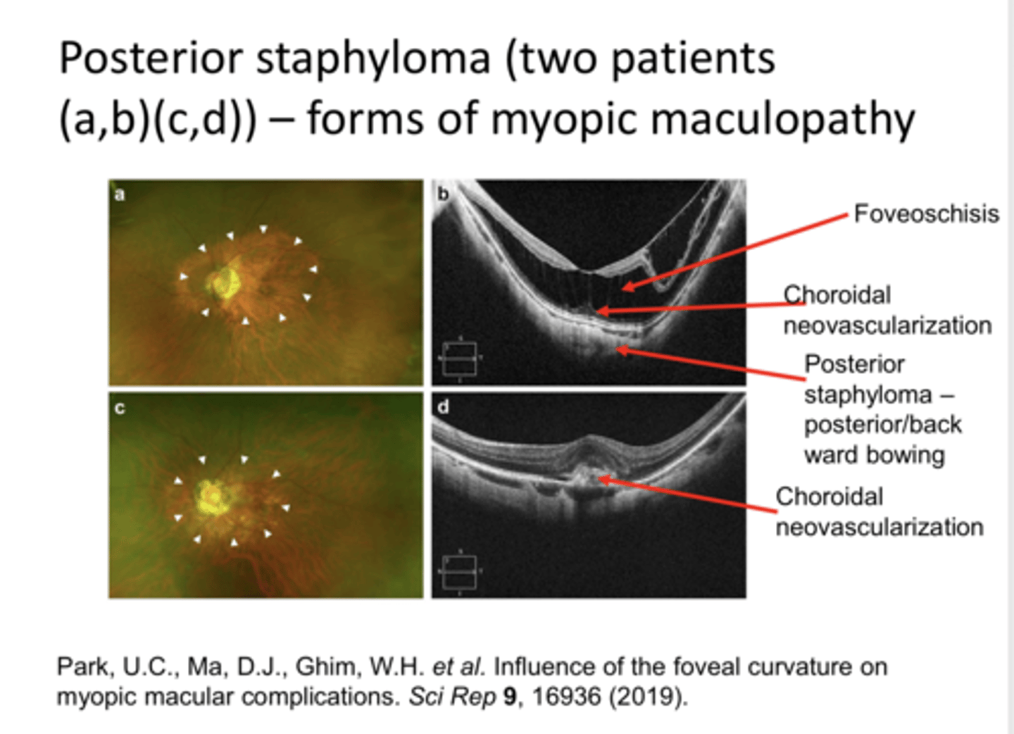
1) Posterior staphyloma
2) Choroidal neovasc
3) Foveoschisis
EXAM QUESTION: What are the 3 components of myopic maculoapthy?

Myopic maculopathy
***NOT retinal detachment like expected
What is the #1 reason that patients with high myopia lose vision?
Choroidal Neovascular Membrane with Hemorrhage in Pathological Myopia (Pic)
Choroidal Neovascular Membrane with Hemorrhage in Pathological Myopia (Pic)
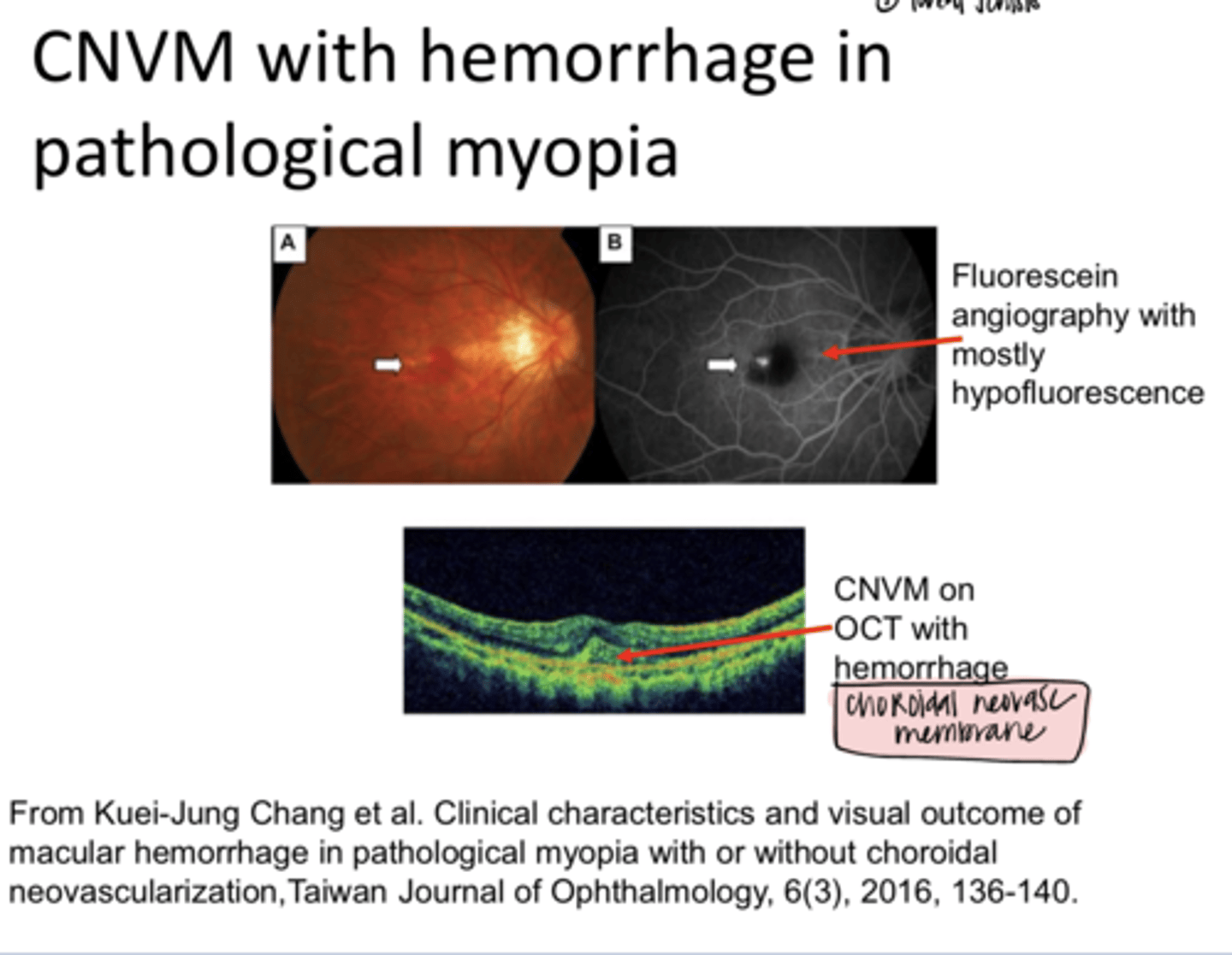
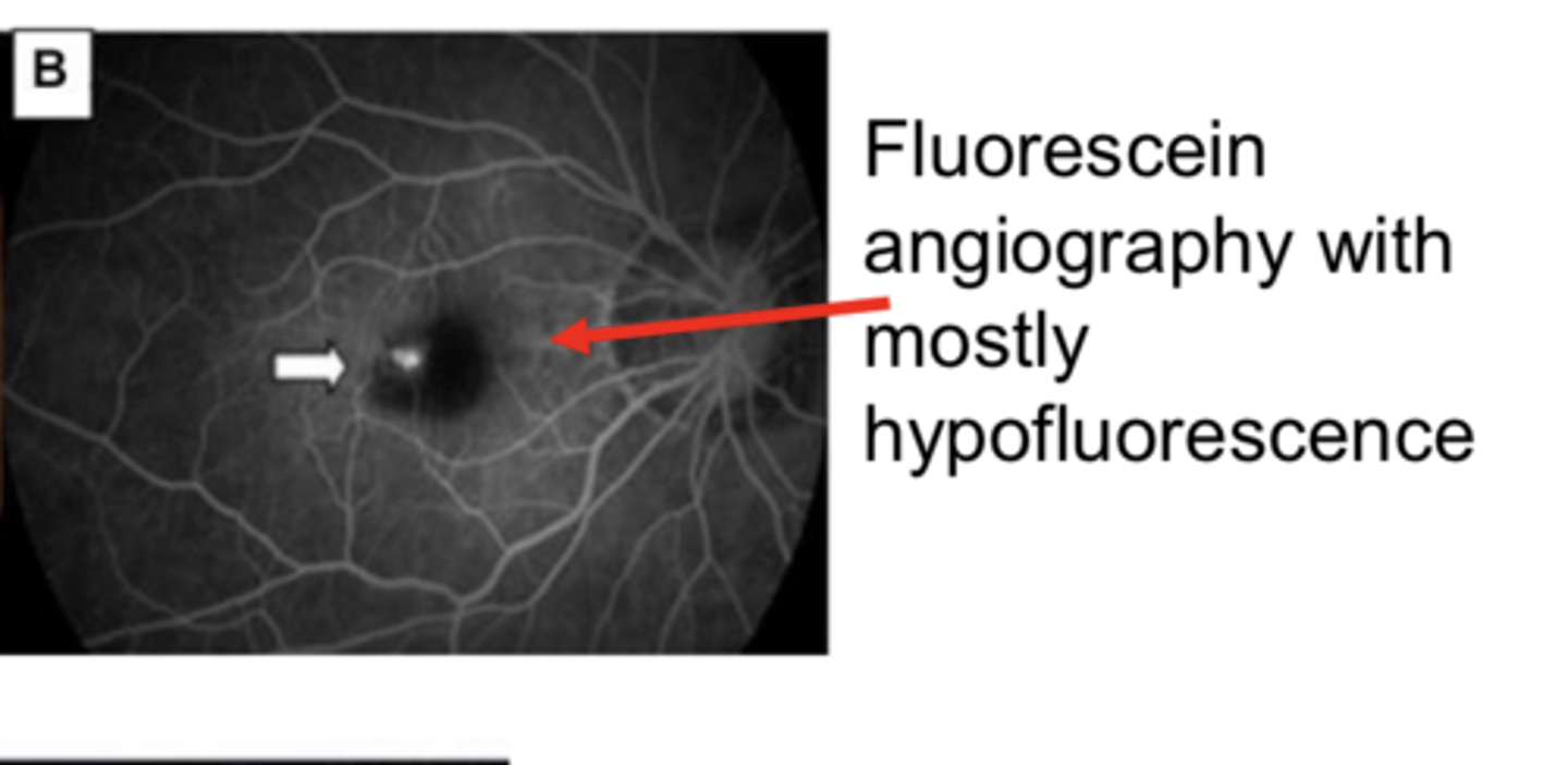
Choroidal Neovascular Membrane with Hemorrhage in Pathological Myopia -- Fluorescein Angiography with Hypofluorescence (Pic)
Choroidal Neovascular Membrane with Hemorrhage in Pathological Myopia -- Fluorescein Angiography with Hypofluorescence (Pic)
Fuch's Spot -- Localized RPE Hyperplasia in Posterior Pole in Someone Who is At Risk for Choroidal Neovasc (Pic)
Fuch's Spot -- Localized RPE Hyperplasia in Posterior Pole in Someone Who is At Risk for Choroidal Neovasc (Pic)
1) Glaucoma?
2) Retinal tear/detachment
3) Choroidal Neovascular Membrane
What is the triad of pathological myopia?
Yes -- >3D has 10x risk
Does someone with higher myopia have an increased risk of retinal detachment?
d/t breaks in Bruch's, choroidal vascular insufficiency
Why is a choroidal neovascular membrane in the triad of pathological myopia?
-protective eyewear to help in preventing retinal detachment
-DFE and OCT q6-12months
-Amsler grid
What is the management of pathological myopia?
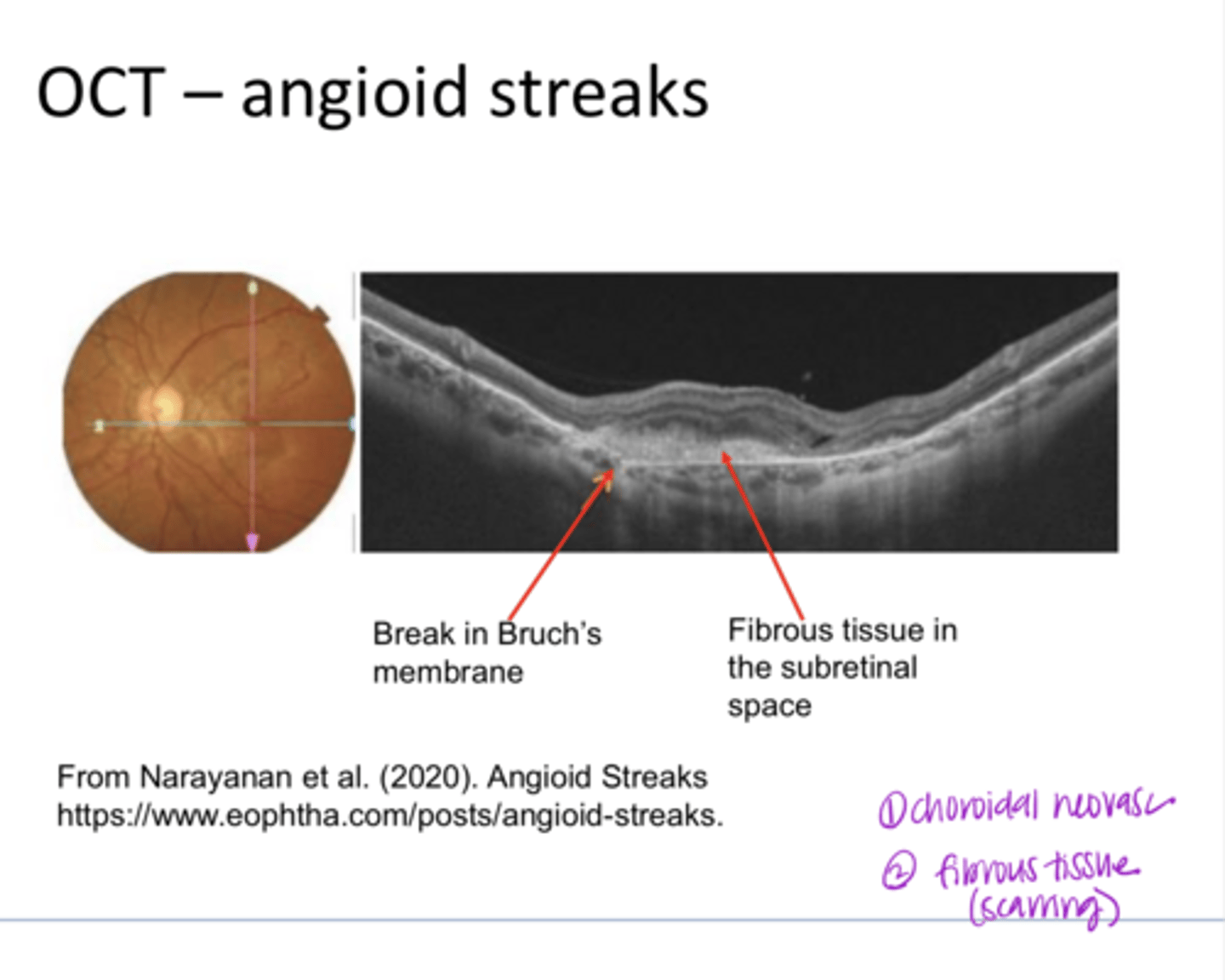
due to the breaks in a thickened, calcified Bruch's membrane; there is then a loss of migration of pigment granules in the RPE
What are angioid streaks d/t?
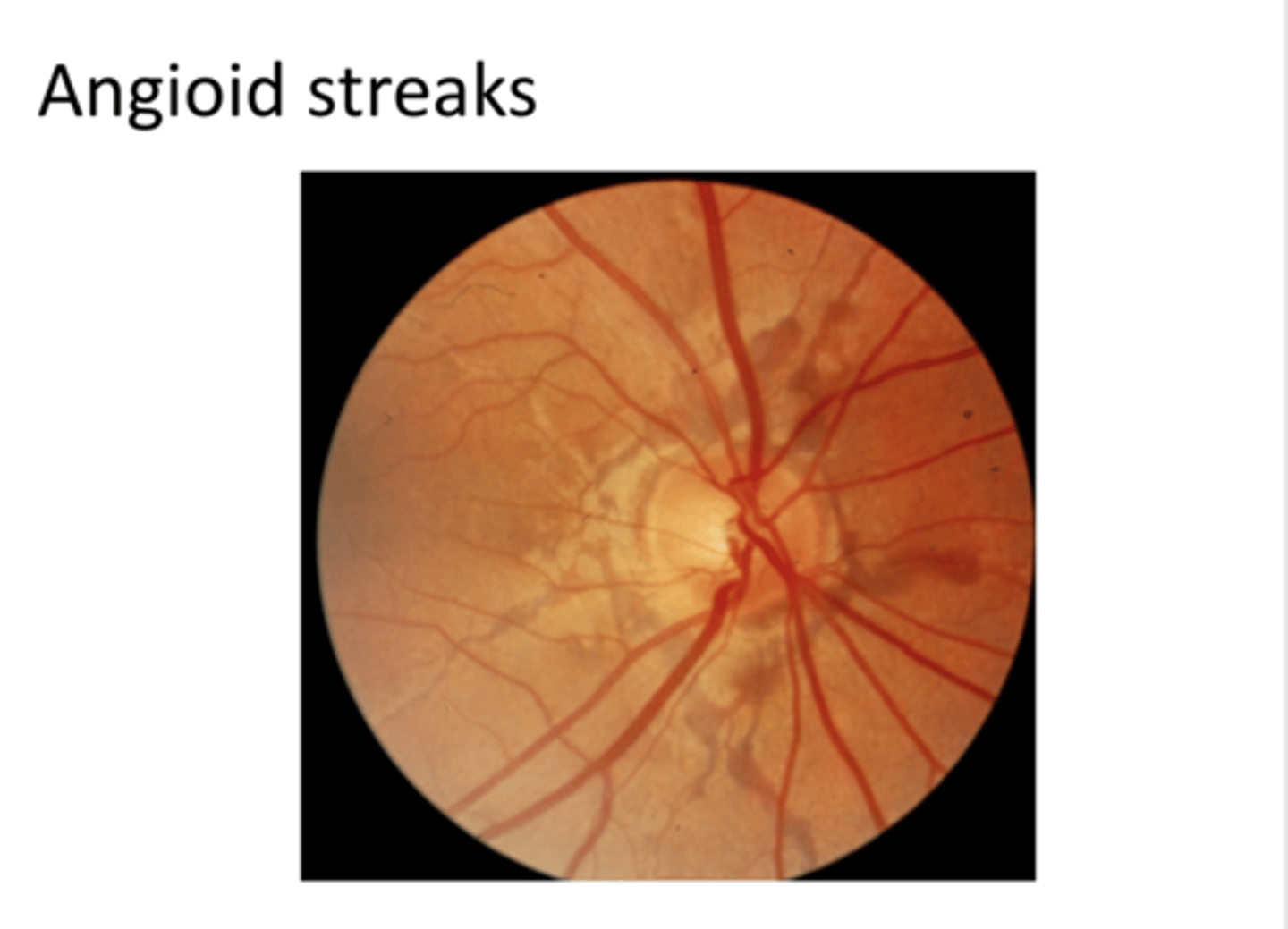
-radiation of spokes from the optic disc
-mottled retina called peau d'orange (esp in pseudoxanthoma elasticum)
What is the common presentation of angioid streaks?

choroidal neovasc (up to 70%), exudative changes
Breaks in Bruch's can result in what?

-PEPSI HAM
-Pseudoxanthoma elasticum
-Ehlers-Danlos
-Paget's disease of bone
-Sickle cell anemia or other hemoglobinopathy (Thalessemias)
-Idiopathic (50%)
-Homocystinuria
-Acromegaly
-Marfan syndrome
Systemic Consequences of Angioid Streaks

Angioid Streaks (Pic)
Angioid Streaks (Pic)

Peau d'orange (Pic)
Peau d'orange (Pic)

1) idiopathic
2) Pseudoxanthoma elasticum
What are the 2 most common causes of Peau d'orange?

OCT Angioid Streaks (Pic)
OCT Angioid Streaks (Pic)
-monitor with home Amsler grid
-examine every 6months with DFE and OCT
-protective spec lenses (polycarb)
What is the management of angioid streaks?

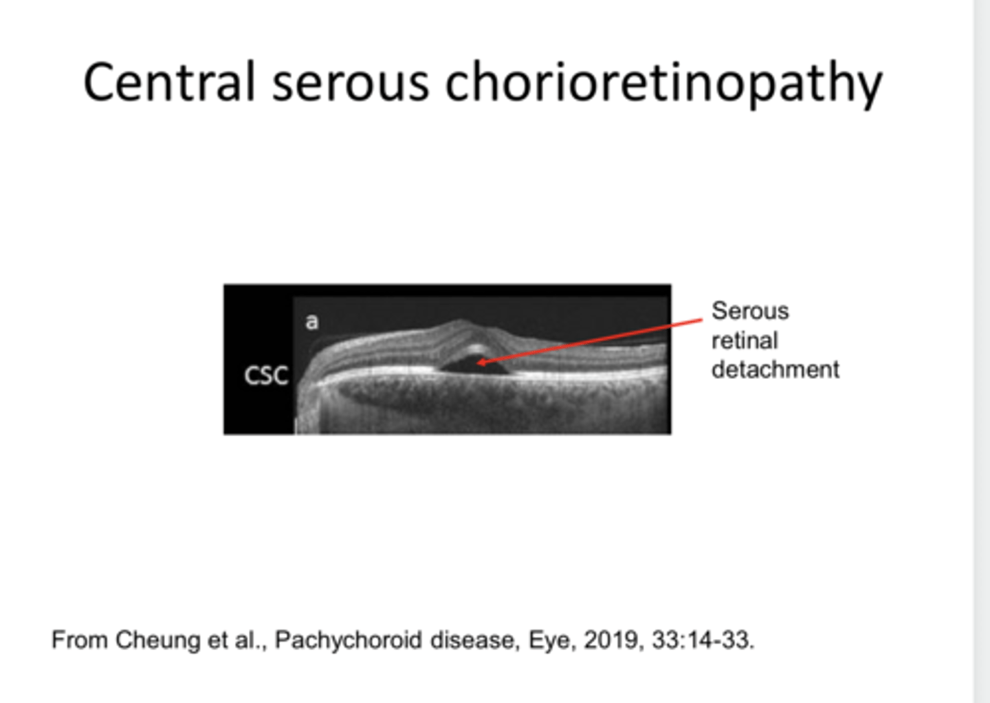
Central Serous Retinopathy
Idiopathic Central Serous Chorioretinopathy AKA
-usually young males, 20-50 years old, but can occur in older patients
-Type A personality (competitive, aggressive, hostile)
-steroid use can precipitate central serous
Who is usually affected by central serous retinopathy?
older
Central Serous is likely to be more chronic in (older/younger) individuals
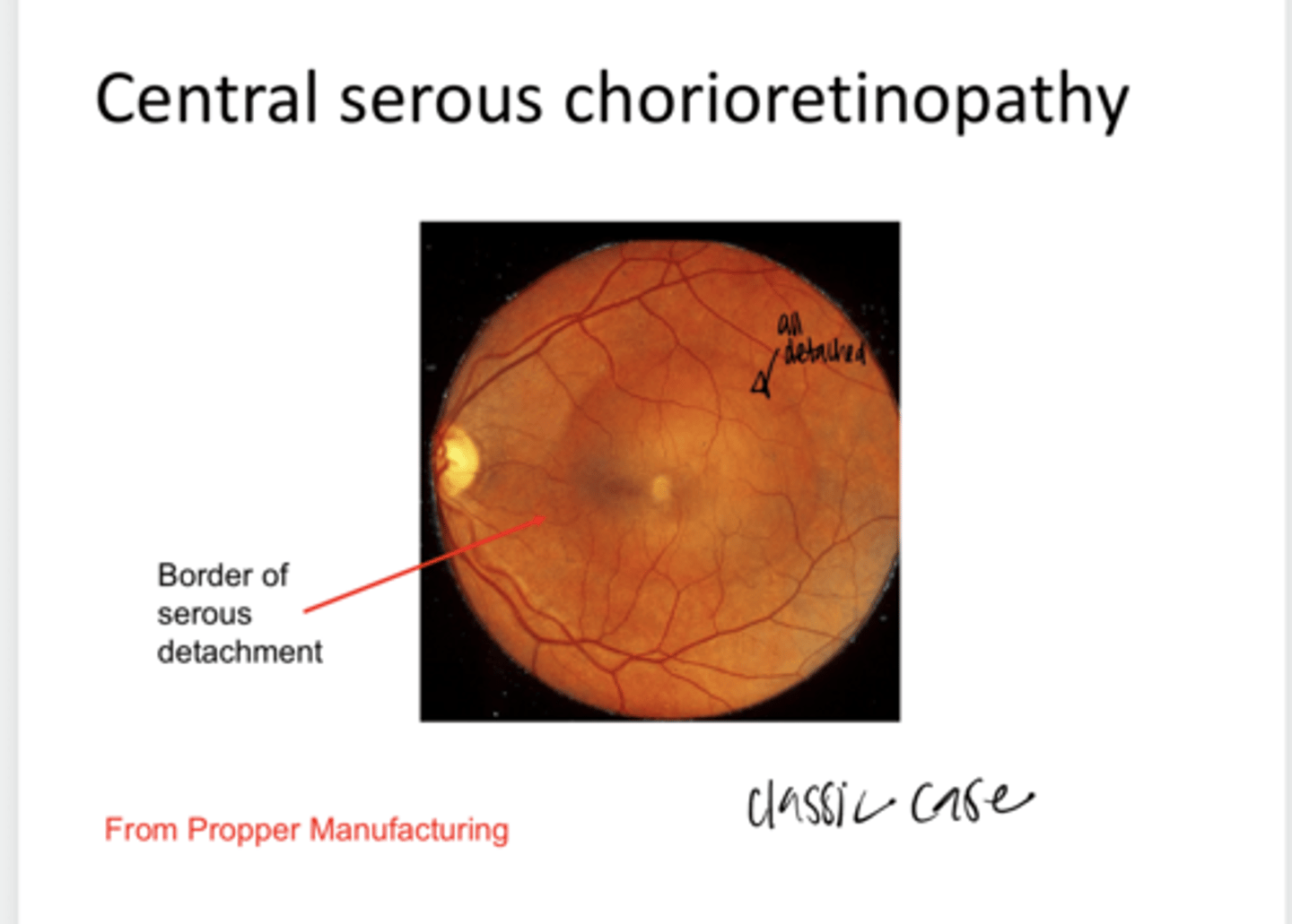
-May be localized breakdown in the choriocapillaris, RPE cells then break down; allows seepage of fluid which may cause sensory (Primarily) retinal detachment, and localized RPE detachment (Dome shaped)
-Possibly too much autonomic innervation
What is central serous retinopathy?

Sudden onset of unilateral visual distortion or slight loss of color vision
What do patients with central serous report?

-may see RPE hyperplasia, pigment migration
-possible loss of foveal light reflex
-old cases may show RPE disturbances (pigmentary changes)
-yellow, lemon drop nodule d/t RPE detachment may occur
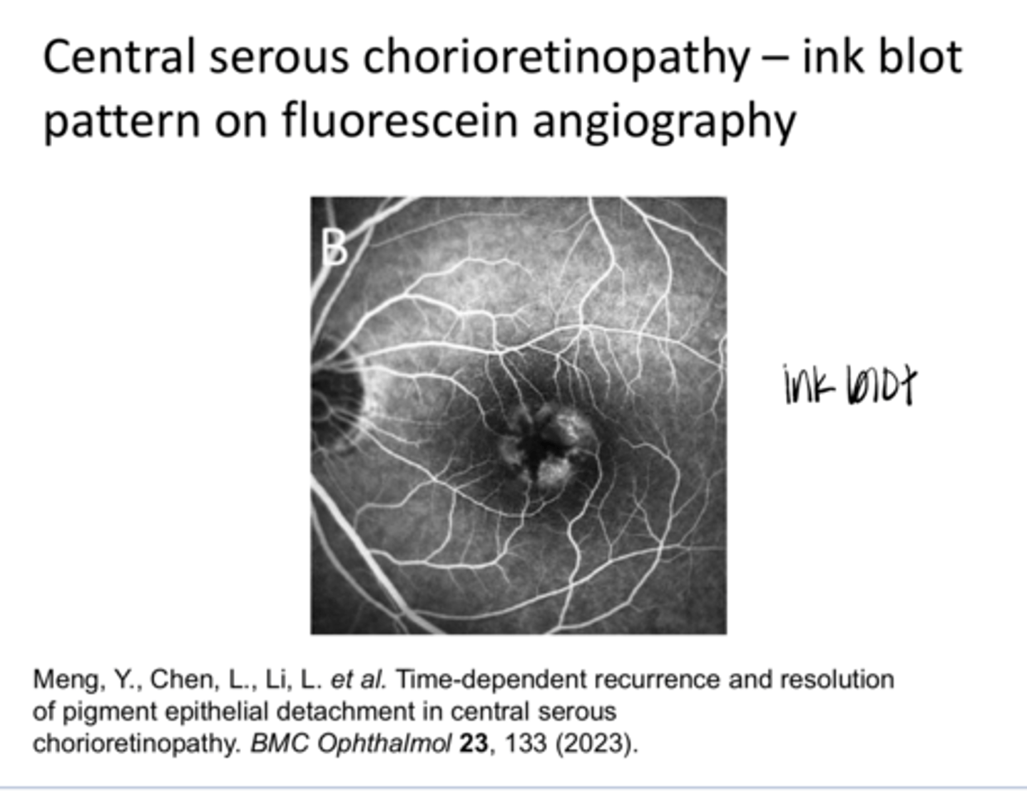
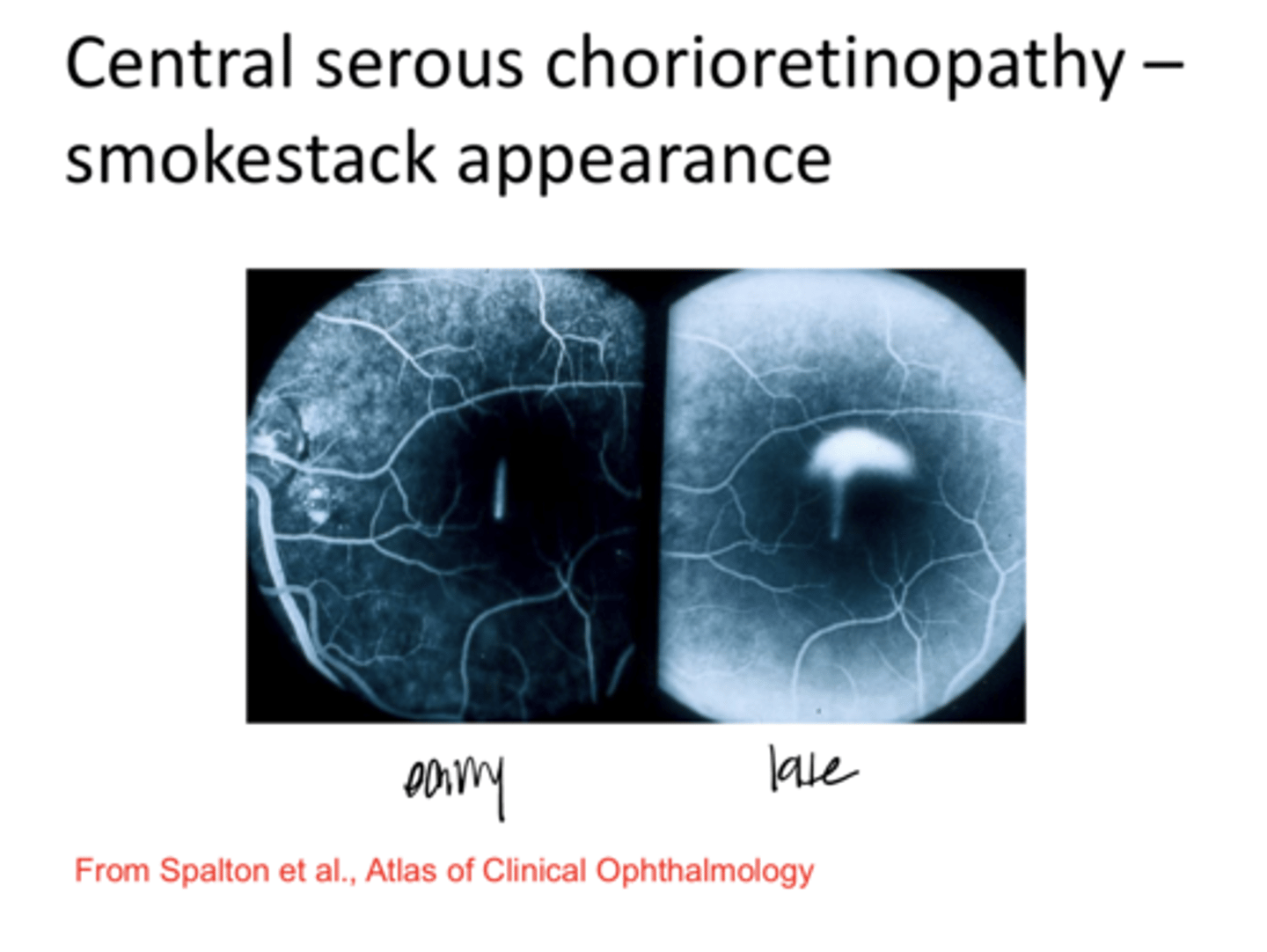
-hyperfluorescence on FANG, "smokestack" FANG
What is the clinical appearance of central serous retinopathy?
hyper
EXAM QUESTION: Central Serous retinopathy is (hyper/hypo) fluorescent on FANG

Ink blot
EXAM QUESTION: What is the #1 FA appearance of central serous retinopathy?

Central Serous Retinopathy (Pic)
Central Serous Retinopathy (Pic)

Central Serous Retinopathy -- Ink Blot Pattern on Fluorescein Angiography (Pic)
Central Serous Retinopathy -- Ink Blot Pattern on Fluorescein Angiography (Pic)
Central Serous Retinopathy -- Smokestack Appearance (Pic)
Central Serous Retinopathy -- Smokestack Appearance (Pic)
No
Is a fluorescein usually done on a central serous patient?
self
Central serous is ____ limiting in most cases
Central Serous Retinopathy -- Macular OCT (Pic)
Central Serous Retinopathy -- Macular OCT (Pic)

Central Serous Retinopathy -- Macular OCT (Pic)
Central Serous Retinopathy -- Macular OCT (Pic)
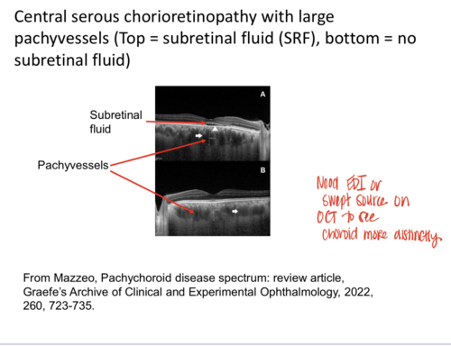
**closer to typical
Central Serous Chorioretinopathy with Large Pachyvessels (Pic)
Central Serous Chorioretinopathy with Large Pachyvessels (Pic)
1-6
Typically, patients will recover from central serous retinopathy in ____ months with no intervention
Yes? Sometimes 20/40ish
Will a patient typically have vision loss from central serous retinopathy?
-Anti VEGF meds
-Laser photocoagulation if subretinal fluid is threatening vision
What are some potential treatments of central serous retinopathy?
-subretinal fluid is threatening vision
-chronic >6m
-significant pigmentary changes (suggestive of damage to the RPE and outer retina)
-obvious choroidal neovasc
When is Laser photocoagulation considered for central serous retinopathy?
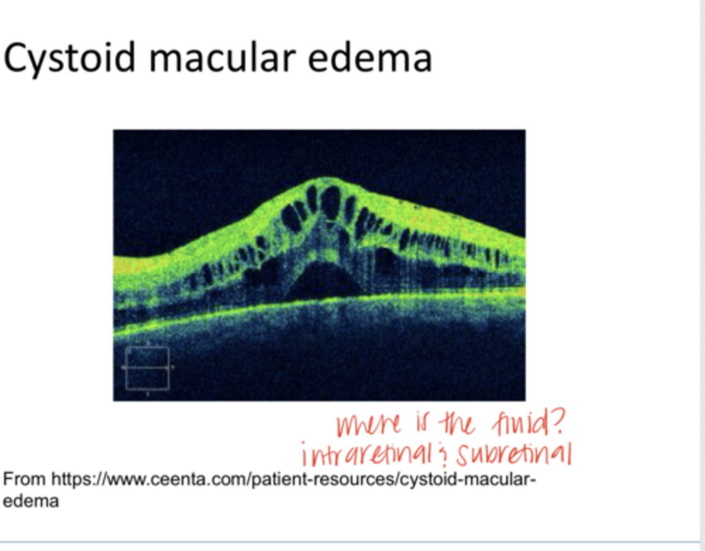
many ocular and systemic conditions
Cystoid Macular Edema is secondary to what?

cataract sx = called Irvine Gas Syndrome
One common association with cystoid macular edema is _______. What is this syndrome called?
6 weeks post op
REVIEW: When does Irvine Gas Syndrome present?
fluid seeps into Henle fibers
Why does Irvine Gas Syndrome present?
-loss of foveal light reflex
-OCT demonstrates dark cystic spaces
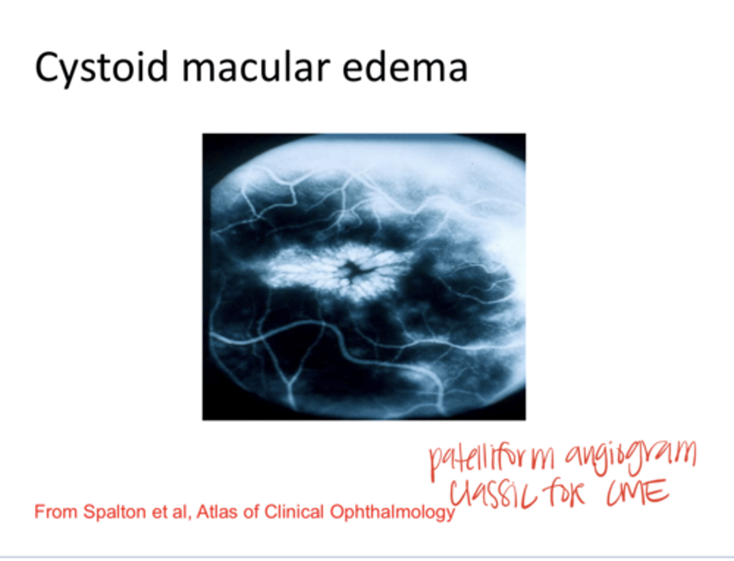
-FA will be useful in the diagnosis, where may see petalliform appearance
What is the clinical presentation of Irvine Gas Syndrome?
Cystoid Macular Edema on OCT (Pic)
Cystoid Macular Edema on OCT (Pic)

Cystoid Macular Edema on FA -- Petalliform Appearance (Pic)
Cystoid Macular Edema on FA -- Petalliform Appearance (Pic)
DEPRIVEN
What is the pneumonic for cystoid macular edema that describes the associations with CME?
-diabetes
-epinephrine (topical glaucoma drops)
-pars planitis (posterior uveitis)
-retinitis pigmentosa
-Irvine-gas Syndrome (intraocular sx)
-Venous occlusion
-E2 prostaglandins
-Nicotinic Acid and Niacin
What does the DEPRIVEN pneumonic stand for?
-treatment depends on the cause
-many patients with the Irvine-Gass Syndrome recover spontaneously within 6 months
-May try oral prostaglandin inhibitors or topical prostaglandin inhibitors
-oral steroids, topical steroids, intravitreal injections of steroids
-carbonic anhydrase inhibitors
-Anti-VEGF injections
-pars plana vitrectomy for chronic cases
What is the management of cystoid macular edema?
Several hours after sungazing, vision drops to the 20/30 to 20/100 range or the patient reports spots or scotomas
What is the onset of solar retinopathy?

RPE dropout and pigmentary clumping
EXAM QUESTION: What is the most common finding in solar retinopathy?

true
True or False:
Visual disruption from solar retinopathy can remain or recover over weeks to months

Yes 00 outer retinal partial thickness or micro home with variable vision of 20/25-20/80
Can you get a micro/partial/full macular hole from solar retinopathy?
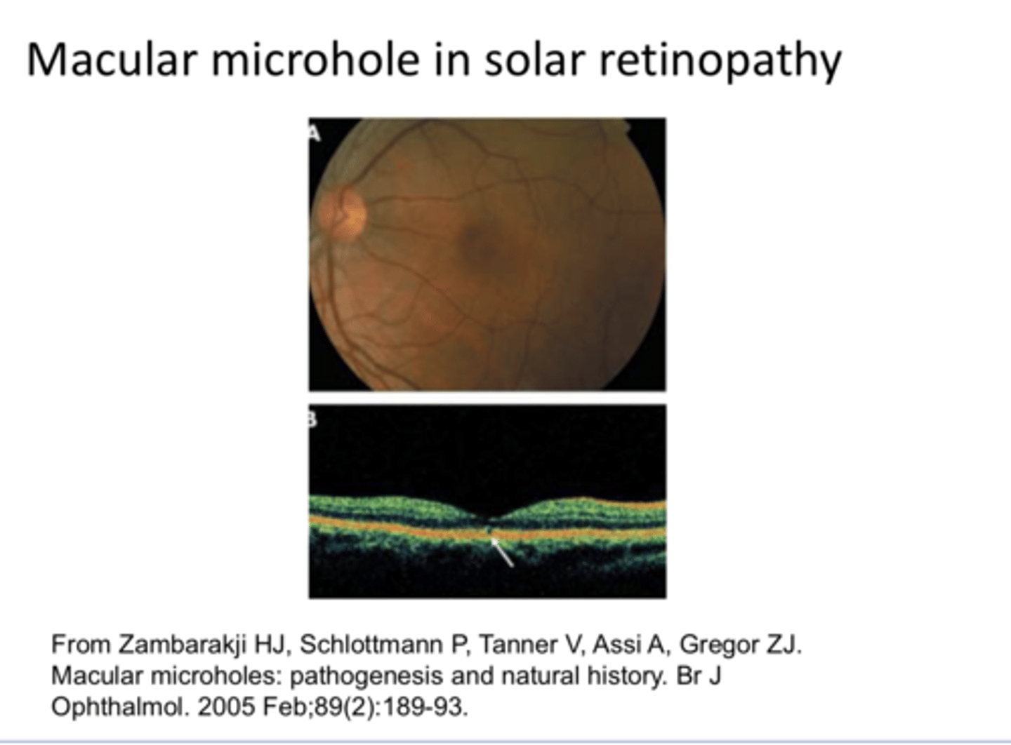
Macular Microhole in Solar Retinopathy (Pic)
Macular Microhole in Solar Retinopathy (Pic)

inner retina (NFL)
What does acute macular neuroretinopathy thought to involve?

unknown -- inflammation in the outer retina
What is the pathophys of acute macular neuroretinopathy?

young women <30
Who does acute macular neuroretinopathy tend to affect?

either
acute macular neuroretinopathy is (unilateral/bilateral)

normal to mildly reduced acuity
What is VA after acute macular neuroretinopathy?
leads to multiple, paracentral scotomas in one or both eyes
What does acute macular neuroretinopathy lead to?

-infection or fibrile illness
-COVID infection and vaccinations
-intravitreal anti-VEGF injections
-oral contraceptive pills
-many other things
What are the possible etiologies of acute macular neuroretinopathy?
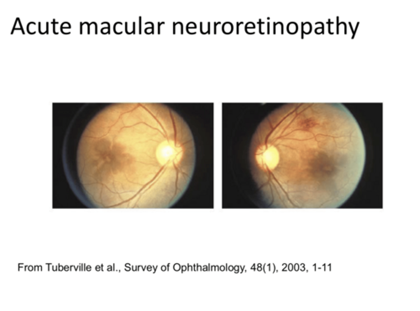
Multiple, well defined, non-elevated wedge-shaped lesions in a flower-petal arrangement around the center of the macula; lesions are reddish-brown
What is the appearance of acute macular neuroretinopathy?

red free filter
acute macular neuroretinopathy is easier to detect with what?

retinal hemes
_____ may occasionally occur with acute macular neuroretinopathy
