Microanatomy of Large Intestine
1/23
There's no tags or description
Looks like no tags are added yet.
Name | Mastery | Learn | Test | Matching | Spaced | Call with Kai |
|---|
No analytics yet
Send a link to your students to track their progress
24 Terms
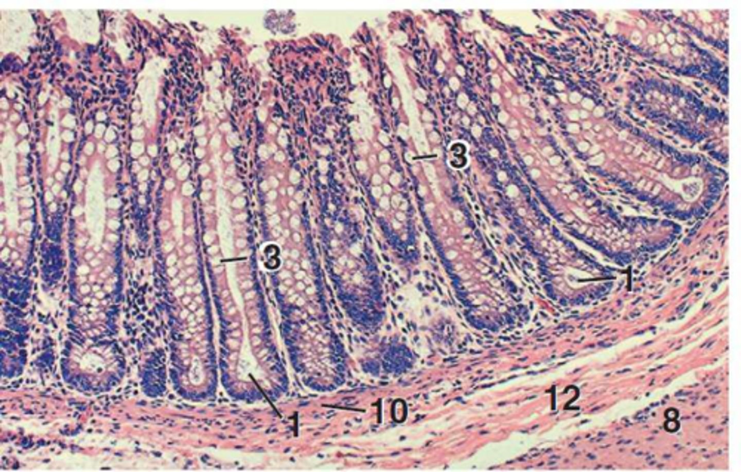
What are the primary functions of the large intestine?
Microbial action on ingesta, absorption of water, vitamins, and electrolytes, and secretion of mucus.
What structural feature is absent in all segments of the large intestine?
Villi.
How do the intestinal glands of the large intestine differ from those of the small intestine?
They are longer, less coiled, simple tubular glands with a higher concentration of goblet cells.
Which specific cell types are notably absent in the large intestine?
Paneth cells.
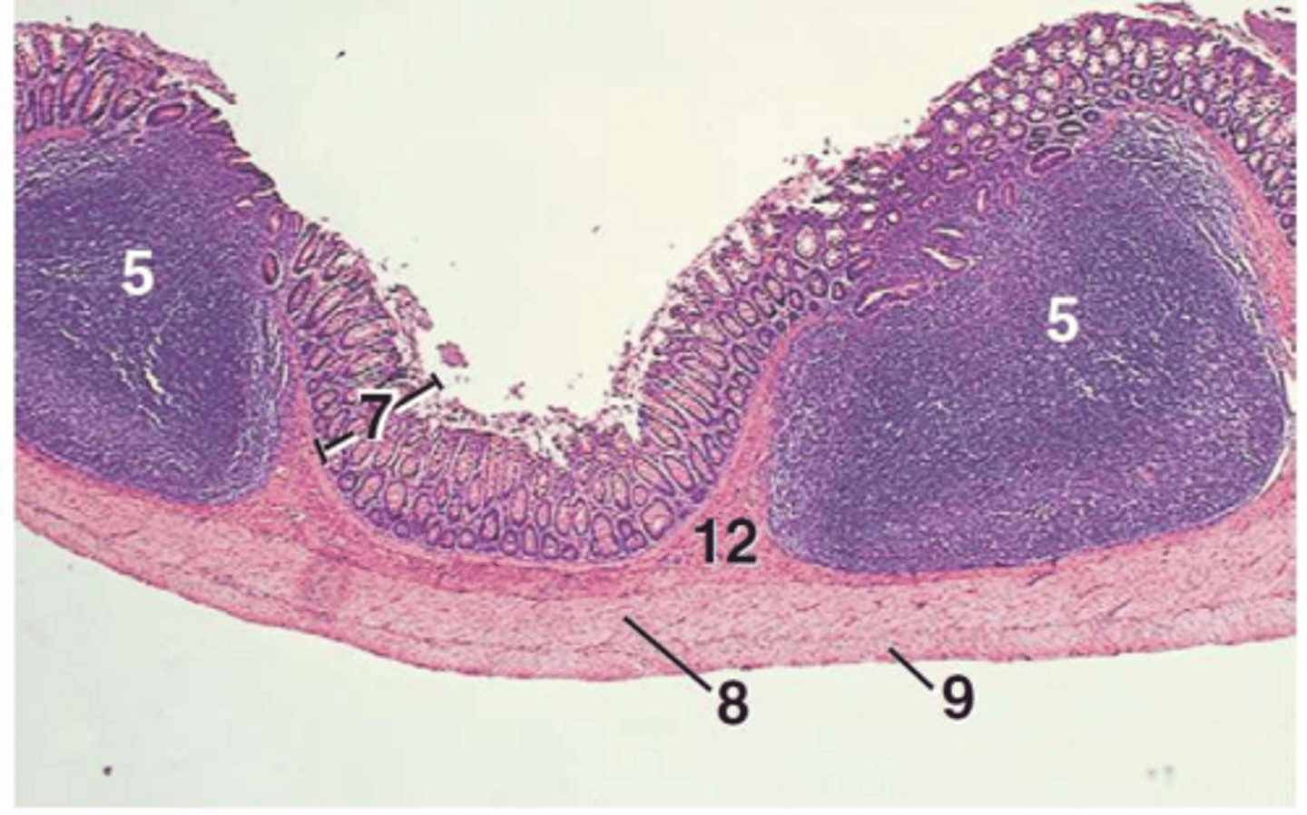
Where are lymphatic nodules primarily concentrated in the cecum of pigs, ruminants, and dogs?
Around the ileal ostium.
Where are lymphatic nodules primarily concentrated in the cecum of horses and cats?
Near the apex of the cecum.
Why is the mucosa of the colon thicker than that of the small intestine?
Due to the increased length of the intestinal glands.
What happens when lymphatic tissue distends the submucosa of the colon?
It disrupts the lamina muscularis, allowing intestinal glands to extend into the submucosa.
What are the taeniae ceci and taeniae coli?
Large, flat muscle bands in the outer longitudinal layer of the tunica muscularis containing numerous elastic fibers.
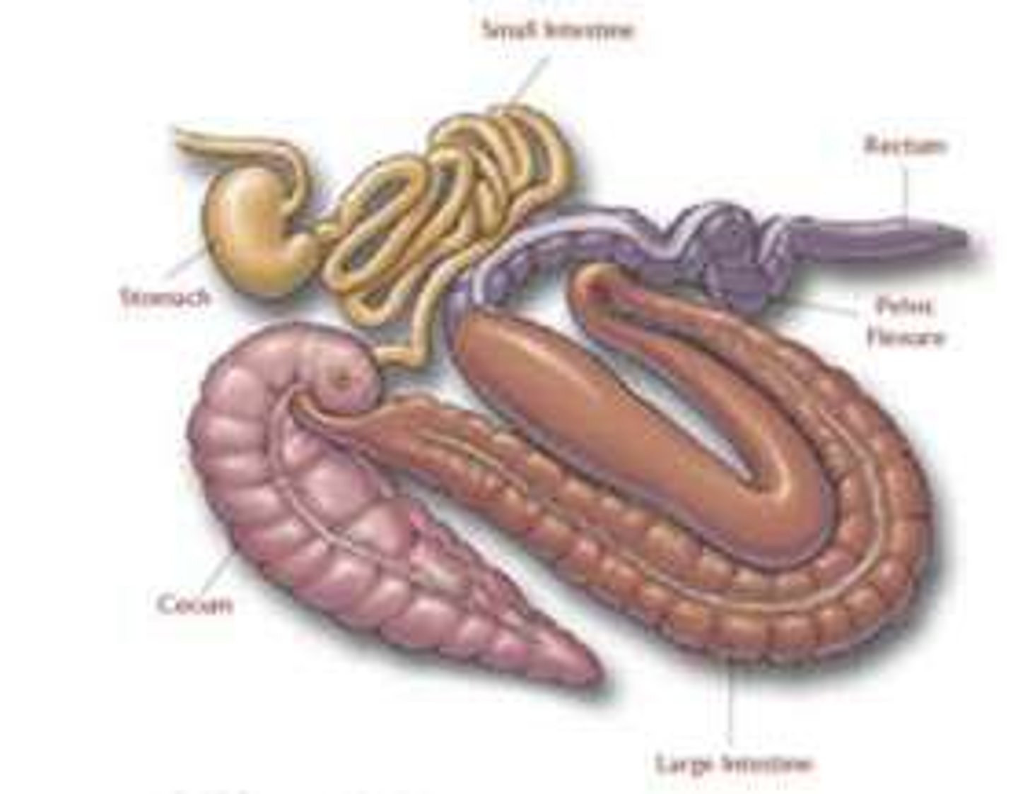
In which species are taeniae coli found?
Pigs and horses.
How does the rectal wall of horses and cattle compare to their colon wall?
The rectal wall is thicker.
Where are elastic fibers most prominent in the rectum of domestic mammals?
In horses and cattle.
What is the difference in coverage between the cranial and retroperitoneal portions of the rectum?
The cranial portion is covered by serosa, while the retroperitoneal portion is surrounded by adventitia.
What are the rectal columns (columnae rectales)?
Longitudinal folds in the rectal mucosa near the junction with the anal canal in ruminants.
What is a prominent feature of the rectum in dogs regarding lymphatic tissue?
Approximately 100 solitary lymphatic nodules visible as rectal pits.
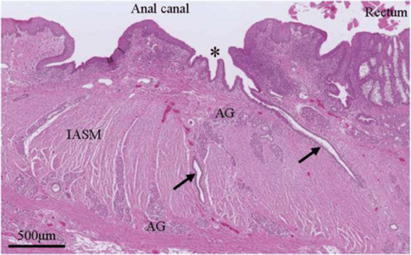
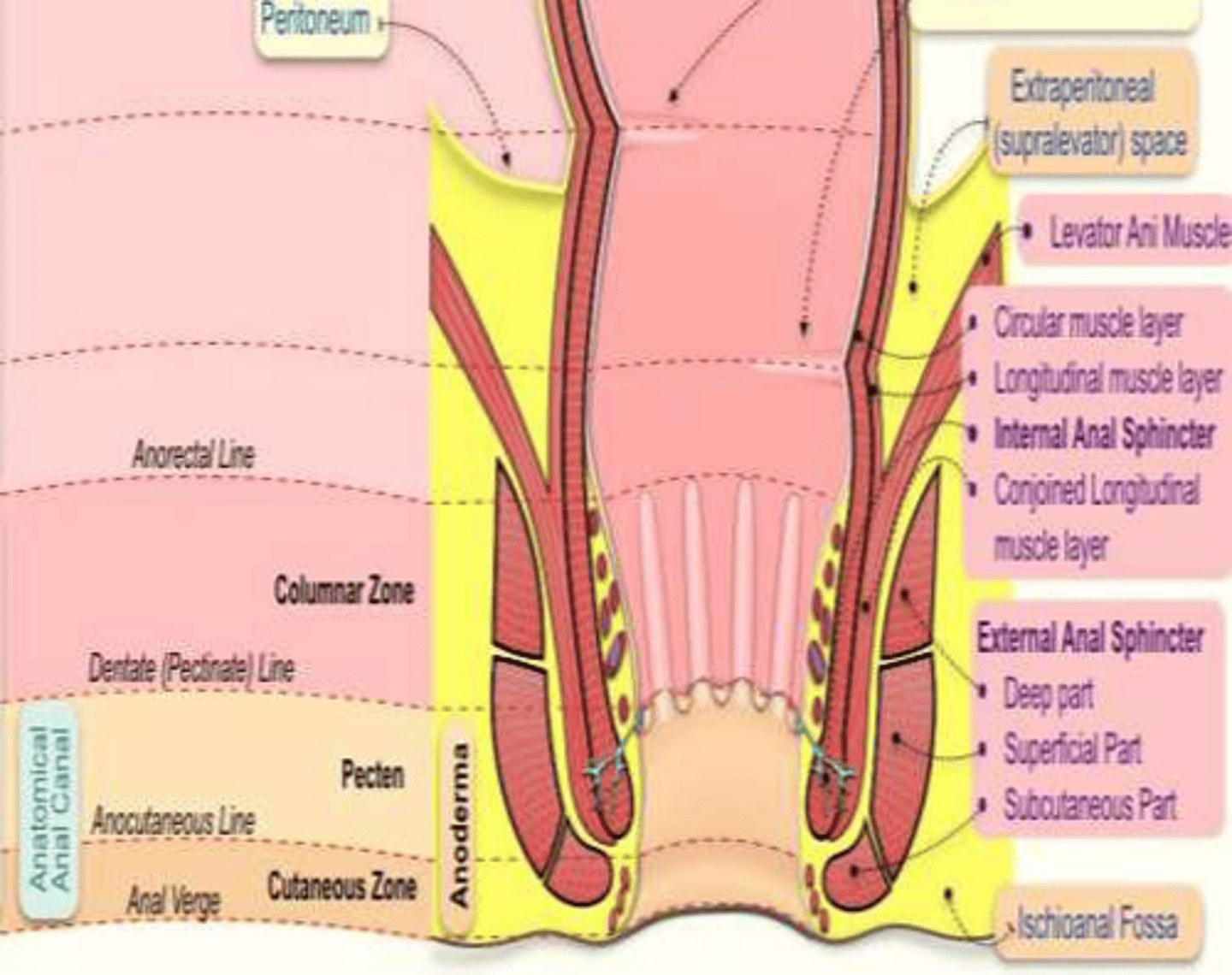
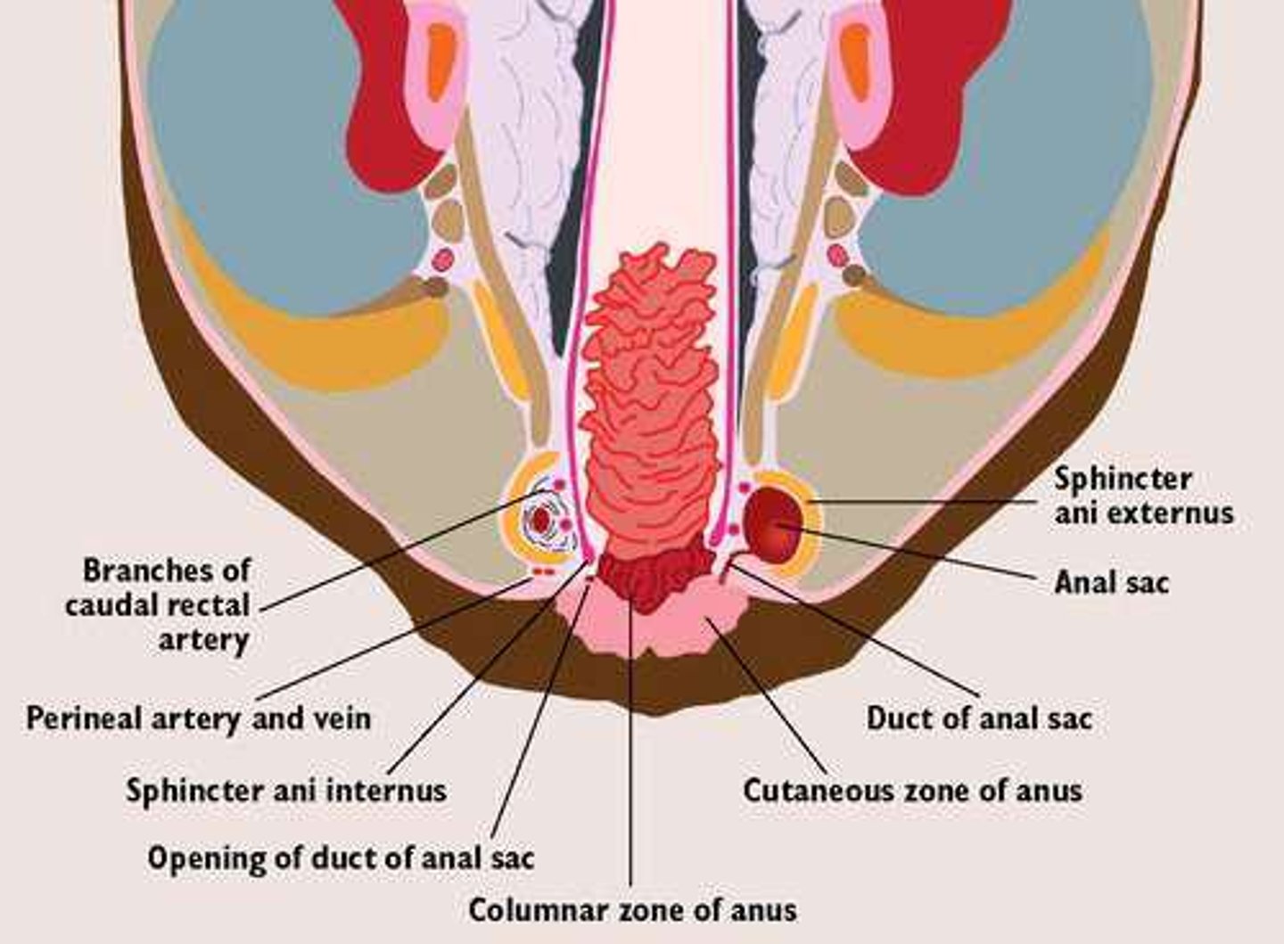
What structure forms the internal anal sphincter?
The continuation of the inner circular layer of the tunica muscularis into the anal canal.
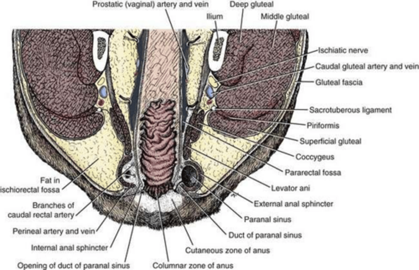
What type of muscle forms the external anal sphincter?
Circularly disposed skeletal muscle.
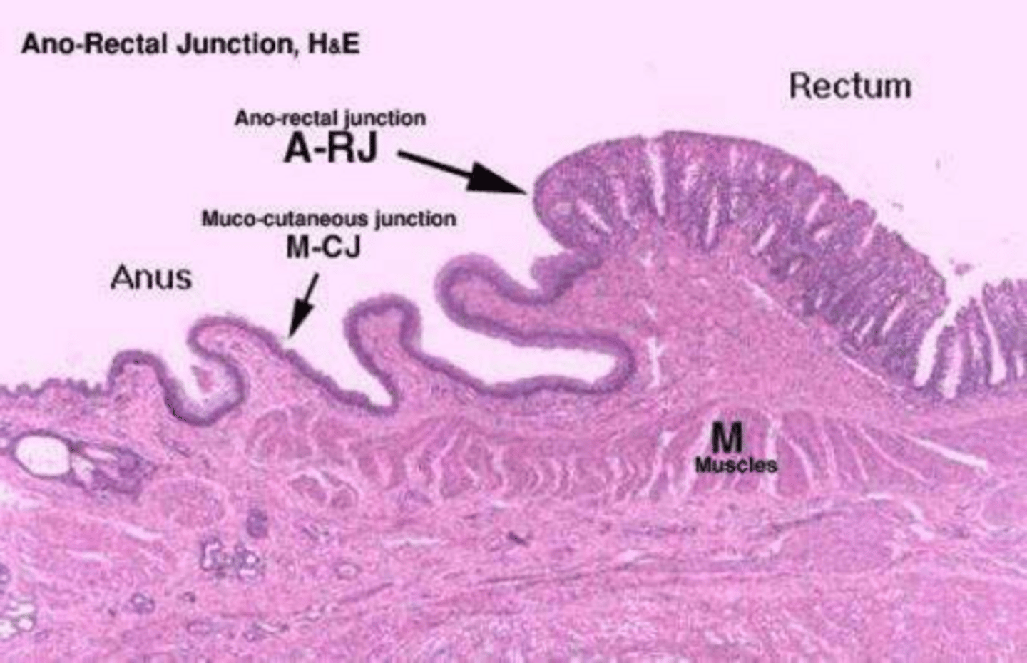
What epithelial change occurs at the anorectal line?
The simple columnar epithelium of the rectum changes abruptly to nonkeratinized stratified squamous epithelium.
What are anal sinuses?
Grooves located between the longitudinal folds (anal columns) in the columnar zone of the anal canal.
What are anal glands and where are they located?
Modified tubuloalveolar sweat glands located in the propria-submucosa of the columnar and intermediate zones.
How does the secretion of anal glands differ between species?
Cats and dogs produce a lipid secretion, while pigs produce a mucous secretion.
What marks the beginning of the cutaneous zone in the anal canal?
The abrupt change from nonkeratinized to keratinized stratified squamous epithelium at the acutaneous line.
What are anal sacs (paranal sinuses)?
Bilateral evaginations of the anal mucosa in carnivores, with ducts opening at the junction of the intermediate and cutaneous zones.
What are circumanal glands?
Large, modified sebaceous glands found in the outermost part of the cutaneous zone in dogs.