LA Pelvic Limb, Urogenital, Head Anatomy
1/152
There's no tags or description
Looks like no tags are added yet.
Name | Mastery | Learn | Test | Matching | Spaced | Call with Kai |
|---|
No analytics yet
Send a link to your students to track their progress
153 Terms
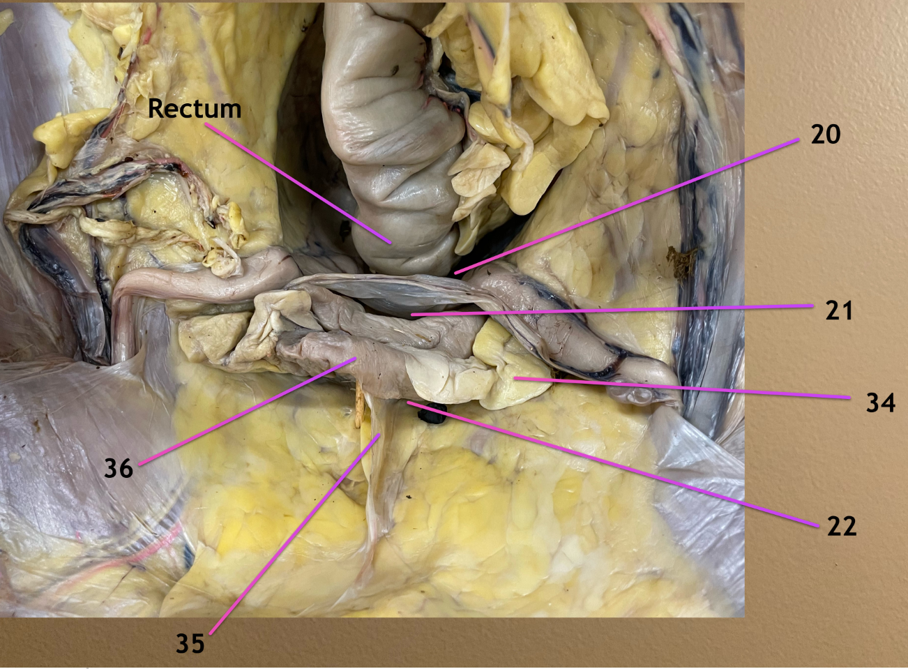
looking into pelvic cavity, cranial to cd view
.
label 20-22
rectogenital pouch
vesicogenital pouch
pubovesical pouch


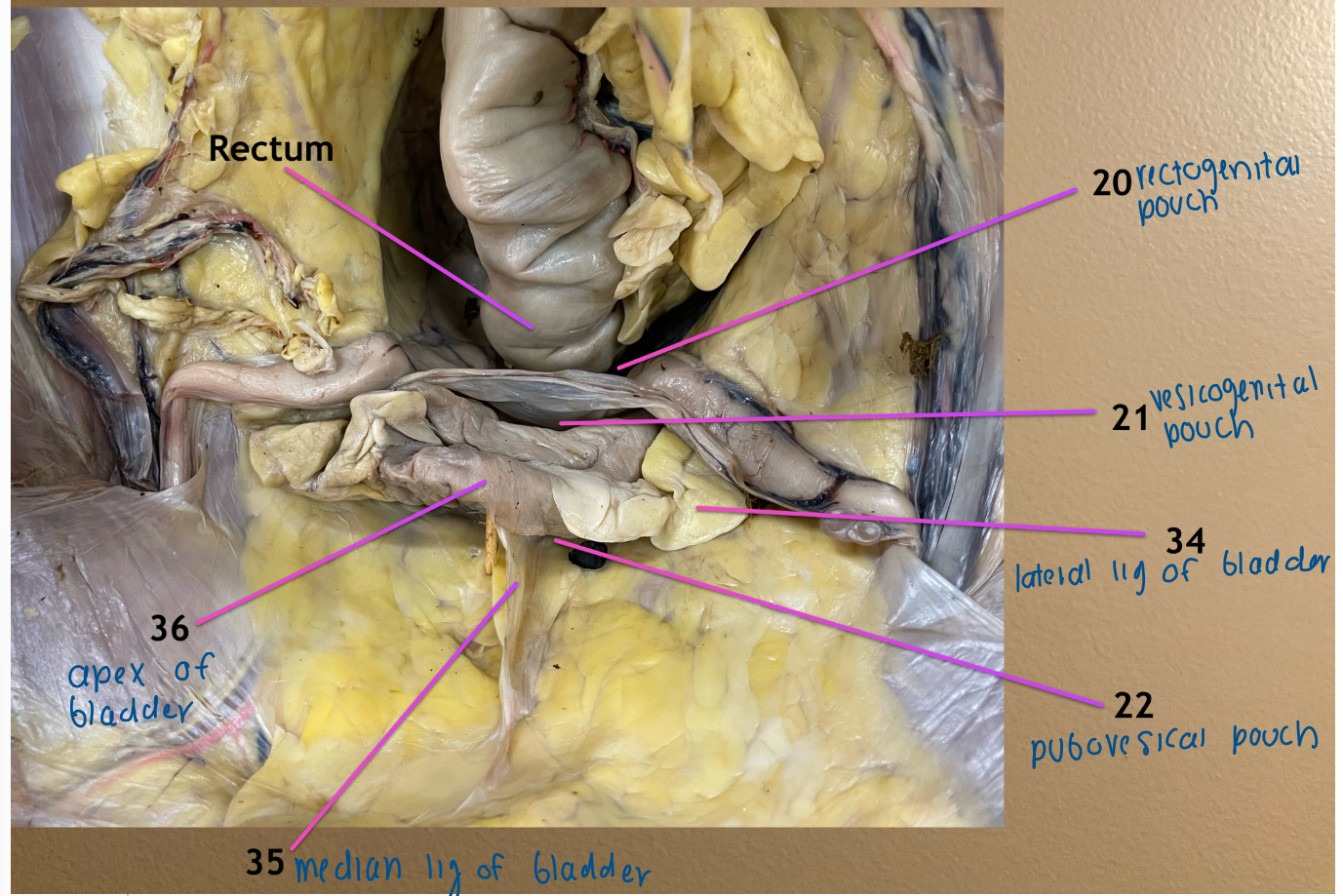
looking into pelvic cavity, cranial to cd view
.
label 34, 35
lateral ligament of the bladder
median ligament of the bladder
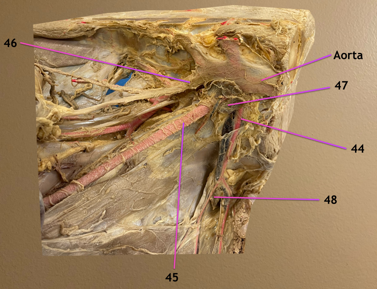
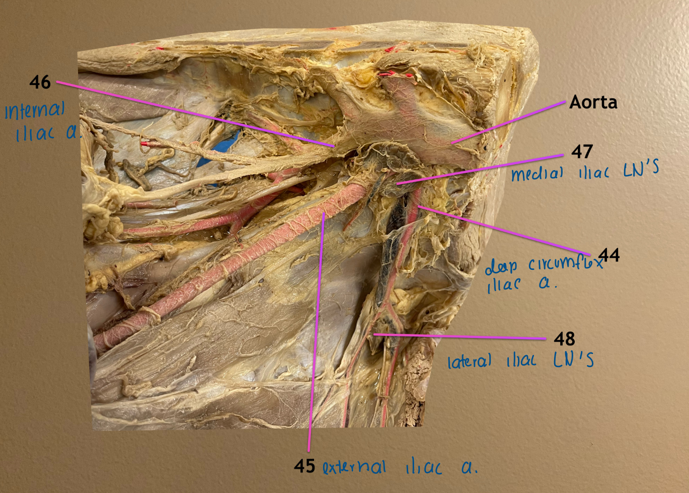
ventral view
.
label 44-48
deep circumflex iliac a.
external iliac a.
internal iliac a.
medial iliac lymph nodes: located at origin of 44
lateral iliac lymph nodes: located where 44 bifurcates
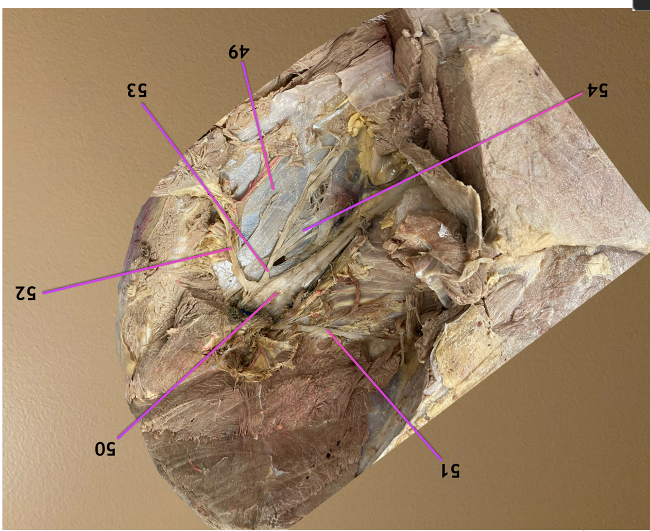
lateral view, head to left, dorsal to top
.
label 49, 54
sacrosciatic ligament: white CT sheet deep to nerves in the lumbosacral plexus
pudendal n.
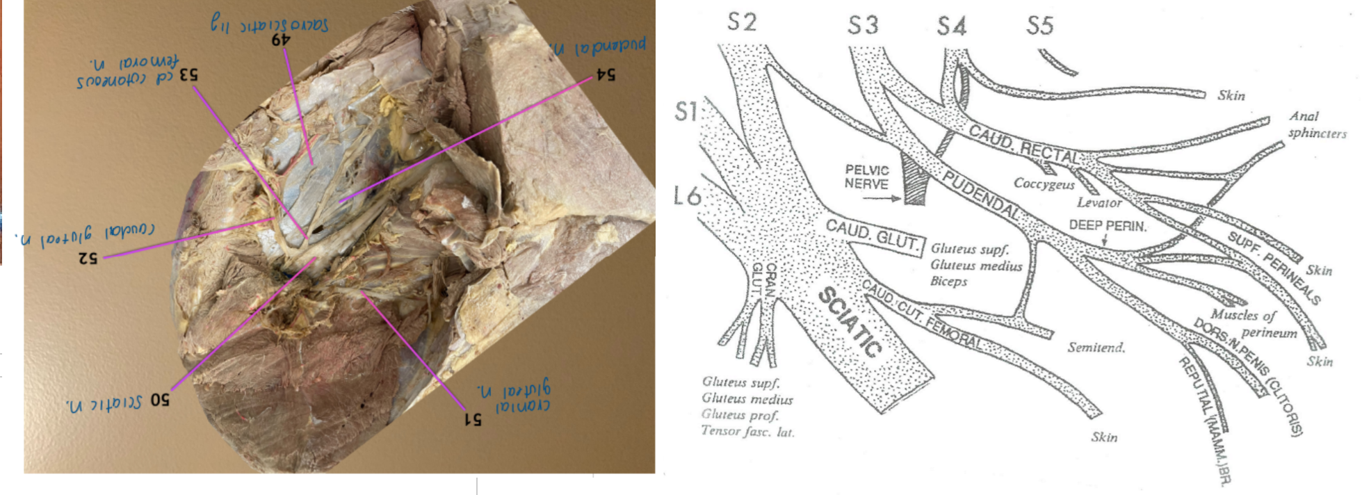
lateral view, head to left
.
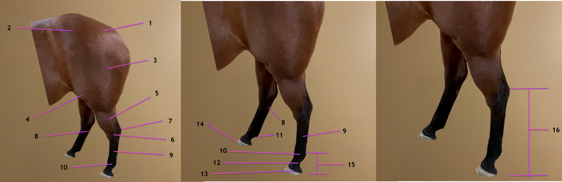
label the topographic regions: 1-4
INCLUDE ALL NAMES FOR 1
rump / croup
point of the hip
thigh
stifle


lateral view, head to left
.
label the topographic regions: 5-8
INCLUDE ALL NAMES FOR 5, 6
crus / leg / gaskin
tarsus / hock
point of the hock
chestnut (remnant of tarsal pad)


lateral view, head to left
.
label the topographic regions: 9-12
INCLUDE ALL NAMES FOR 9, 10
metatarsus / cannon
fetlock / ankle / metatarsophalangeal joint
ergot (remnant of metacarpal pad)
pastern


lateral view, head to left
.
label the topographic regions: 13-16
coronet
hoof
digit
pes
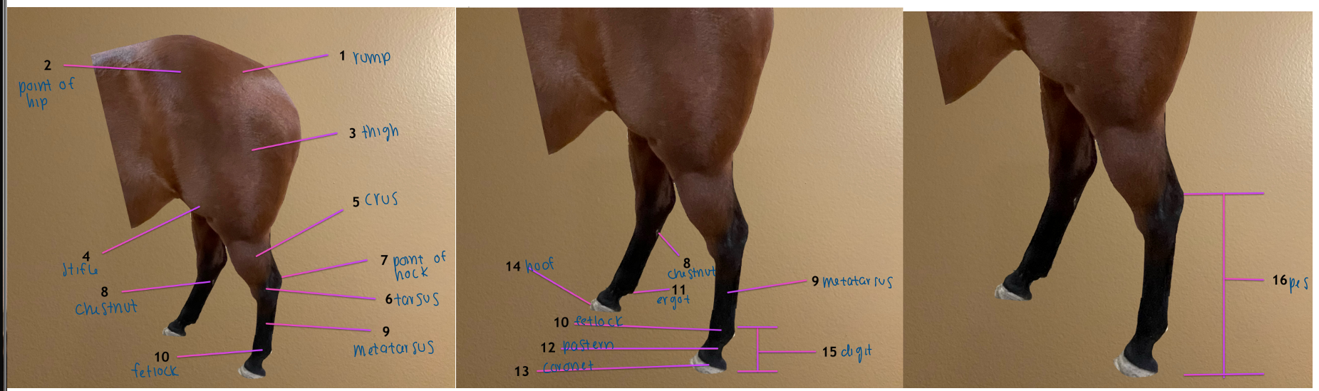

pic 1: cranial view
pic 2: caudal view
pic 3: lateral view
.
name this bone: 17
label 17a, 18, 19
femur
17a: head of femur
fovea capitis
greater trochanter


pic 1: cranial view
pic 2: caudal view
pic 3: lateral view
.
label 20-23
cranial part of greater trochanter
caudal part of greater trochanter
third trochanter
lesser trochanter
.
bone is the femur

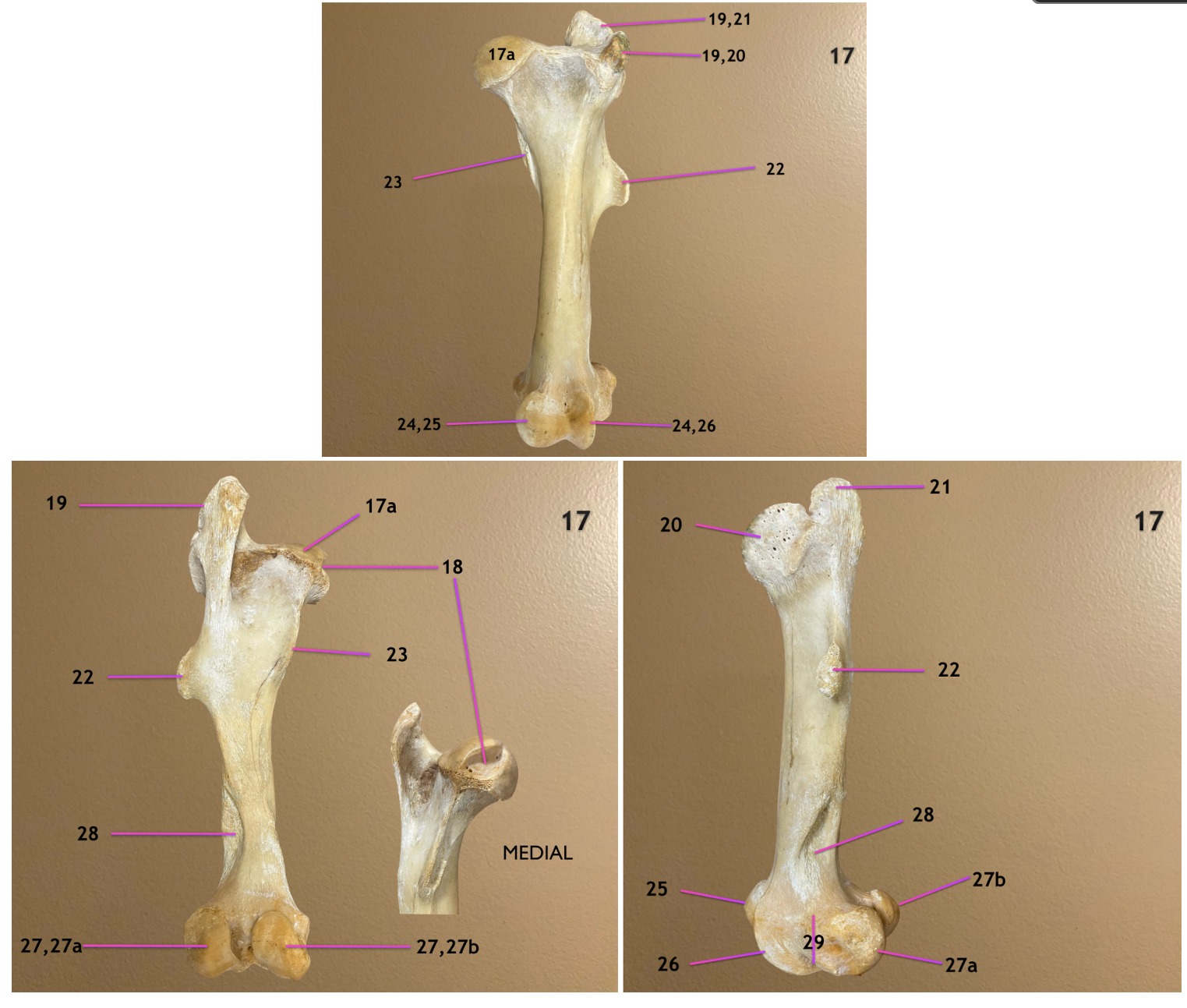
pic 1: cranial view
pic 2: caudal view
pic 3: lateral view
.
label 24-27
trochlea of femur
medial trochlear ridge
lateral trochlear ridge
condyle of femur
.
bone is the femur


pic 1: cranial view
pic 2: caudal view
pic 3: lateral view
.
label 27a, 27b, 28, 29
27a: lateral condyle
27b: medial condyle
supracondyloid fossa
extensor fossa
.
bone is the femur
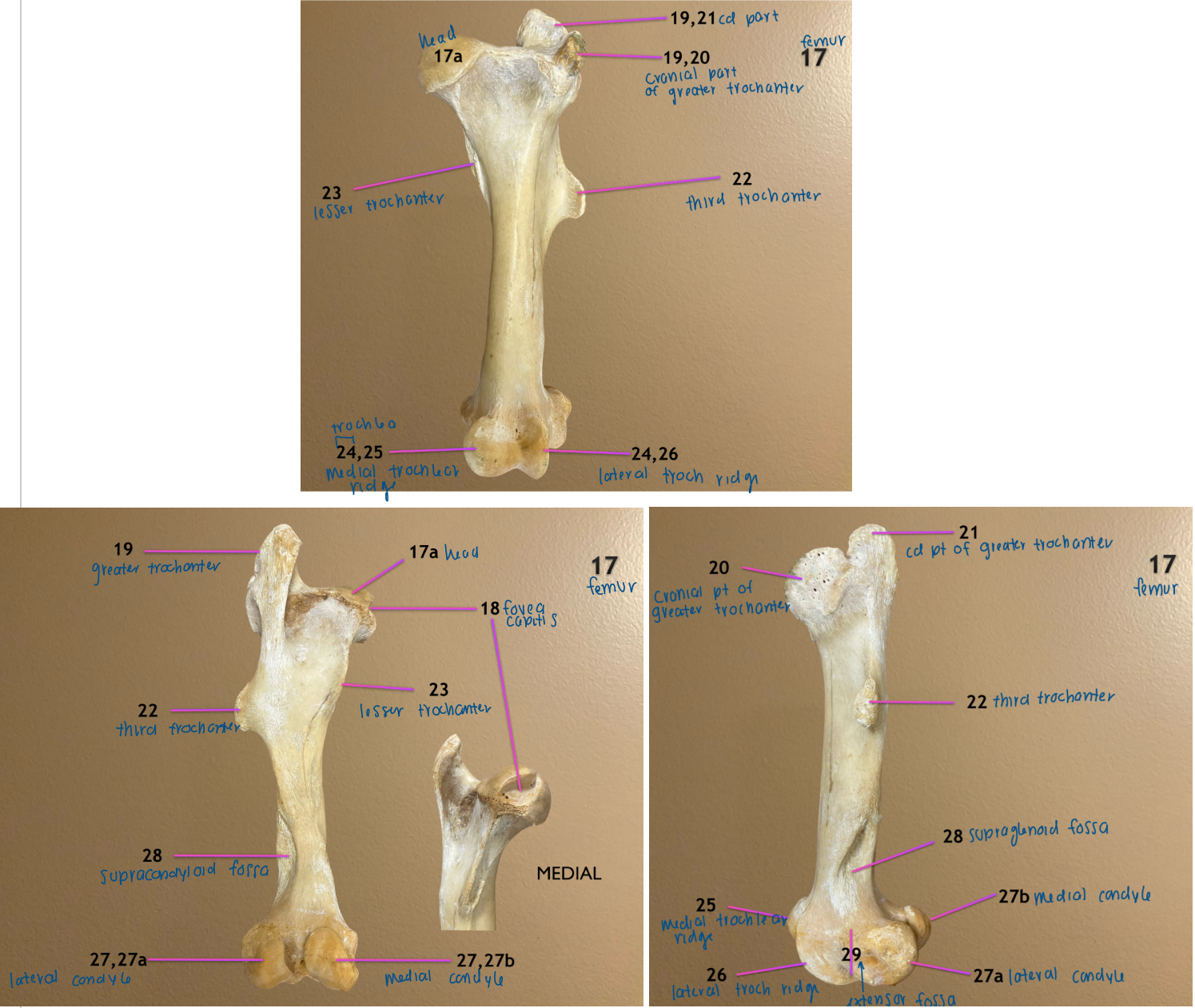
cranial view
.
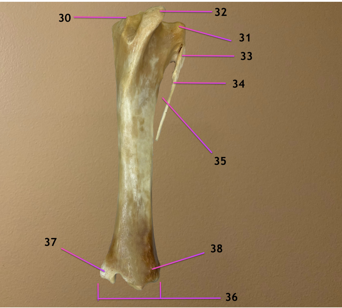
what bone is this
.
label 30-33
tibia (left, medial) & fibula (rt, lateral, not fully developed in equine)
.
medial condyle of tibia
lateral condyle of tibia
tibial tuberosity
head of fibula


cranial view
.
label 34-38
body of fibula
crural interosseous space
cochlea
medial malleolus
lateral malleolus
.
bone is tibia & fibula, fibula is lateral
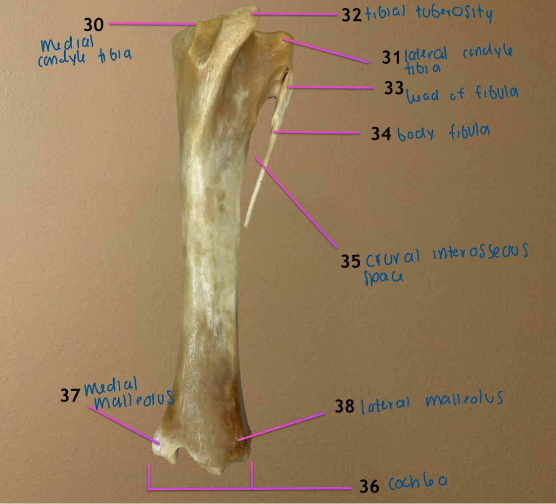
plantar view
.
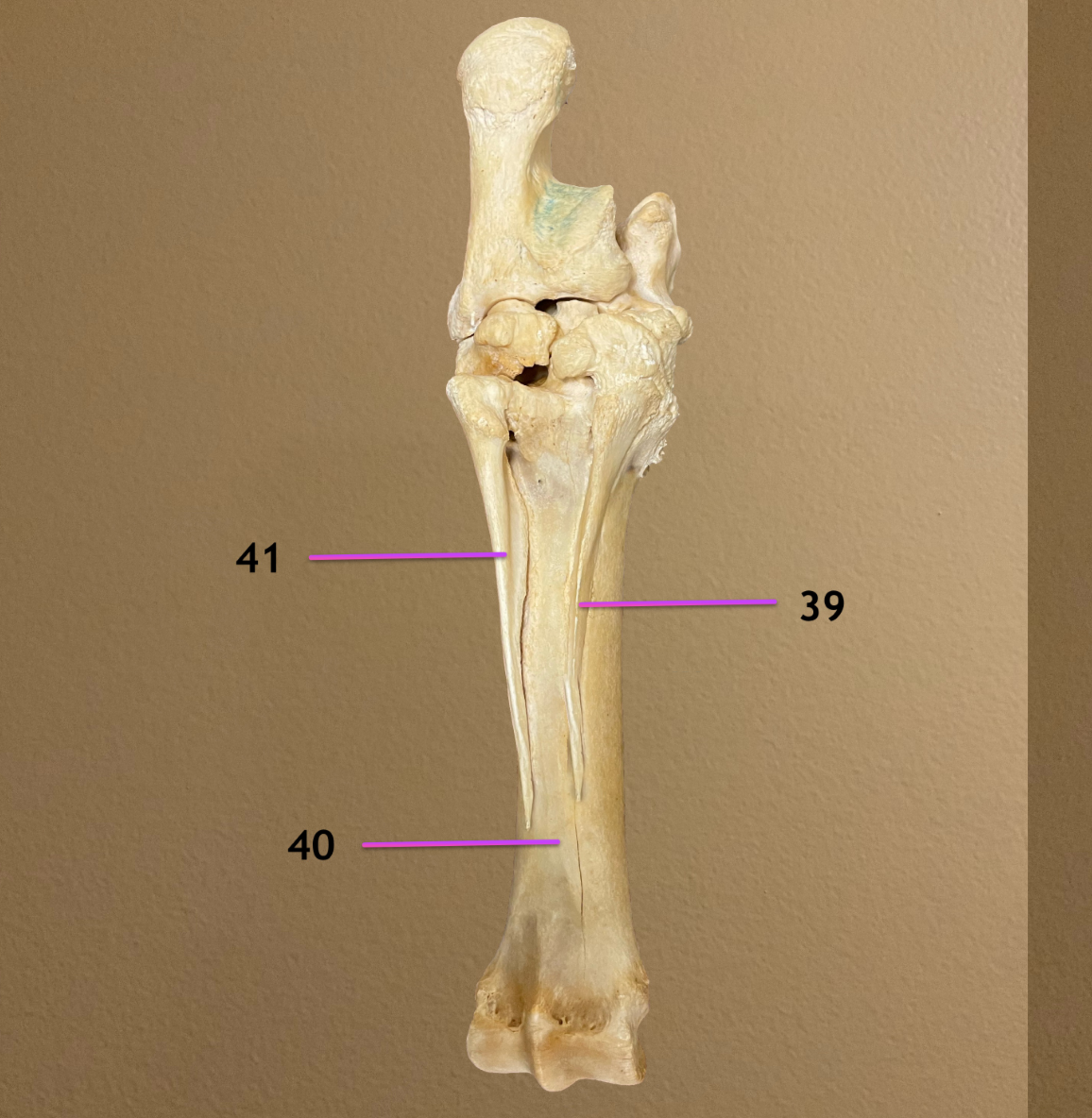
label 39-41
INCLUDE ANATOMICAL & COMMON NAME FOR ALL
metatarsal bone II / medial splint bone
metatarsal bone III / cannon bone
metatarsal bone IV / lateral splint bone
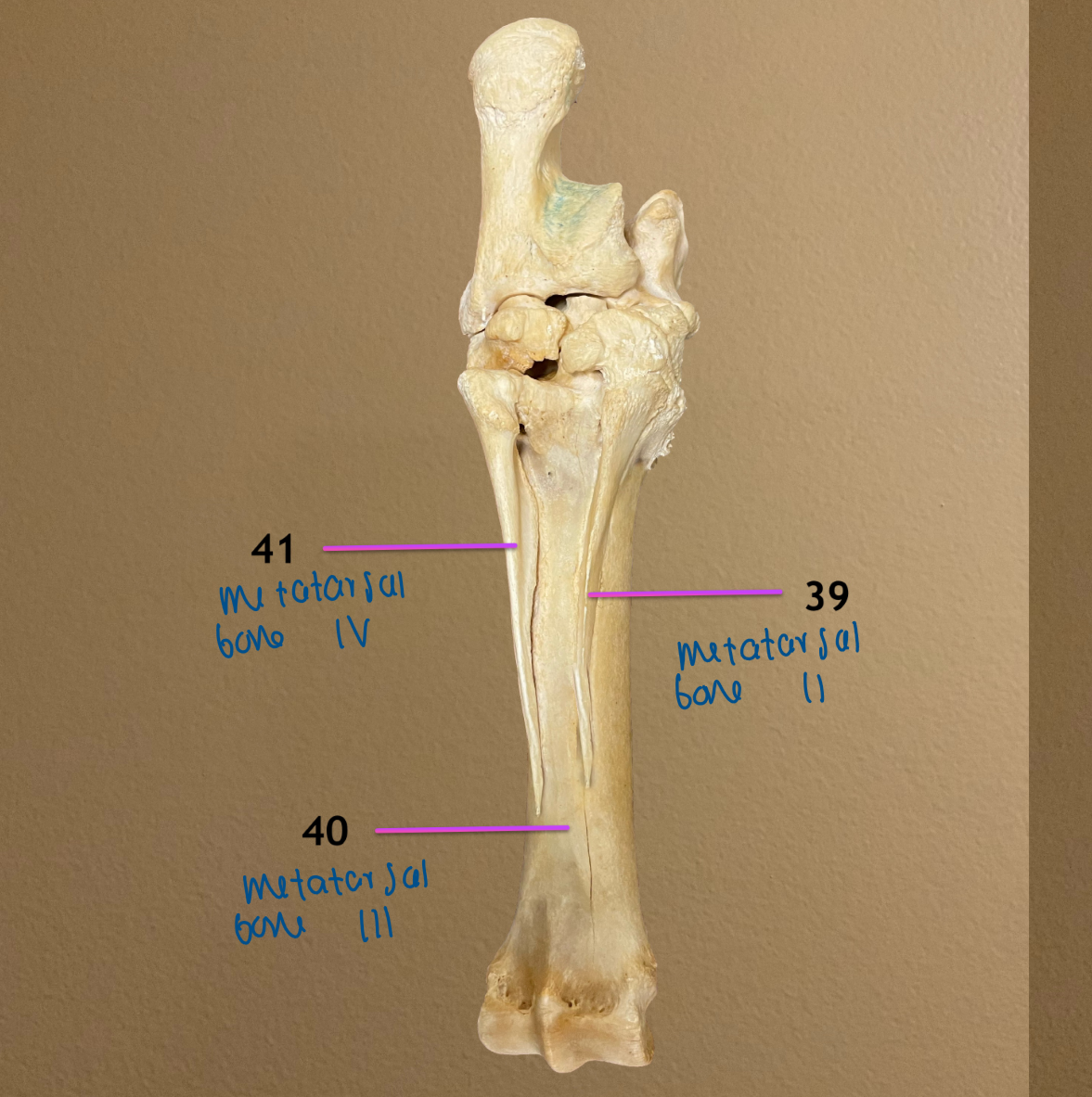

left pic: dorsal view
rt pic: plantar view
.
label 42-44
include all 3 names for 44
medial proximal sesamoid bone
lateral proximal sesamoid bone
proximal phalanx / PI / long pastern bone
.
note: this is the exact same as thoracic limb
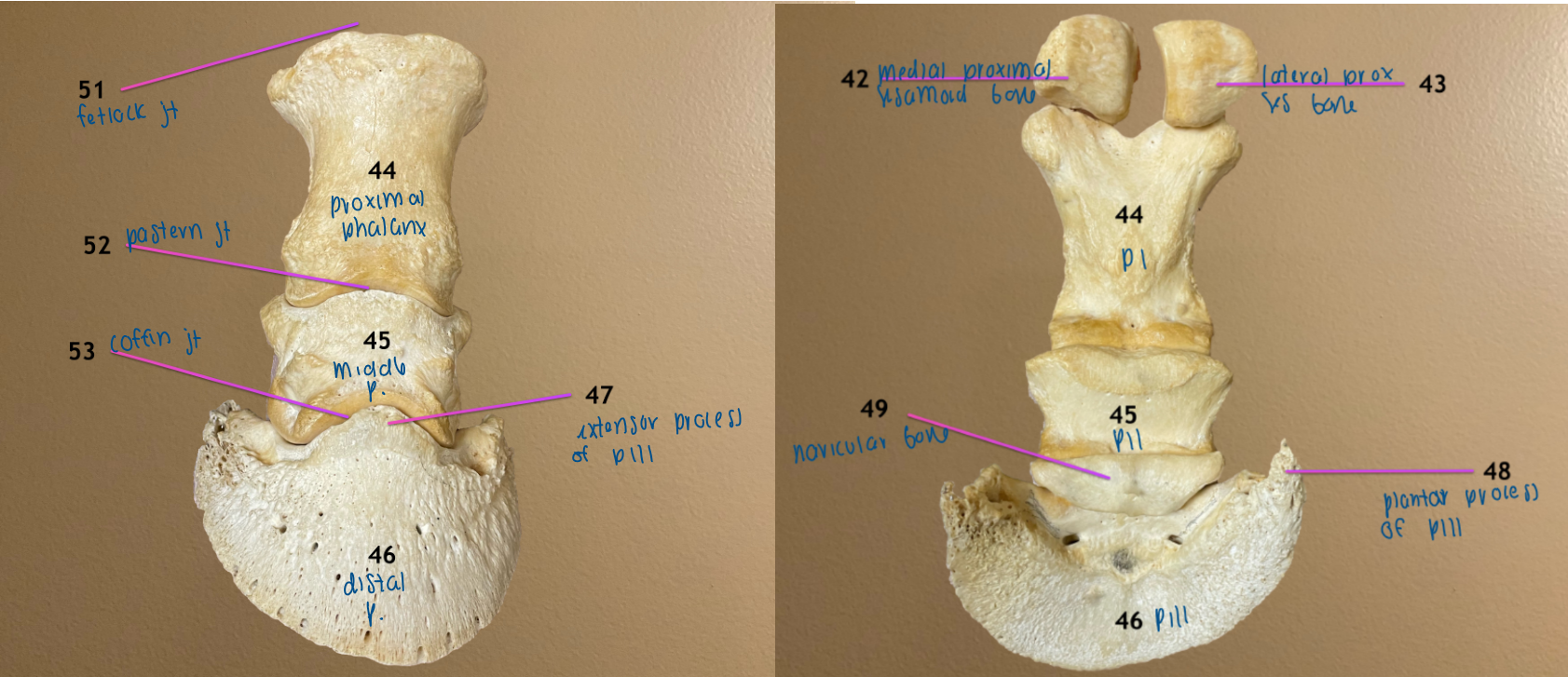
left pic: dorsal view
rt pic: plantar view
.
label 45-48
include all 3 names for 45, 46
middle phalanx / PII / short pastern bone
distal phalanx / PIII / coffin bone
extensor process of PIII
plantar process of PIII
.
note: this is the exact same as thoracic limb


left pic: dorsal view
rt pic: plantar view
.
label 49, 51-53
include anatomical & common name for 51-53
navicular bone
metatarsophalangeal / fetlock joint
proximal interphalangeal / pastern joint
distal interphalangeal / coffin joint
.
note: this is the exact same as thoracic limb


all are lateral view of hip & thigh, cranial to rt
pic 2: 57 & 60 reflected caudally
pic 3: 54 reflected
pic 4: 55 & 56 reflected
.
label 54-57
middle gluteal m.
middle gluteal accessory head
biceps femoris m.
semitendinosus
.
note: all are hip extensors

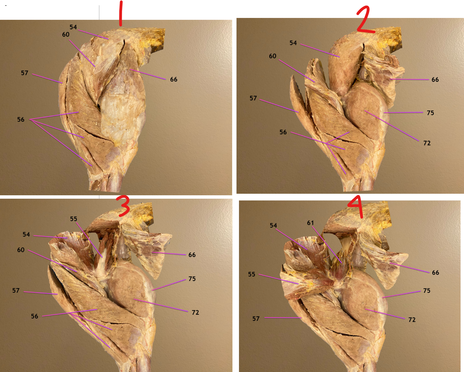
all are lateral view of hip & thigh, cranial to rt
pic 2: 57 & 60 reflected caudally
pic 3: 54 reflected
pic 4: 55 & 56 reflected
.
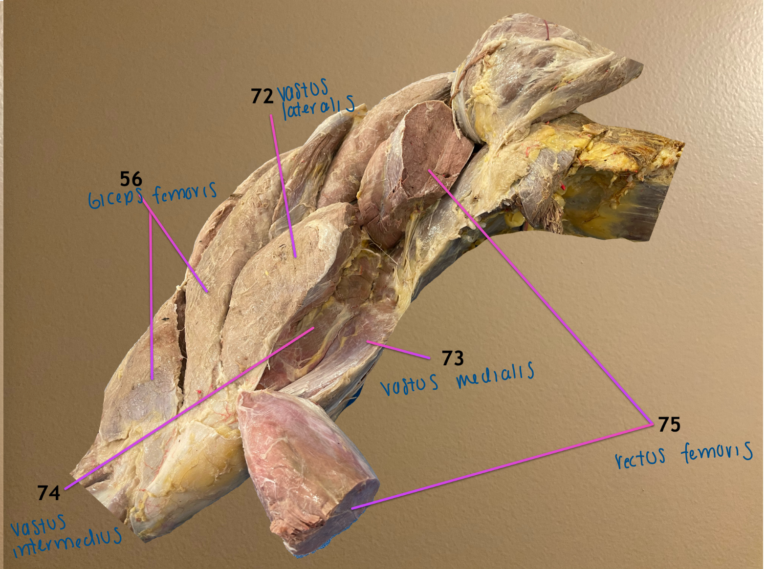
label 60, 61, 66, 75
superficial gluteal m.: hip abductor
deep gluteal m.: hip abductor
tensor fasciae latae: hip flexor
rectus femoris: hip flexor & stifle extensor
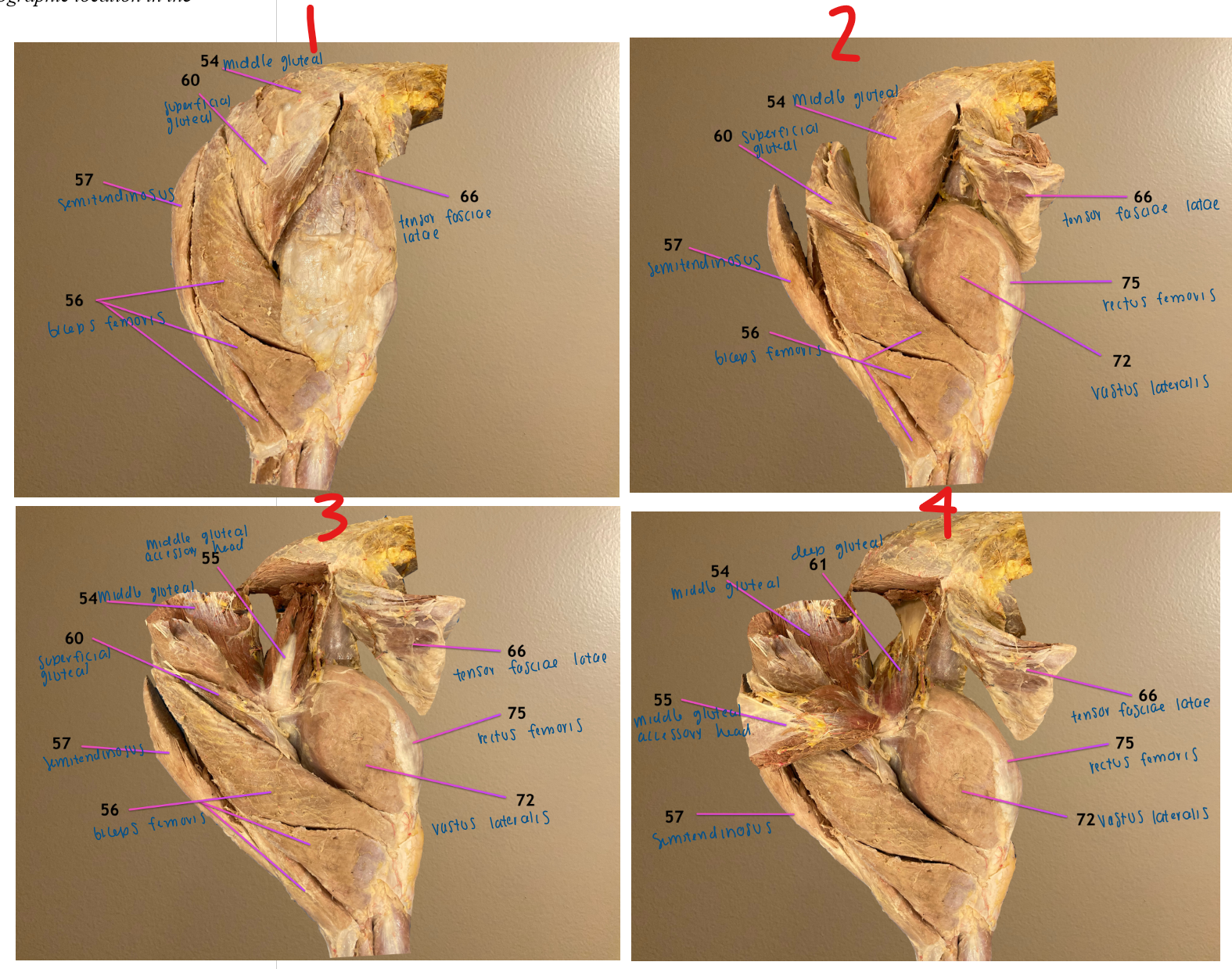
lateral view, cranial to rt
.
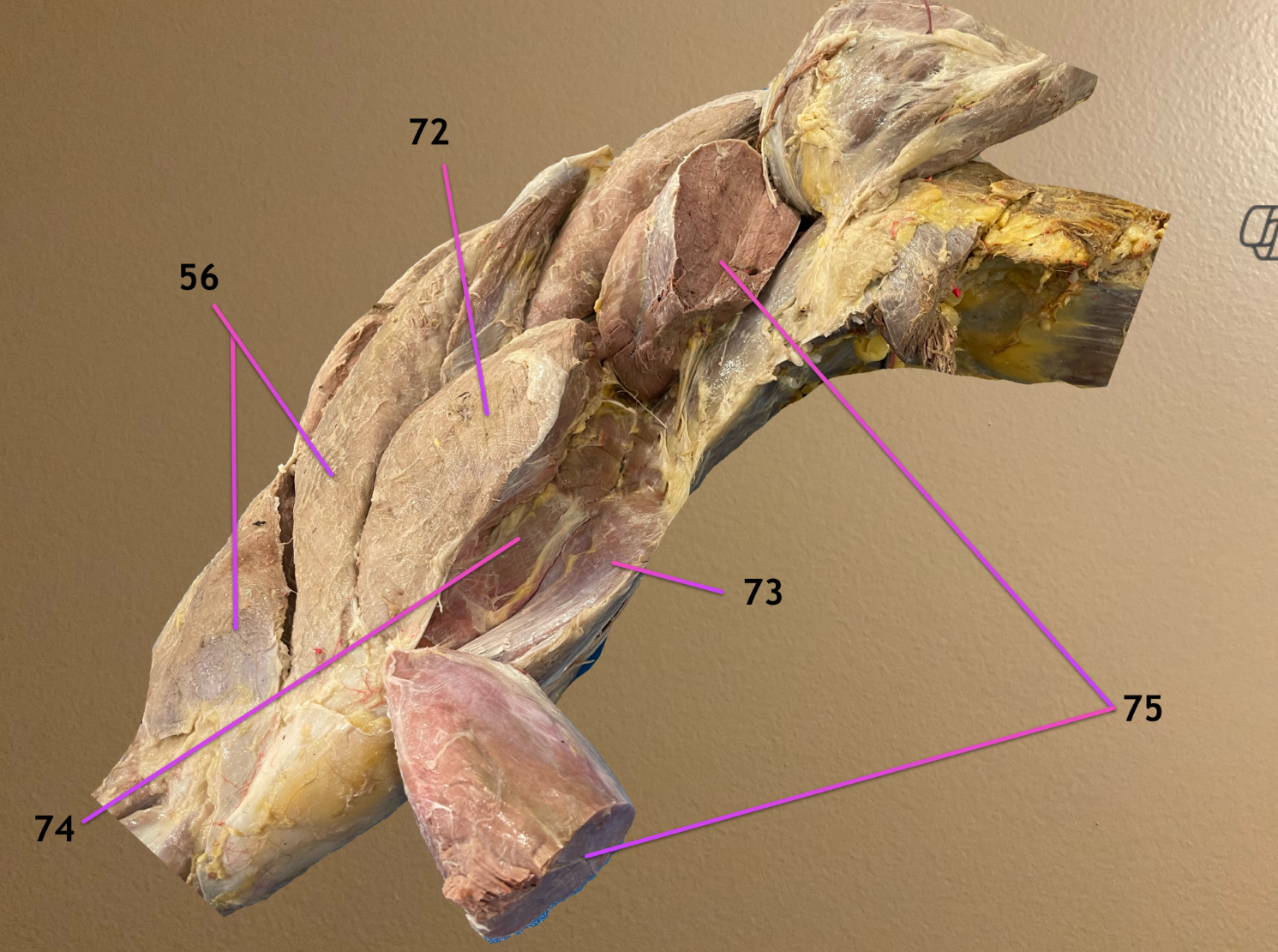
label the parts of the quadriceps femoris: 72-74
vastus lateralis
vastus medialis
vastus intermedius
.
note: all are stifle extensors; rectus femoris (75) is also part of quad fem

all are medial view of thigh, cranial to rt
pic 3: 62 & 68 reflected ventrally to reveal deeper structures
.
label 58, 62, 68, 69
semimembranosus m.: hip extensor
sartorius m.: hip flexor
gracilis m.: hip adductor
adductor m.: hip adductor
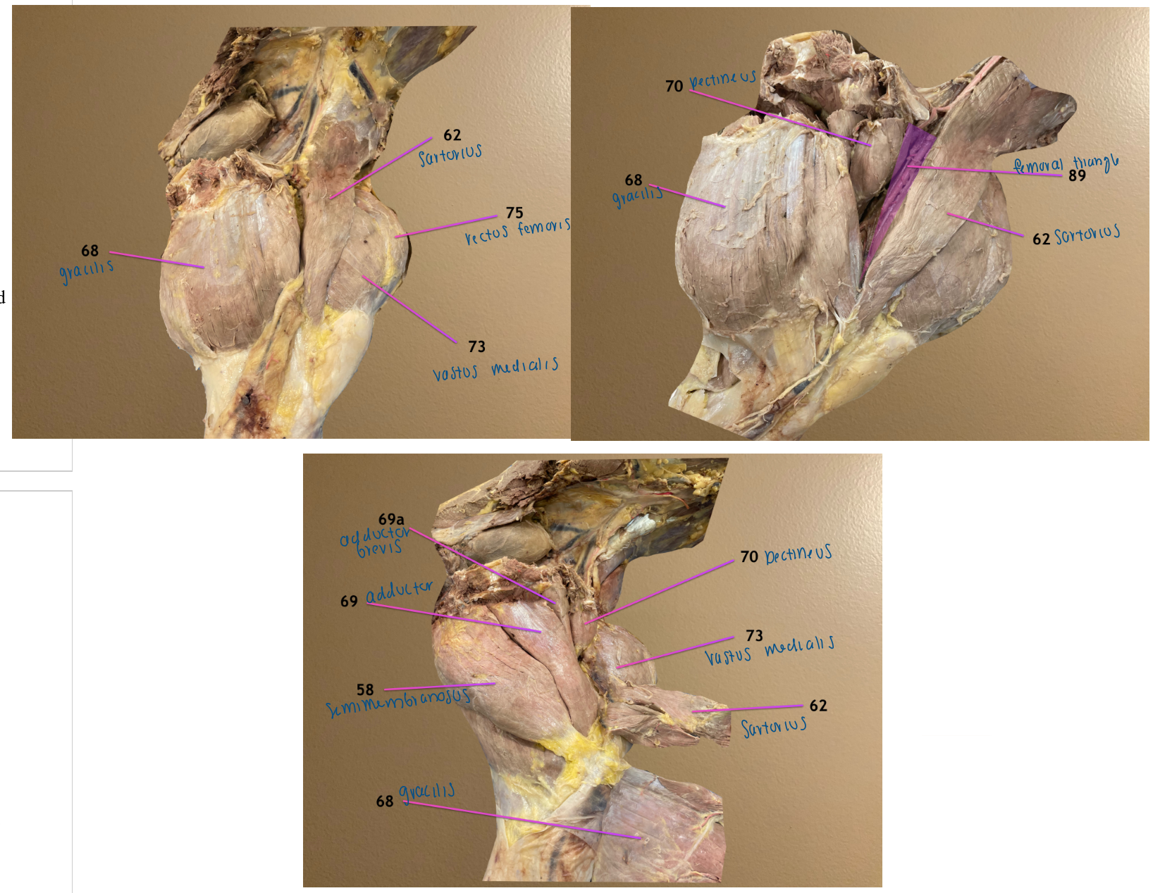
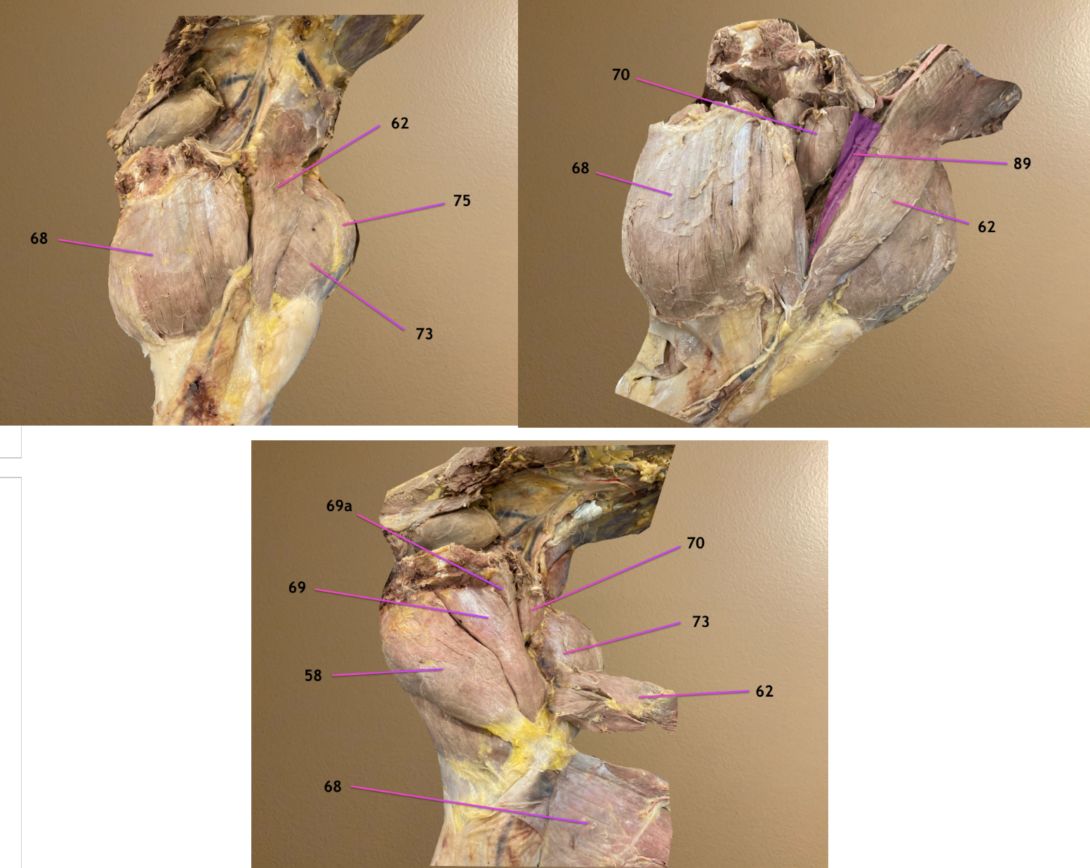
all are medial view of thigh, cranial to rt
pic 3: 62 & 68 reflected ventrally to reveal deeper structures
.
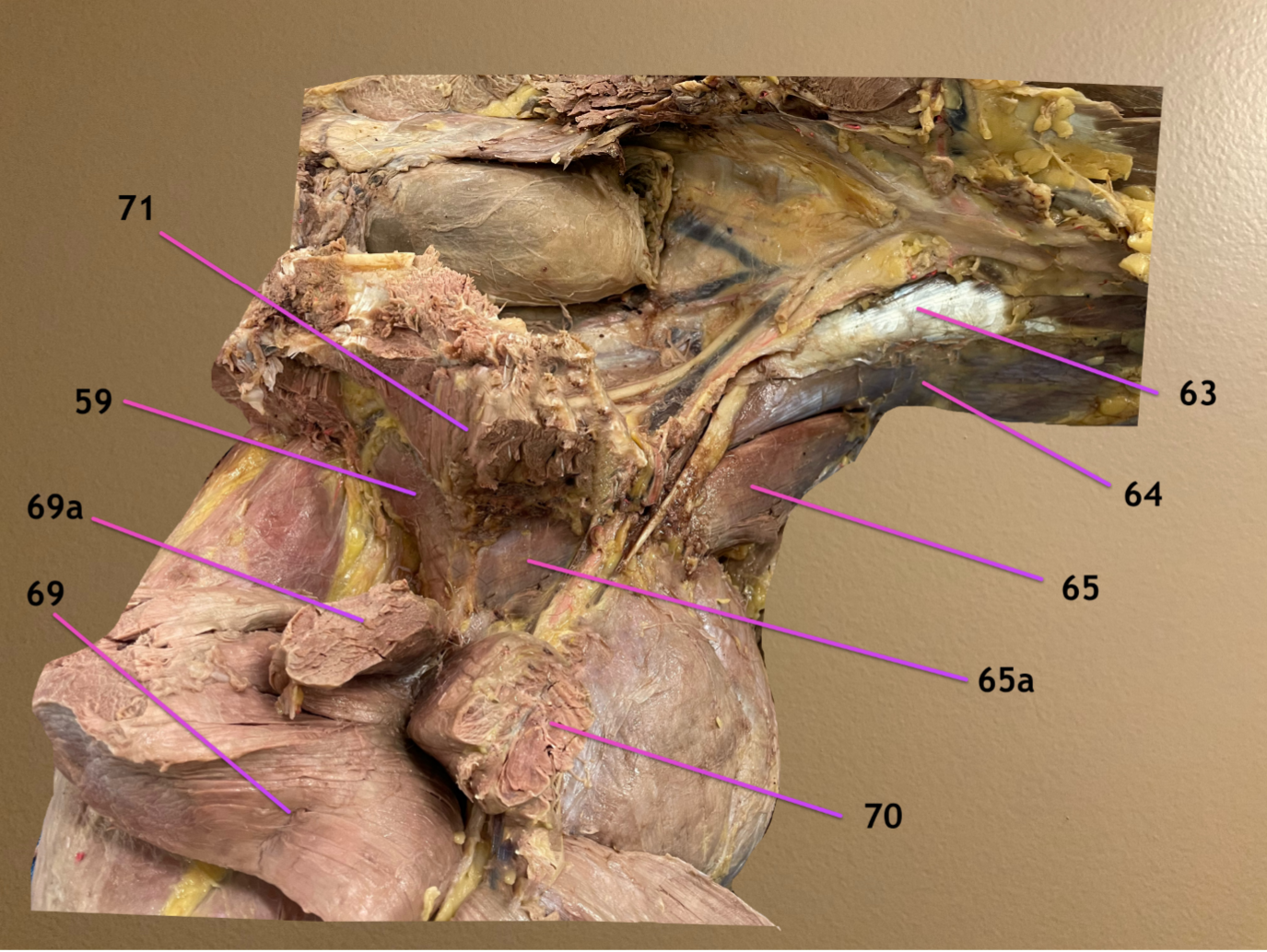
label 69a, 70, 89
69a: adductor brevis: hip adductor
pectineus m.: hip adductor
femoral triangle: area between sartorius (62) cranially & pectineus (70) caudally


ventral view of hip, horse laying on back, cranial to rt, left side of body toward bottom
.
label 59, 63, 64
quadratus femoris m.: hip extensor
psoas minor m.: hip flexor
psoas major m.: hip flexor
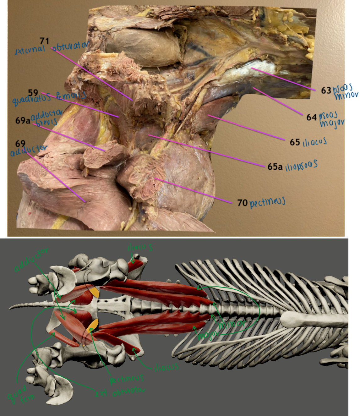
ventral view of hip, horse laying on back, cranial to rt, left side of body toward bottom
.
label 65, 65a, 71
iliacus m.: hip flexor
65a: iliopsoas m.: hip flexor
external obturator m.: hip adductor


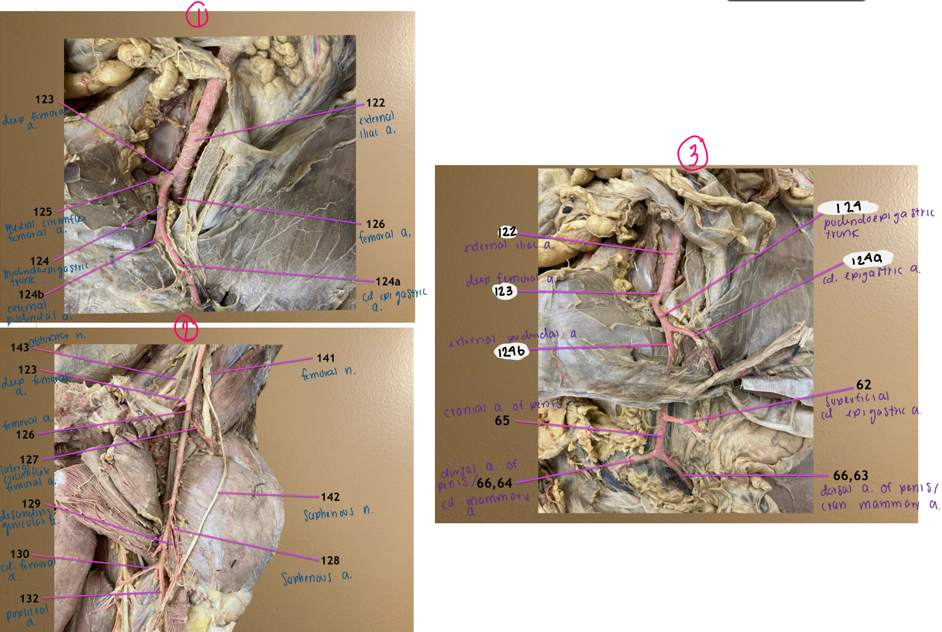
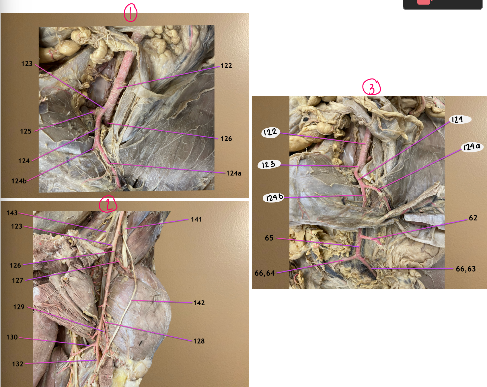
medial view of blood flow near inguinal canal
pic 2 follows 126
pic 3 follows 123
.
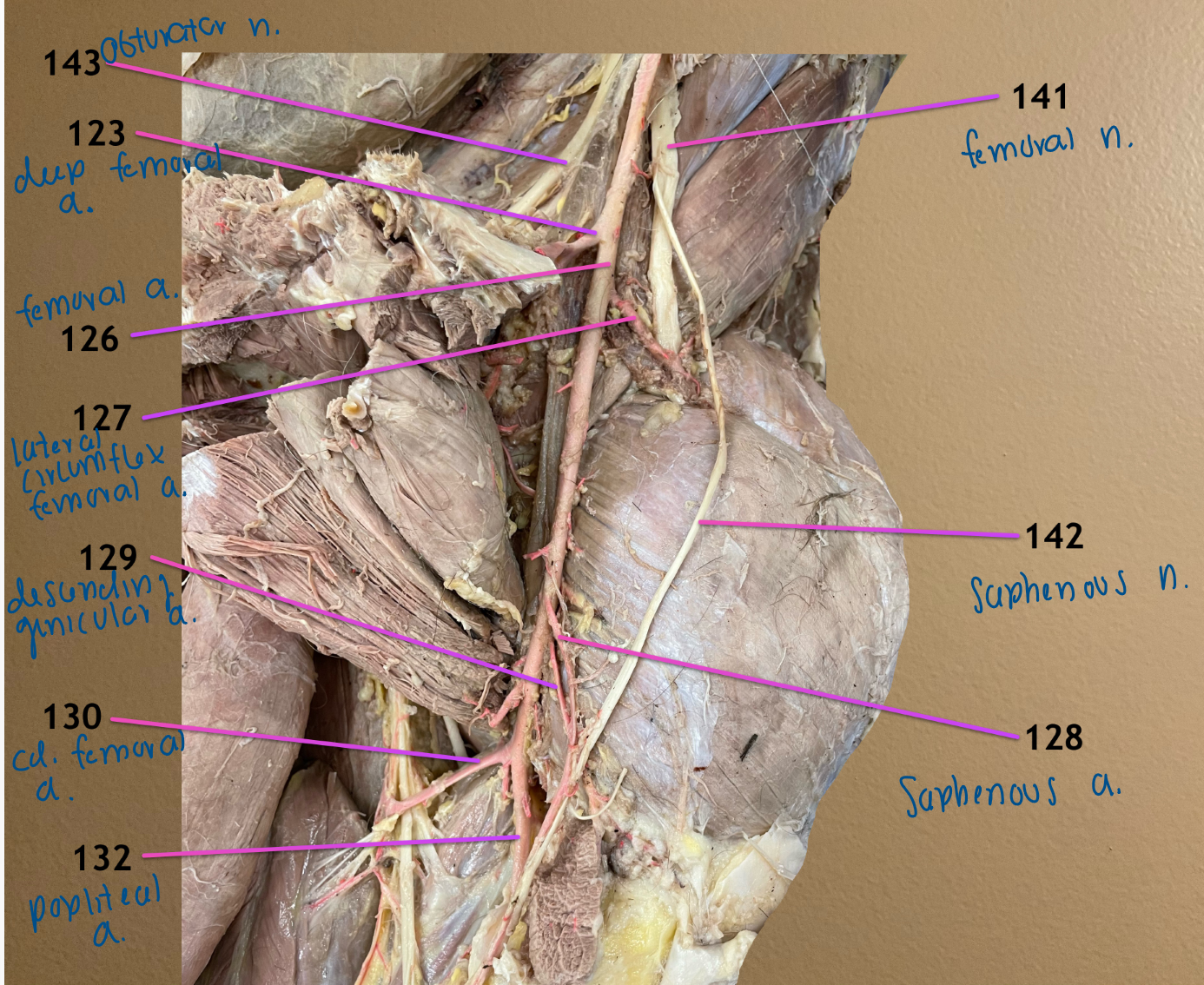
label 122-124, 124a, 124b
external iliac a.
deep femoral a.
pudendoepigastric trunk
124a: caudal epigastric a.
124b: external pudendal a.

medial view of blood flow near inguinal canal
pic 2 follows 126
pic 3 follows 123
.
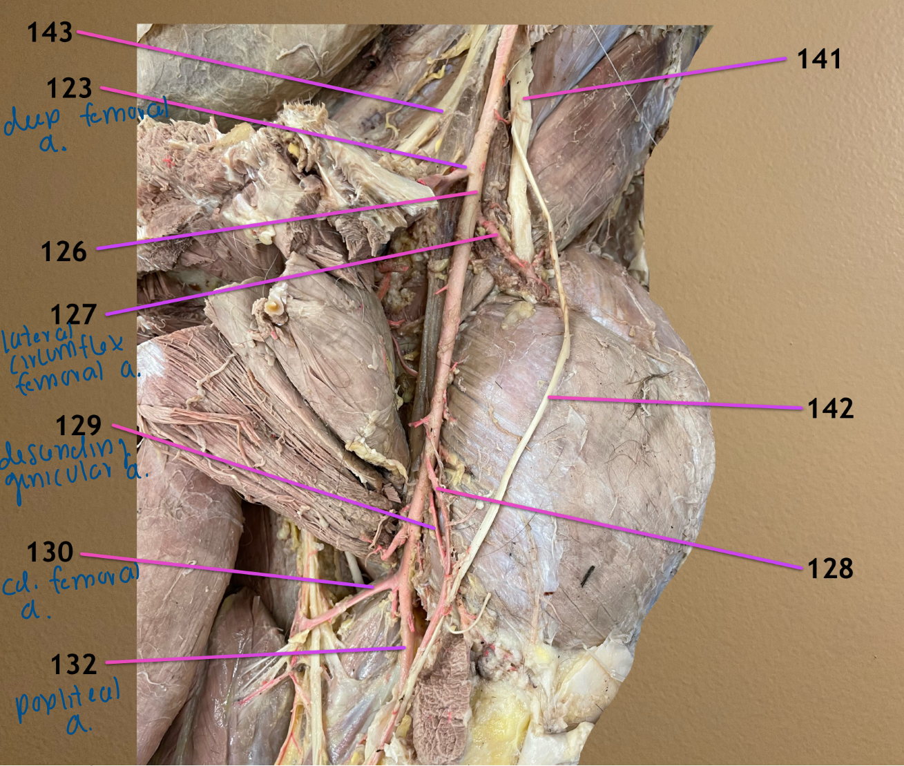
label 125-128
medial circumflex femoral a.
femoral a.
lateral circumflex femoral a.
saphenous a.

medial view of blood flow near inguinal canal
pic 2 follows 126
pic 3 follows 123
.
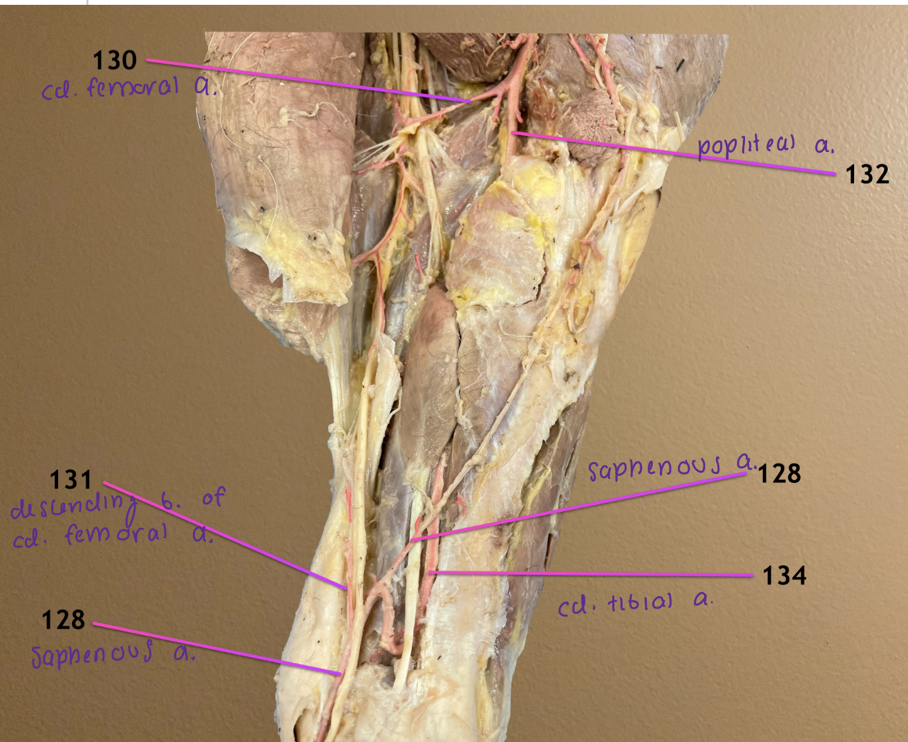
label 129, 130, 132
descending genicular a.
caudal femoral a.
popliteal a.


medial view of blood flow near inguinal canal
pic 2 follows 126
pic 3 follows 123
.
label 62, 65, 63/66, 64/66
superficial caudal epigastric a.; branch of external pudendal a. after it passes thru inguinal canal; supplies prepuce & abdominal wall
cranial artery of the penis: in male, external pudendal changes name to this after 62 comes off (in female, ext pud just bifurcates into 63/64)
63/66. cranial mammary a. / dorsal artery of the penis
64/66. caudal mammary a. / dorsal artery of the penis

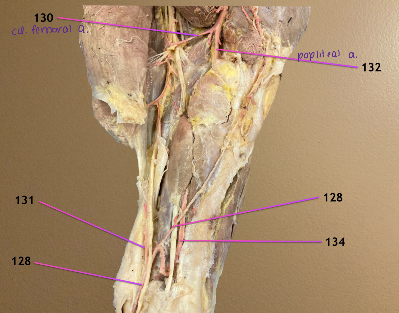
medial view of pelvic limb where deep femoral a. changes name to popliteal a.
.
label arteries at the level of the hock: 128, 131, 134
saphenous a.
descending branch of caudal femoral a.
caudal tibial a.: after cd femoral a. comes off, poplital splits into this & cranial tibial a.
.
these form the s-shaped anastomosis & saphenous will continue down the limb

left pic: dorsolateral view of crus & metatarsus, cranial to left
rt pic: same view but of metatarsus & hoof
.
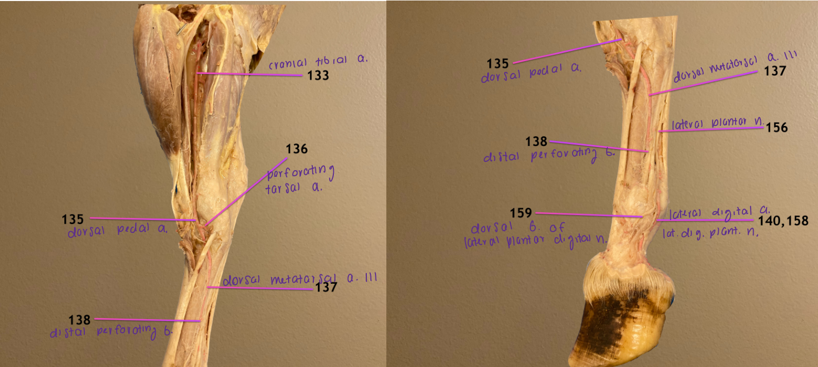
label 133, 135, 136
cranial tibial a.: deep to cranial tibial m., popliteal a. branches into this & caudal tibial a.
dorsal pedal a.: ^ changes name to this at the hock
perforating tarsal a.: branch of ^, will pass thru tarsal canal to reach plantar surface


left pic: dorsolateral view of crus & metatarsus, cranial to left
rt pic: same view but of metatarsus & hoof
.
label 137, 138, 140
dorsal metatarsal artery III: dorsal pedal a. changes name to this after it passes tarsal jt; located between cannon & lateral splint bone
distal perforating branch: ^ changes name to this when it dives between cannon & lateral splint bone; proximal to fetlock, this bifurctes into medial & lateral digital a.
lateral digital a.

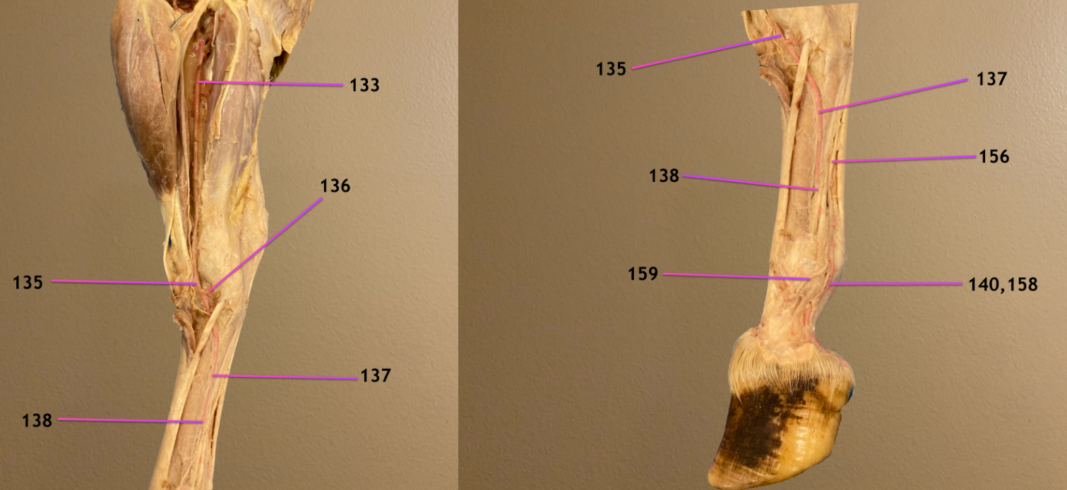
left pic: dorsolateral view of crus & metatarsus, cranial to left
rt pic: same view but of metatarsus & hoof
.
label 156, 158, 159
lateral plantar n.
lateral plantar digital n.: ^ changes name to this at level of fetlock
dorsal branch of lateral plantar digital n.
medial view of blood flow near inguinal canal
.
label 141-143
femoral n.: innervates quadriceps femoris, gives rise to 142
saphenous n.: passes distally to provide sensory to leg
obturator n.: innervates all limb adductors
left pic: lateral view of lumbosacral plexus
rt pic: caudal view
.
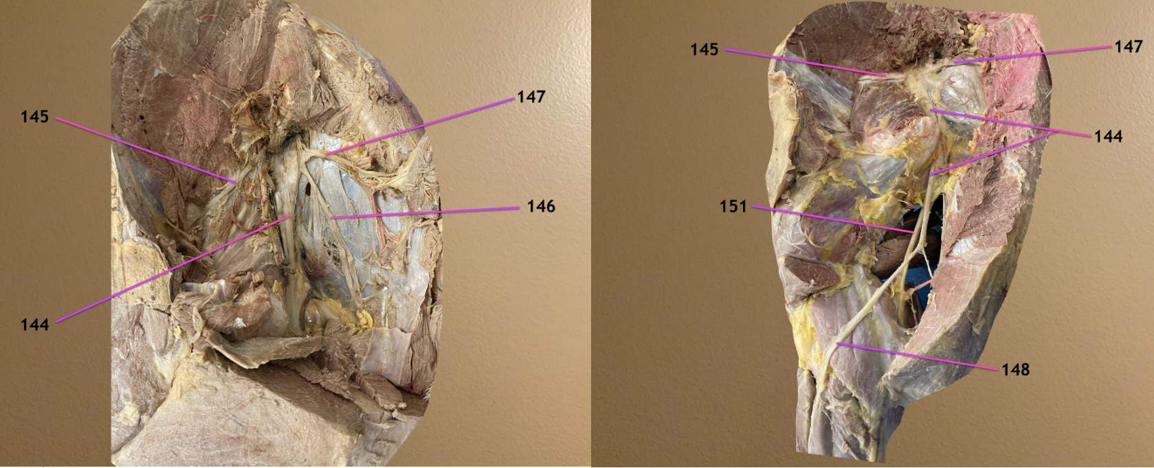
label 144-147
sciatic n.
cranial gluteal n.: branches off 144, in ventral part of ligament
caudal cutaneous femoral n.: branches off 144, in between 145 & 147
caudal gluteal n.: branches off 144, always in dorsal part of sacrosciatic ligament
.
after 145 & 147 come off, sciatic curves around ischial arch & bifurcates into its 2 main branches → common peroneal & tibial n.
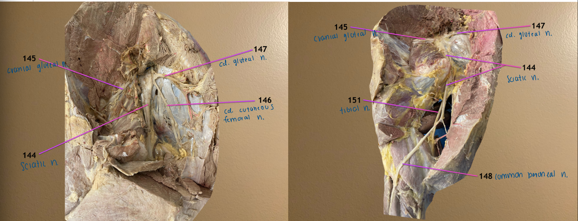

left pic: lateral view of lumbosacral plexus
rt pic: caudal view
.
label 148, 151
common peroneal n.: one of 2 main branches that sciatic n. bifurcates into
tibial n.: same ^

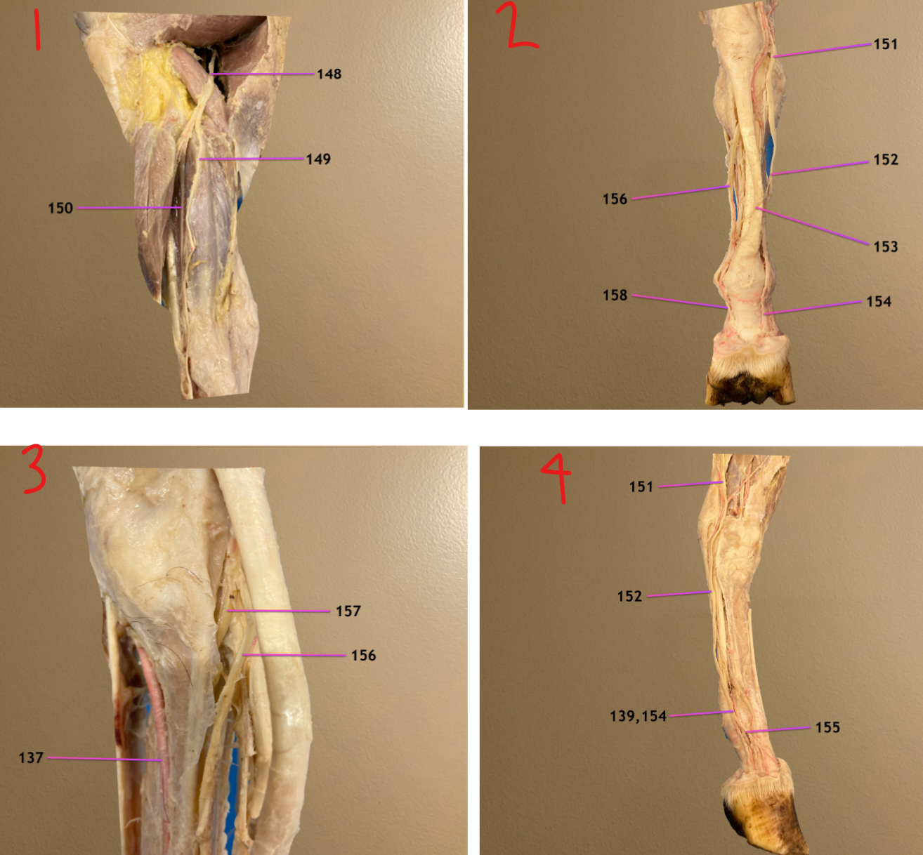
pic 1: cranial view of crus, transected muscle at top is vastus lateralis
pic 2: plantar view of tarsus to hoof showing branches of 151
pic 3: zoomed in lateral view
pic 4: medial view of tarsus to hoof, cranial to rt
.
label 148-151
common peroneal n.: bifurcates into 149 & 150
superficial peroneal n.
deep peroneal n.
tibial n.: bifurcates into medial & lateral plantar nn.


pic 1: cranial view of crus, transected muscle at top is vastus lateralis
pic 2: plantar view of tarsus to hoof showing branches of 151
pic 3: zoomed in lateral view
pic 4: medial view of tarsus to hoof, cranial to rt
.
label 152-155
medial plantar n.: comes from tibial n.; changes name to 154 at fetlock
communicating branch: located at mid to distal metatarsal region; connects medial & lateral plantar nn.
medial plantar digital n.
dorsal branch of medial plantar digital n.


pic 1: cranial view of crus, transected muscle at top is vastus lateralis
pic 2: plantar view of tarsus to hoof showing branches of 151
pic 3: zoomed in lateral view
pic 4: medial view of tarsus to hoof, cranial to rt
.
label 156, 157
lateral plantar n.: comes from tibial n.; changes name to lateral plantar digital a
deep branch of lateral plantar n.: branches off 156; supplies deep structures in metacarpus
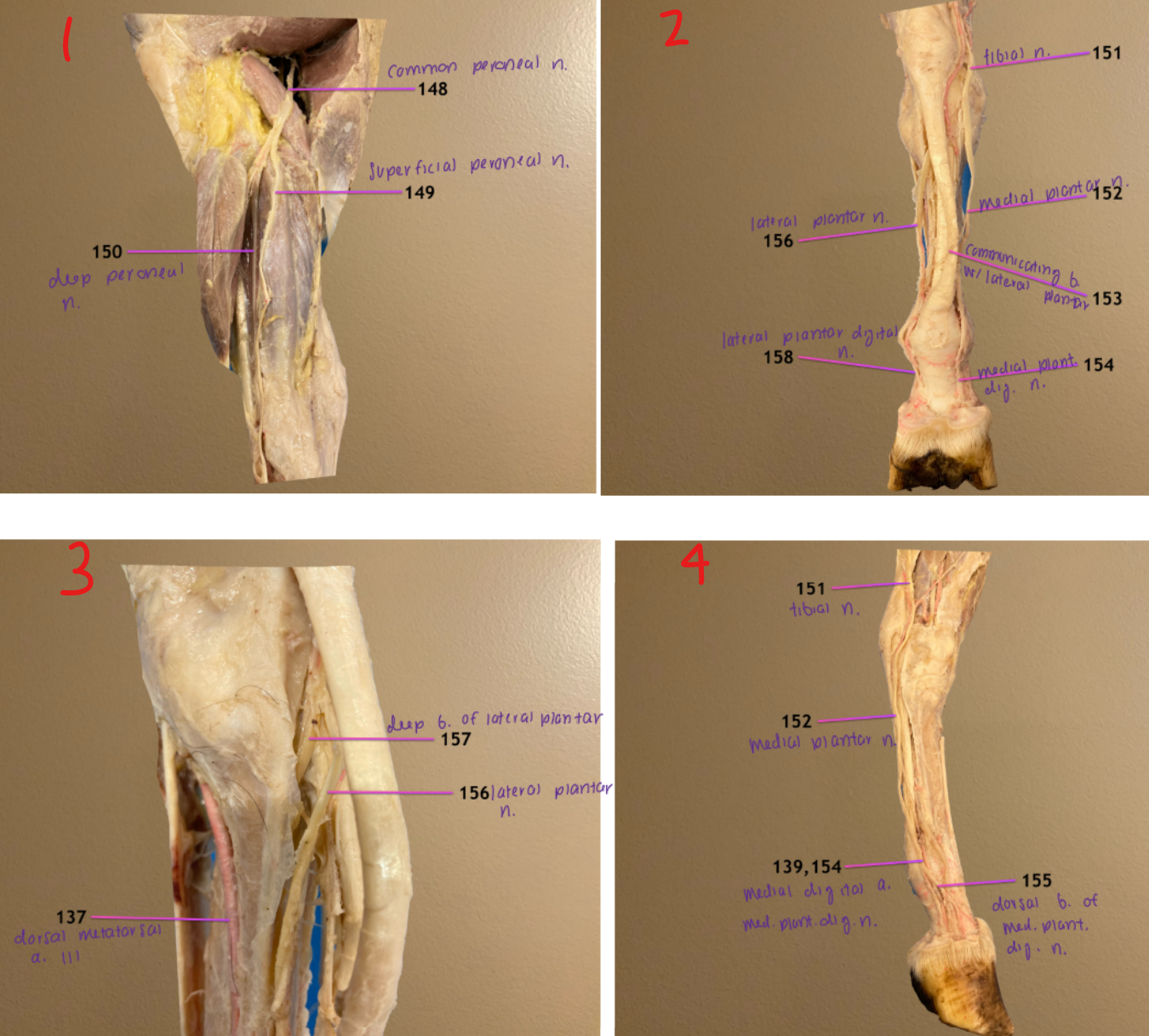
ventral view
.
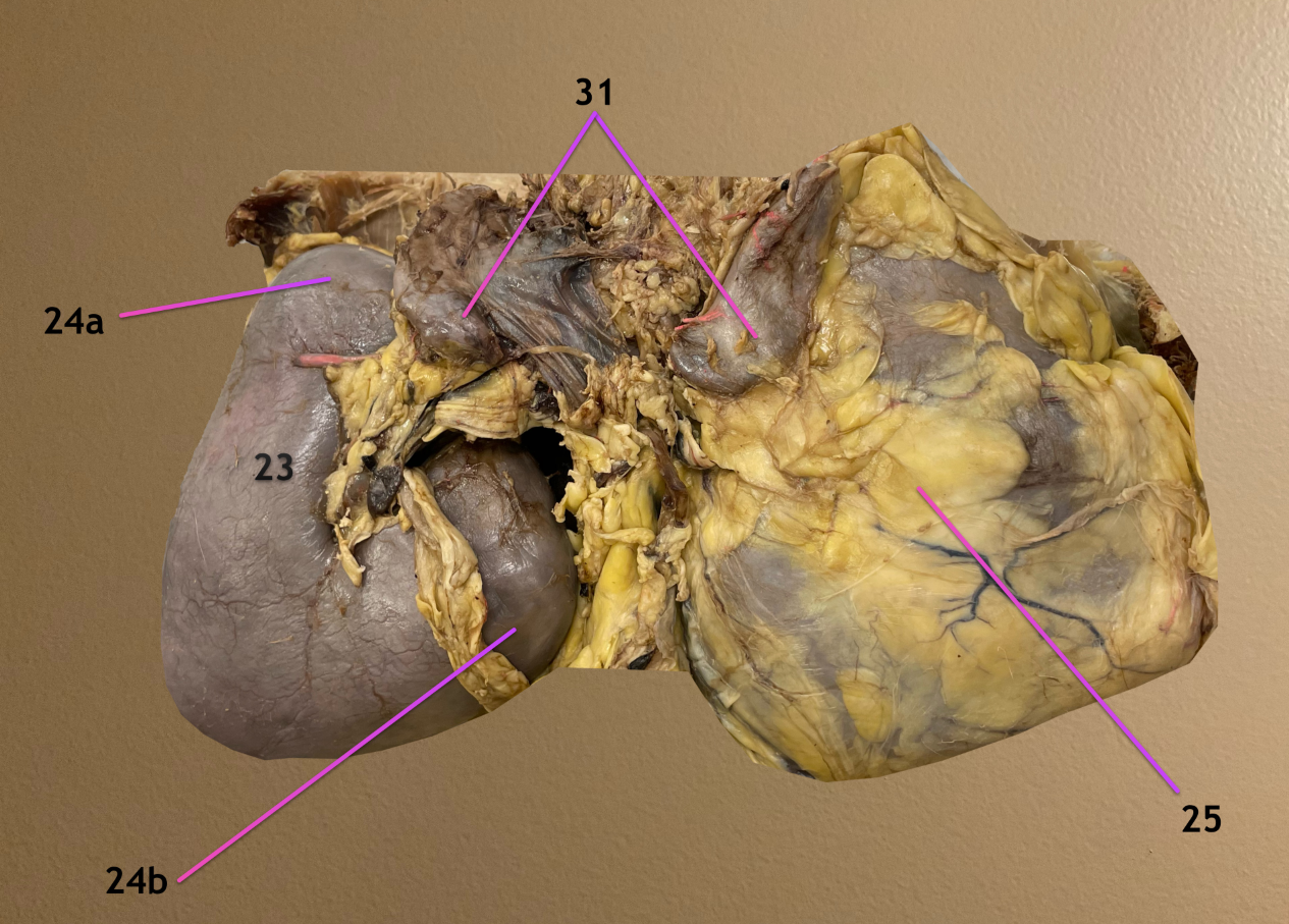
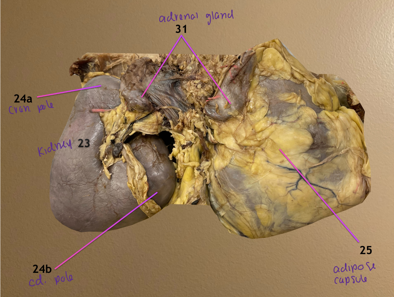
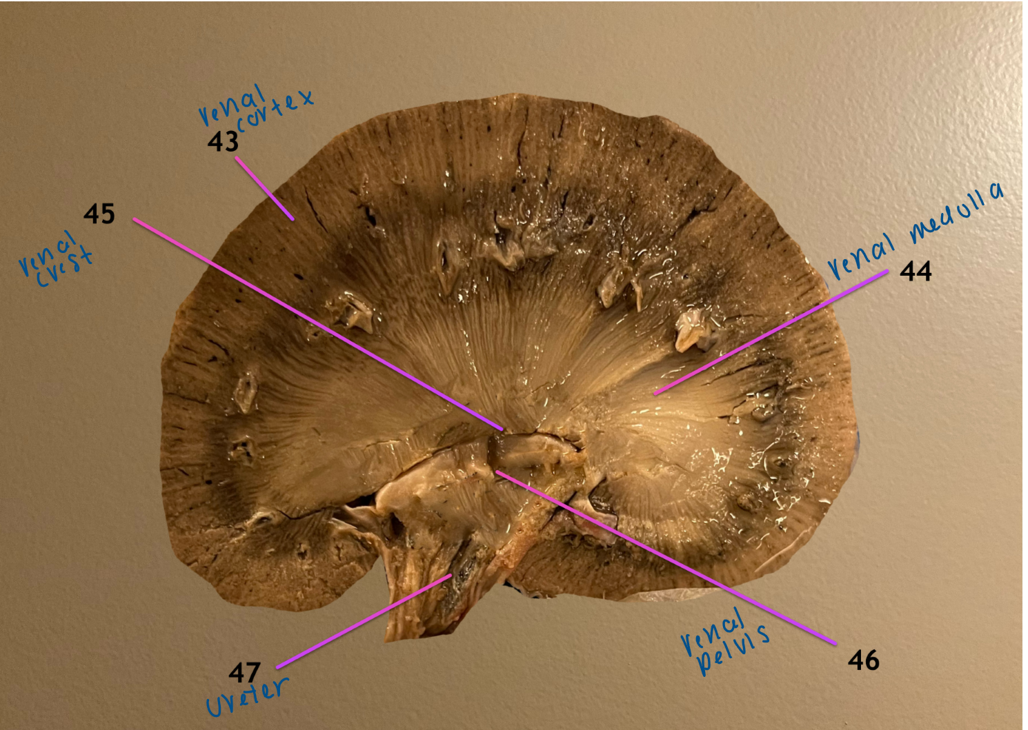
label 23, 24a, 24b, 25, 31
kidney
24a: cranial pole of kidney
24b: caudal pole of kidney
adipose capsule
adrenal gland: medial to each kidney
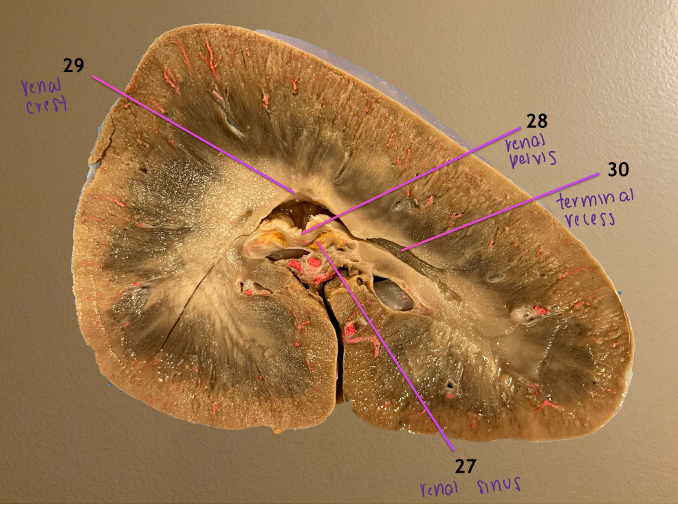
longitudinal section of kidney
.
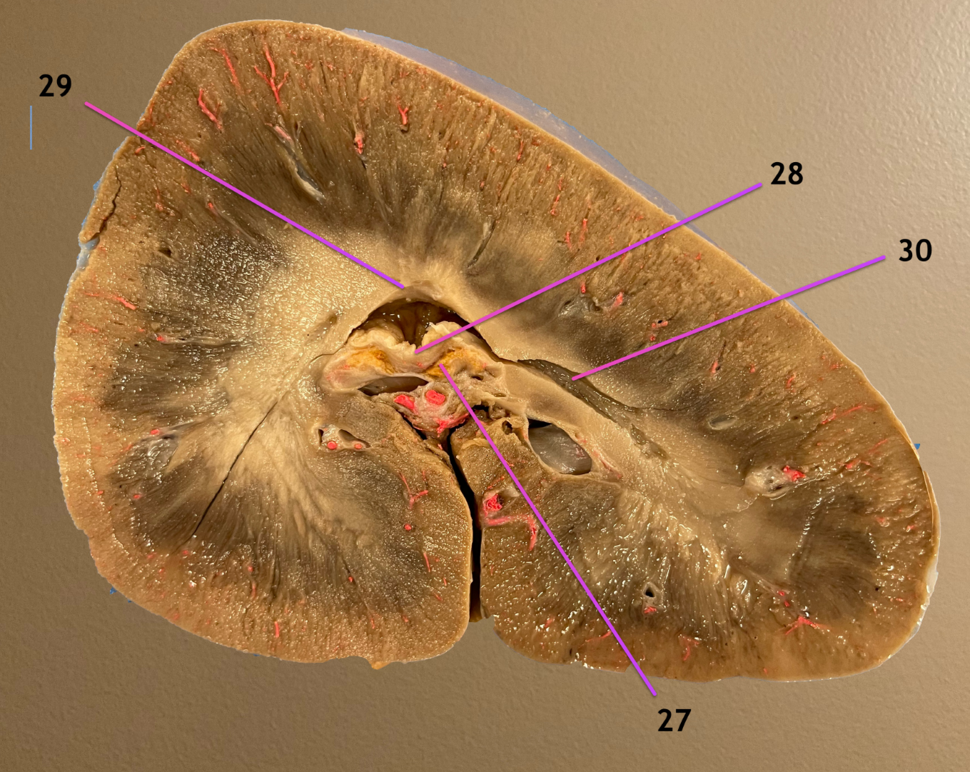
label 27-30
name the topographic region that 27 & 28 sit in
renal sinus: filled w/ fat & surrounds pelvis
renal pelvis
renal crest: drains urine from center of kidney, flows into 28
terminal recess: extension of pelvis that collects urine from the poles
.
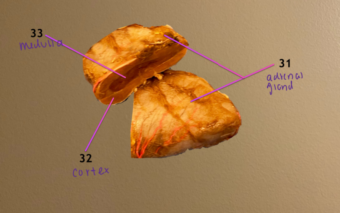
renal hilus
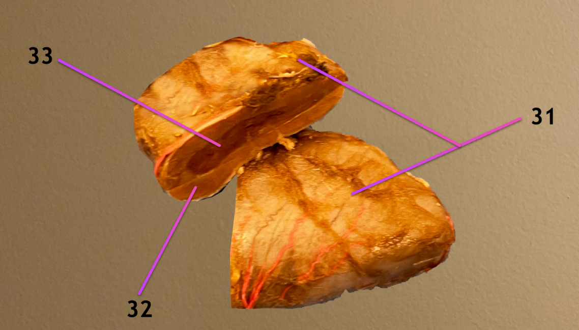
what structure is this: 31
label the areas of it we can see on cross section: 32, 33
adrenal gland
cortex of adrenal gland
medulla of adrenal gland
UB is opened, cranial to rt
.
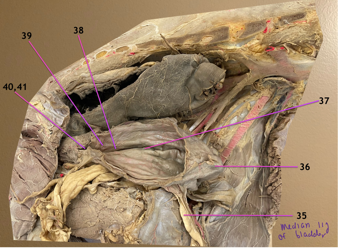
label the topographic regions of the UB: 36-39
.
label 40 & its opening 41
apex of bladder
body of bladder
neck of bladder
trigone of bladder
.
urethra
internal urethral orifice
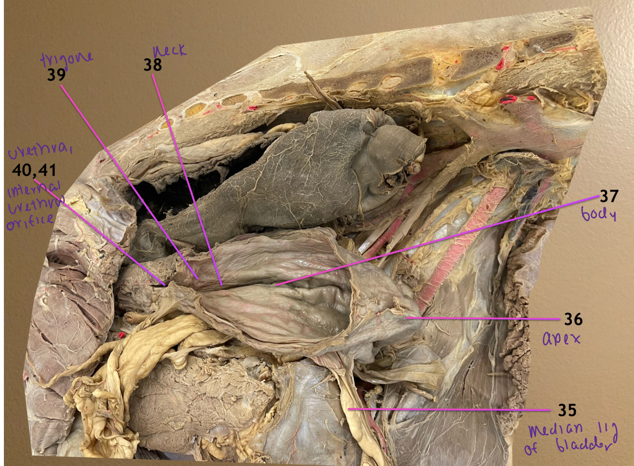

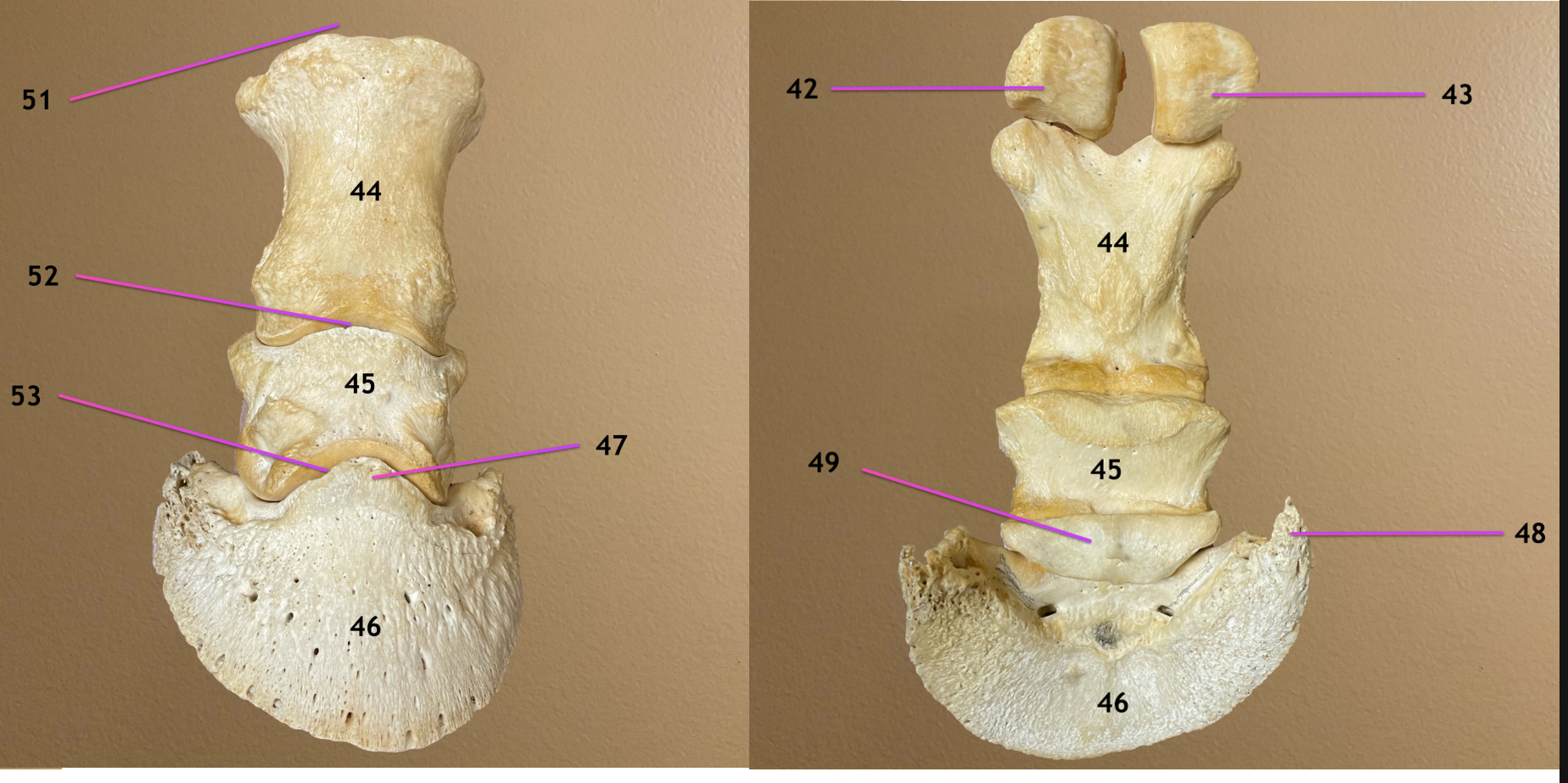
pic 1: cranial to cd view of pelvis & lumbar spine
pic 2/3: caudolateral view of pelvis
.
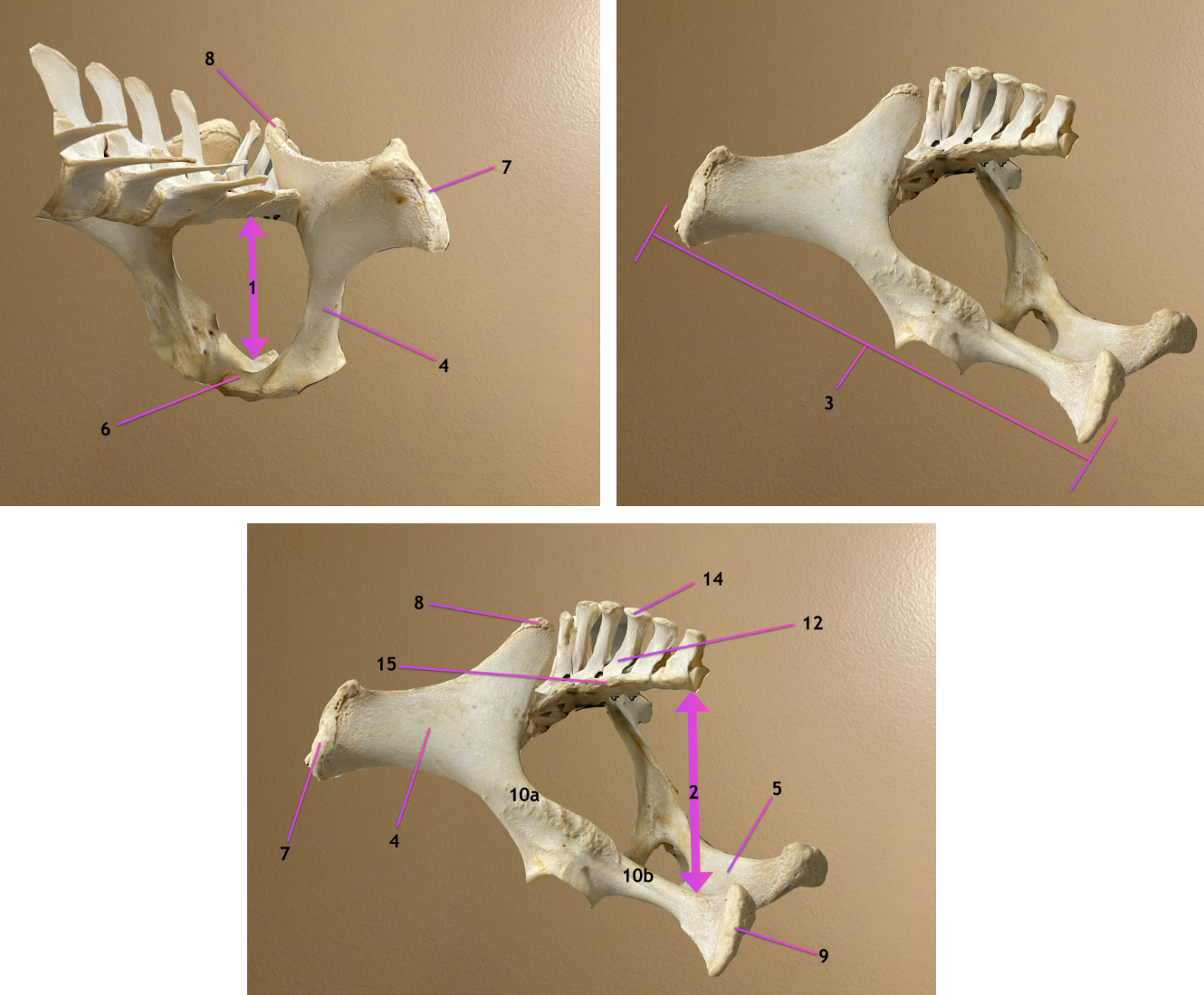
label the topographic region 1, 2
label 3, 4
pelvic inlet
pelvic outlet
os coxae
ilium

pic 1: cranial to cd view of pelvis & lumbar spine
pic 2/3: caudolateral view of pelvis
.
label 5-8
ischium
pubis
tuber coxae
tuber sacrale

pic 1: cranial to cd view of pelvis & lumbar spine
pic 2/3: caudolateral view of pelvis
.
label 9, 10a, 10b
ischiatic tuberosity
10a: greater sciatic notch
10b: lesser sciatic notch

left pic: dorsal view
rt pic: ventral view
.
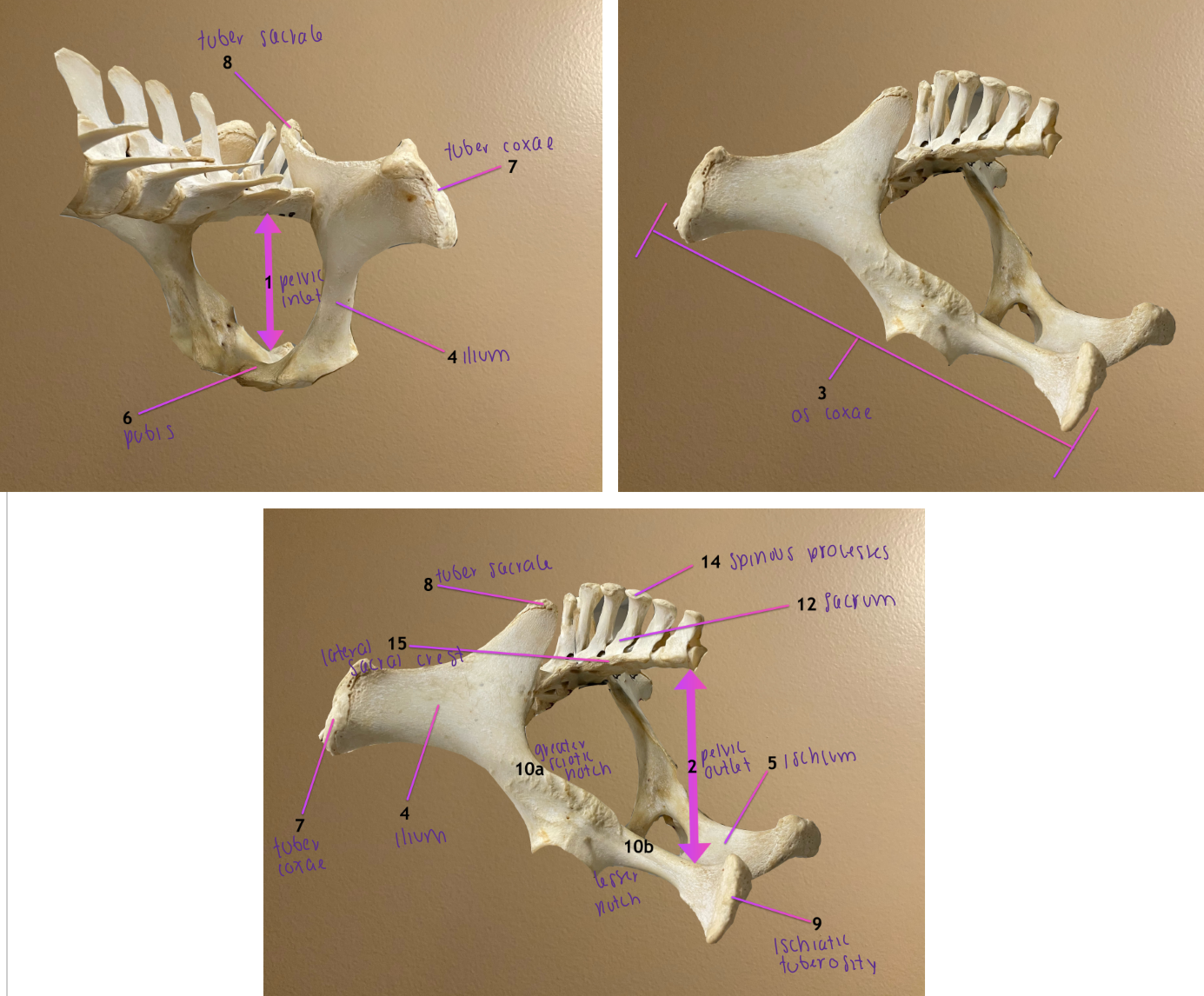
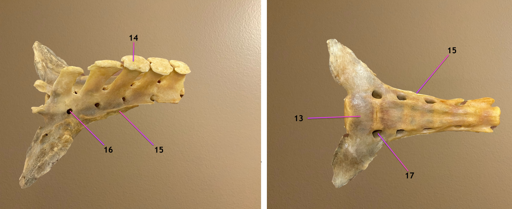
what bone is this
.
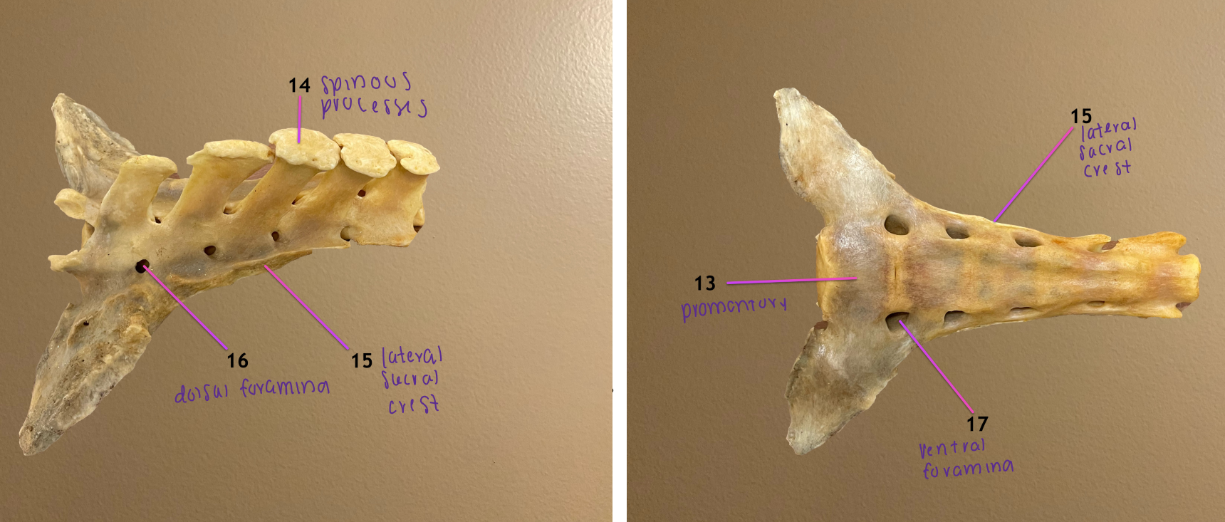
label the parts of it: 13-17
sacrum
.
promontory of sacrum
spinous processes of sacrum
lateral sacral crest: formed by transverse processes
dorsal foramina
ventral foramina
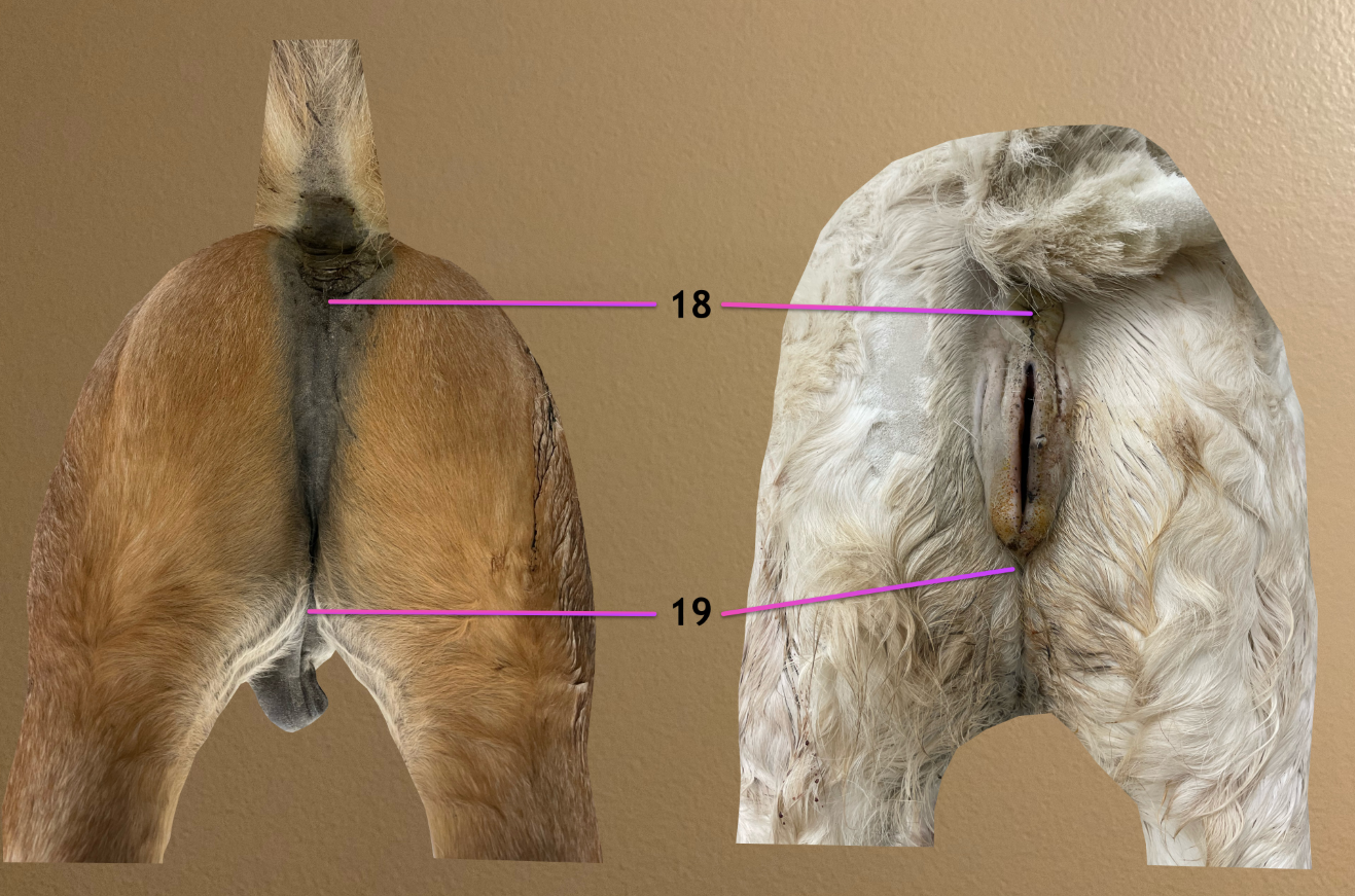
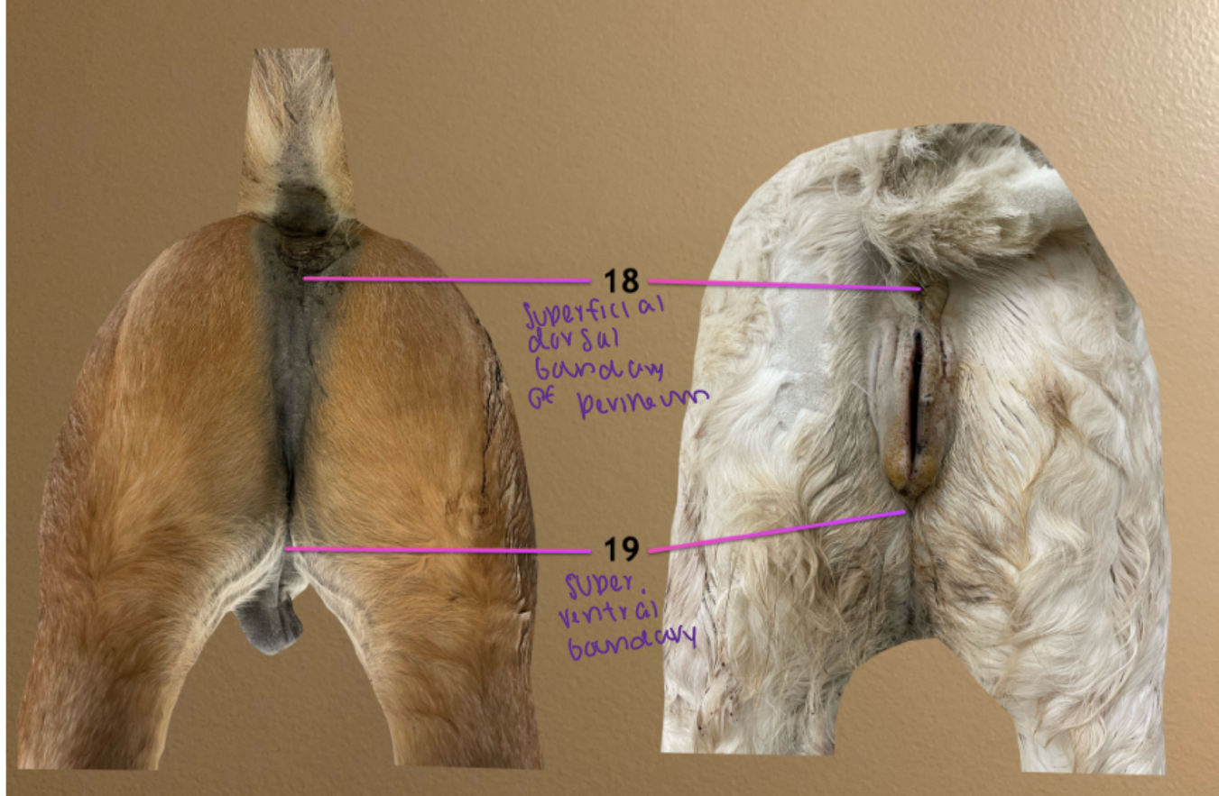
label the topographic regions of the perineal region: 18, 19
superficial dorsal boundary of perineum
superficial ventral boundary of perineum
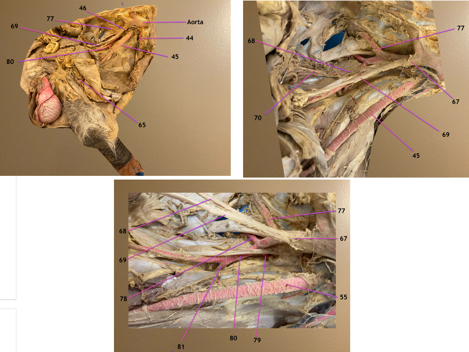
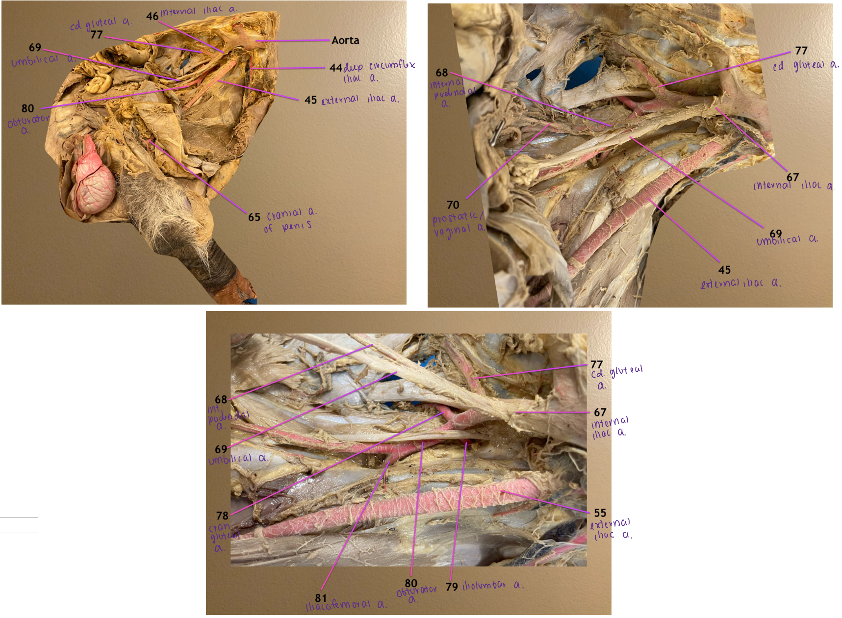
pic 1: medial view of left pelvic wall, cranial to rt
pic 2: zoomed in on branches of aorta
pic 3: zoomed in on branches of internal iliac (67)
.
label 68-70, 77
name 70 in male & female
.
note: 45 & 55 are external iliac a.
internal pudendal a.
umbilical a.
prostatic / vaginal a.: branch of 68, will terminate as a. of the penis
caudal gluteal a.


pic 1: medial view of left pelvic wall, cranial to rt
pic 2: zoomed in on branches of aorta
pic 3: zoomed in on branches of internal iliac (67)
.
label 78-81
.
note: 45 & 55 are external iliac a.
cranial gluteal a.
iliolumbar a.
obturator a.: branch of 78, passes thru obturator foramen & travels w/ obturator n.
iliacofemoral a.

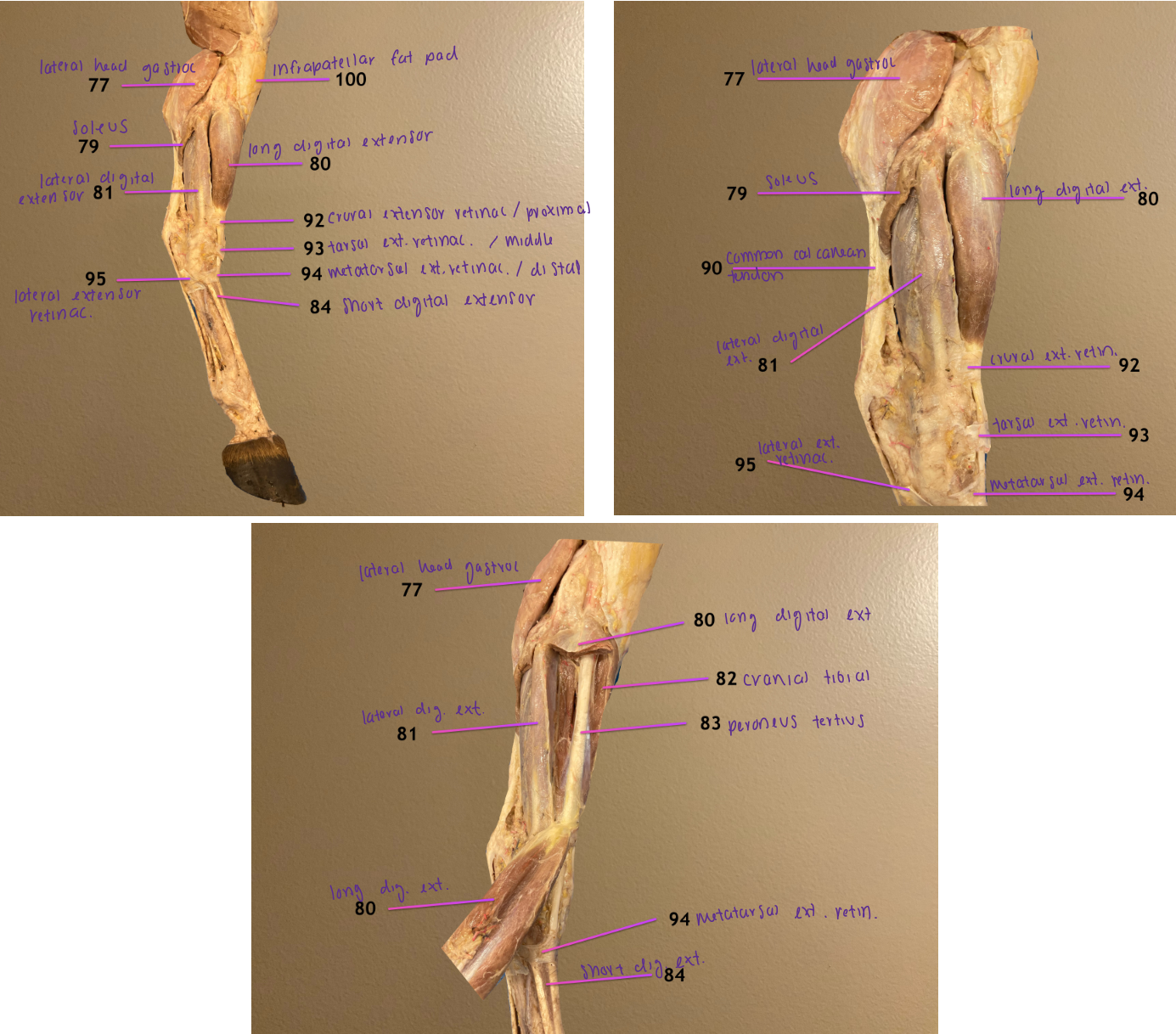
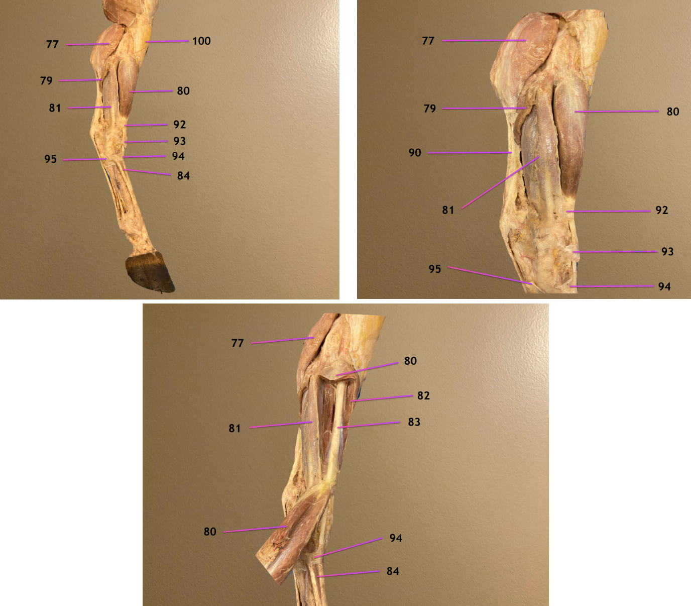
pic 1: lateral view of crus, cranial to rt
pic 2/3: same but zoomed in & some muscles cut
.
label 77, 79-81
lateral head of gastrocnemius m.: one of 3 heads of triceps surae; extends tarsus
soleus m.: vestigial; one of 3 heads of triceps surae; extends tarsus
long digital extensor m.: flexes tarsus
lateral digital extensor m.: flexes tarsus

pic 1: lateral view of crus, cranial to rt
pic 2/3: same but zoomed in & some muscles cut
.
label 83, 84, 90, 100
peroneus tertius: cord-like structure, part of reciprocal apparatus; sits superficial to cranial tibial m.; flexes tarsus
short digital extensor m.: only m. in metatarsus; extends digit
common calcanean tendon
infrapatellar fat pad: fills infrapatellar space

pic 1: lateral view of crus, cranial to rt
pic 2/3: same but zoomed in & some muscles cut
.
label 92-95
crural/proximal extensor retinaculum
tarsal/middle extensor retinaculum
metatarsal/distal extensor retinaculum
lateral extensor retinaculum


pic 1: medial view of crus, cranial to left
pic 2: zoomed in on crus & 78 reflected
pic 3: zoomed in on cannon region
.
label 76, 78, 82, 85
popliteus m.: flexes stifle
medial head of gastrocnemius m.: one of 3 heads of triceps surae; extends tarsus
cranial tibial m.: flexes tarsus
superficial digital flexor m.: flexes digit
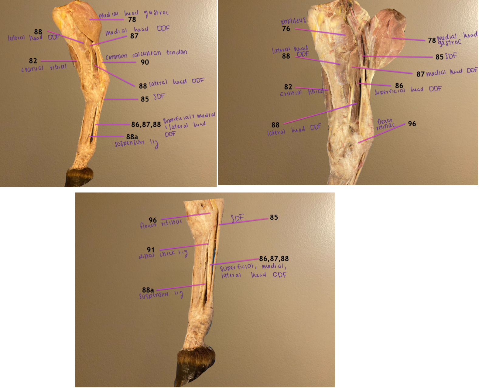
pic 1: medial view of crus, cranial to left
pic 2: zoomed in on crus & 78 reflected
pic 3: zoomed in on cannon region
.
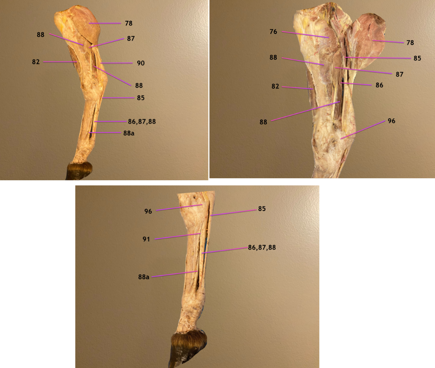
label 86-88, 88a
give both names for 86
superficial head of deep digital flexor / caudal tibial m.
medial head of deep digital flexor
lateral head of deep digital flexor
88a: suspensory ligament
.
all flex the digit


pic 1: medial view of crus, cranial to left
pic 2: zoomed in on crus & 78 reflected
pic 3: zoomed in on cannon region
.
label 91, 96
distal check ligament
flexor retinaculum

plantar view of distal limb
.
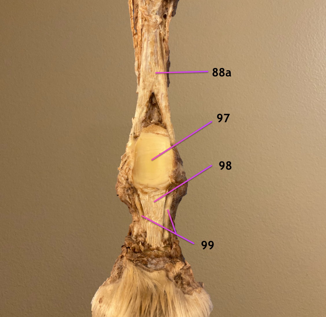
label 88a, 97-99
88a: suspensory ligament
plantar ligament
straight sesamoidean ligament
oblique sesamoidean ligament
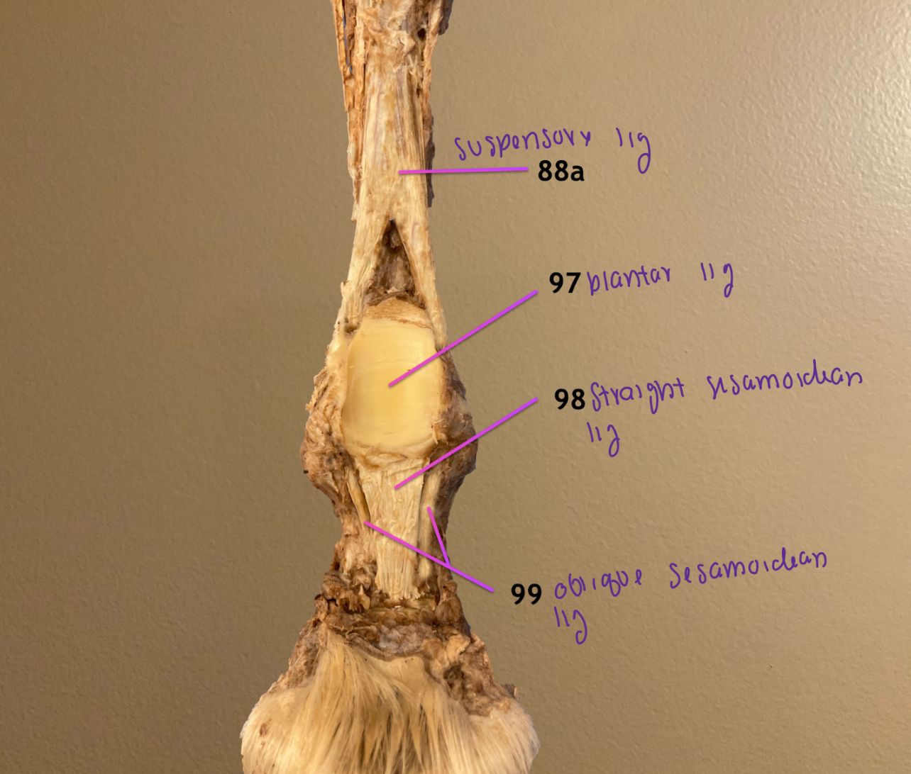
distal limb
.
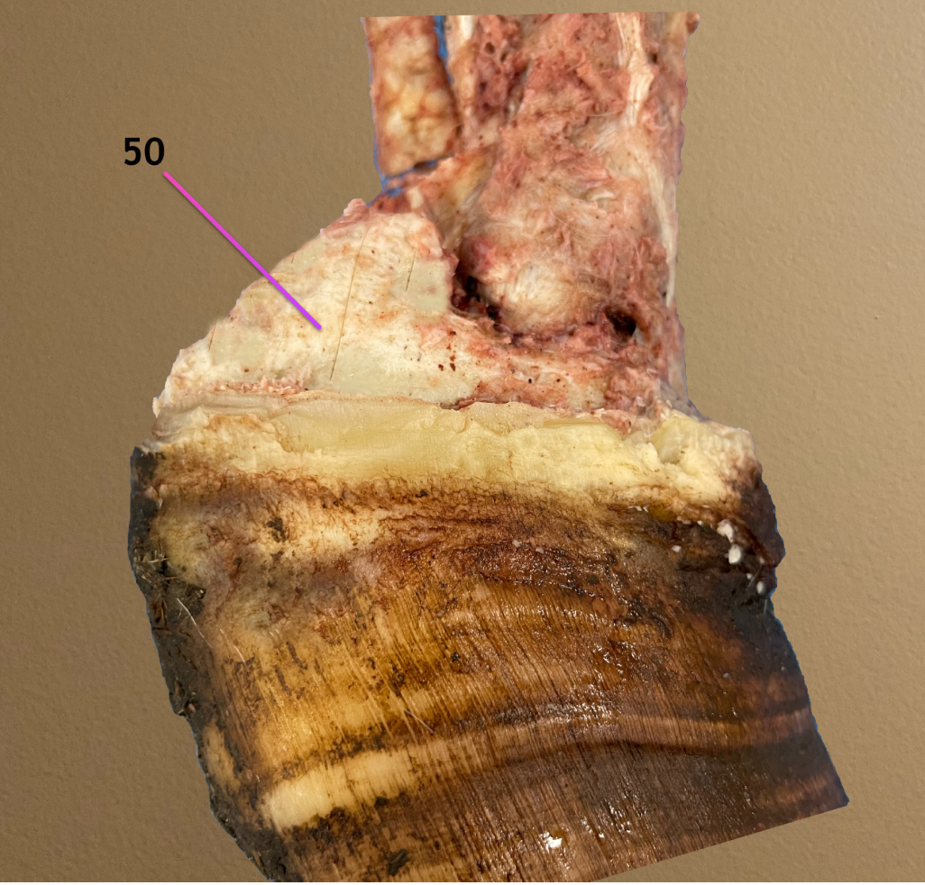
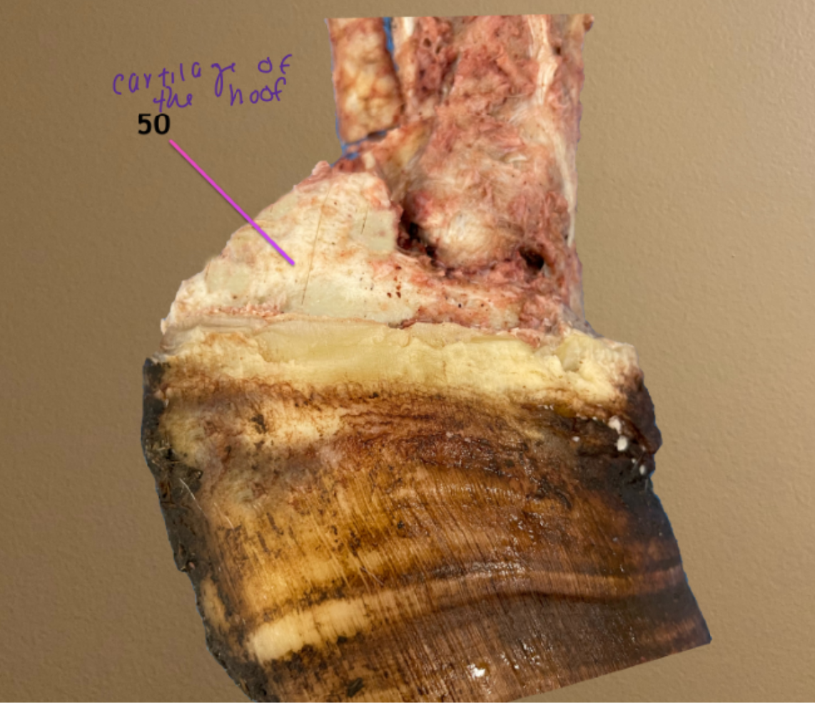
label 50
cartilage of the hoof
cranial view of stifle jt
.
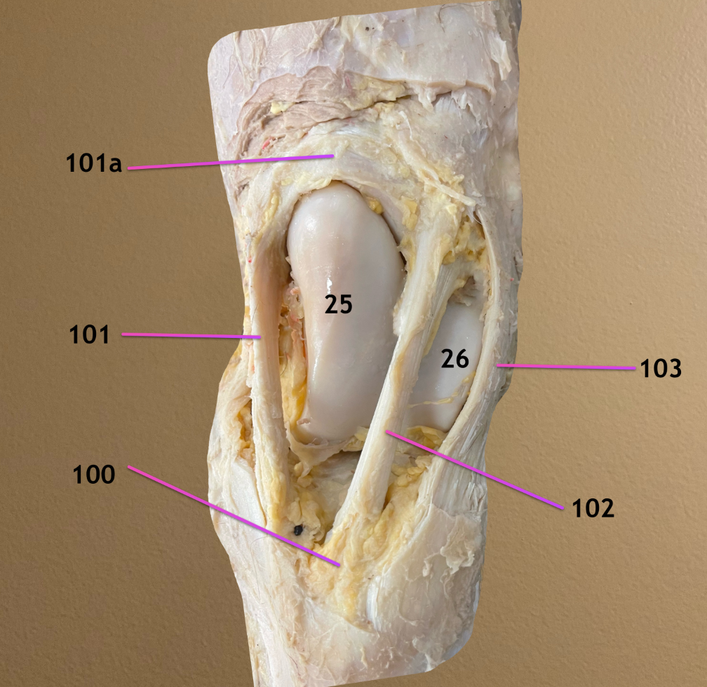
label 100, 101a, 101-103
.
note: 25 is medial trochlear ridge, 26 is lateral troch ridge
infrapatellar fat pad
101a. patellar fibrocartilage: hooks behind an enlargment on dorsal aspect of medial troch ridge to lock patella in place
medial patellar ligament: this & 102 form the patellar loop as they pass around medial troch ridge
intermediate patellar ligament
lateral patellar ligament

pic 1/2: craniolateral view of tarsus
pic 3: caudal view of tarsus
.
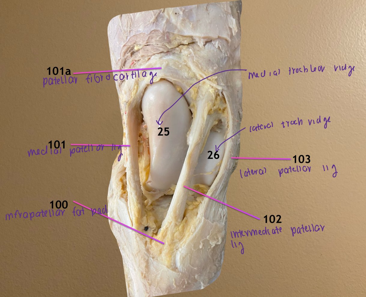
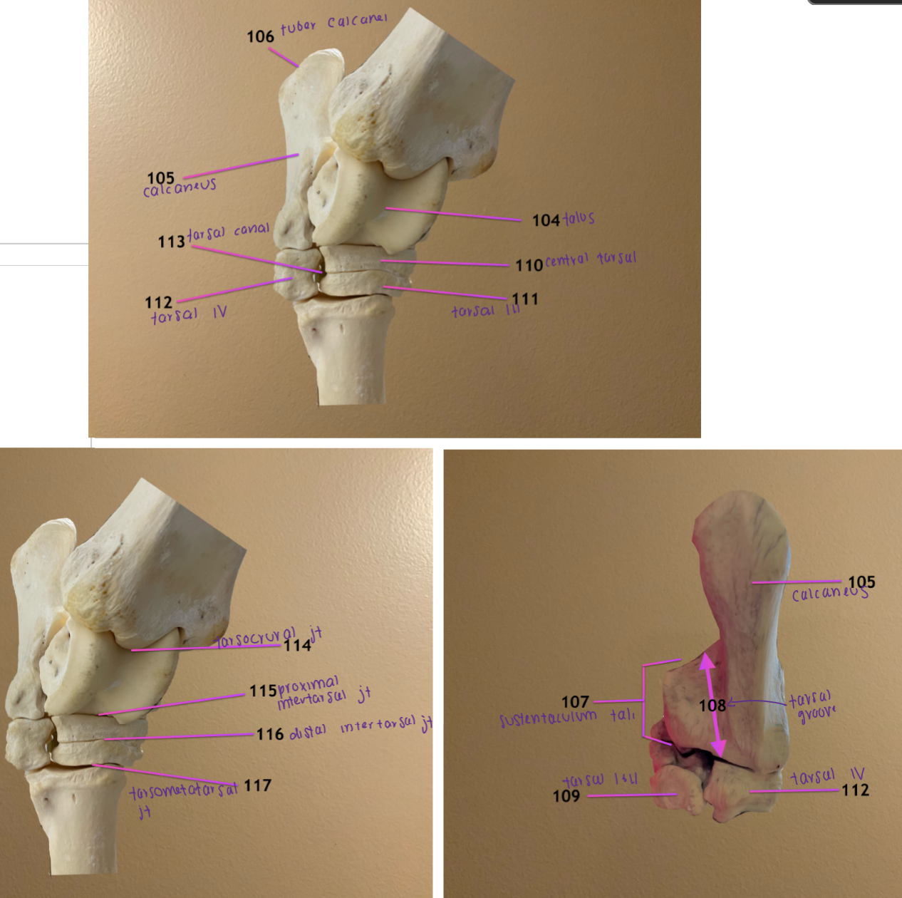
label 104-107
talus
calcaneus: located on lateral side of joint & proximal to tarsal I/II & IV
tuber calcanei
sustentaculum tali: medially projecting process coming off calcaneus

pic 1/2: craniolateral view of tarsus
pic 3: caudal view of tarsus
.
label 108-111
tarsal groove: curved caudal surface of sustentaculum tali
fused tarsal bone I & II
central tarsal bone
tarsal bone III


pic 1/2: craniolateral view of tarsus
pic 3: caudal view of tarsus
.
label 112-114
tarsal bone IV: positioned laterally
tarsal canal: formed by central tarsal, tarsal III, & tarsal bone IV
tarsocrural joint


pic 1/2: craniolateral view of tarsus
pic 3: caudal view of tarsus
.
label 115-117
proximal intertarsal joint
distal intertarsal joint
tarsometatarsal joint
dorsal view of tarsus
.
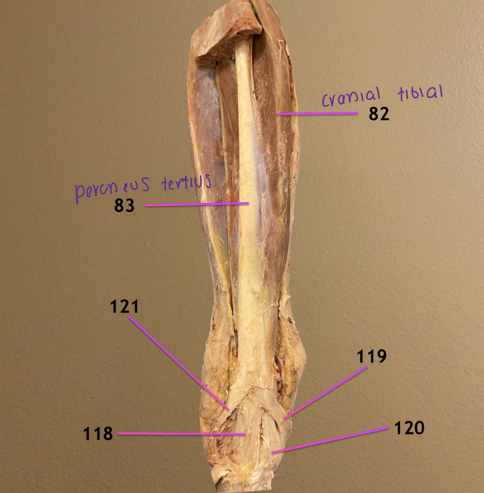
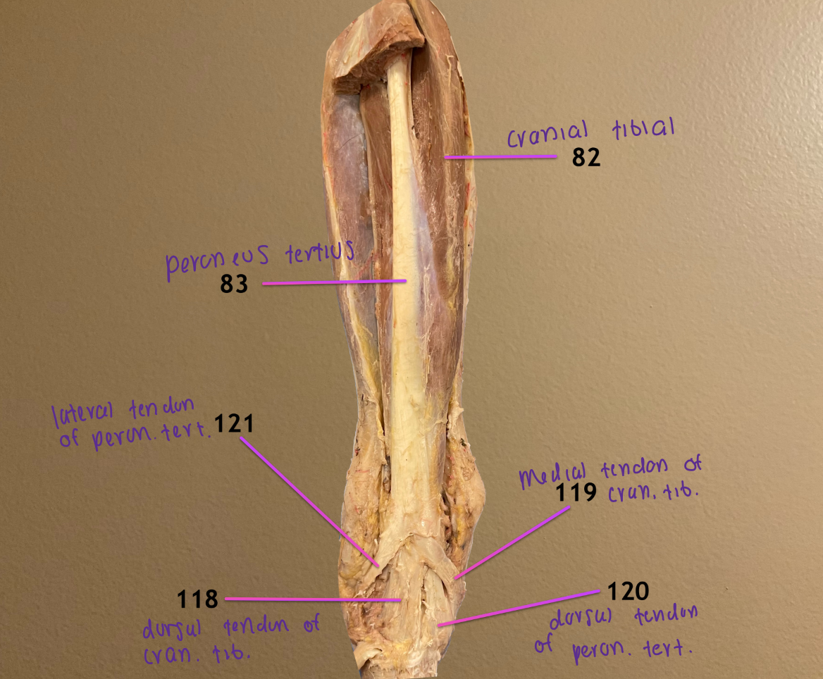
label the tendons 118-121
dorsal tendon of cranial tibial m.
medial tendon of cranial tibial m.
dorsal tendon of peroneus tertius m.
lateral tendon of peroneus tertius m.
.
trick: from lateral to medial → 121, 118, 120, 119

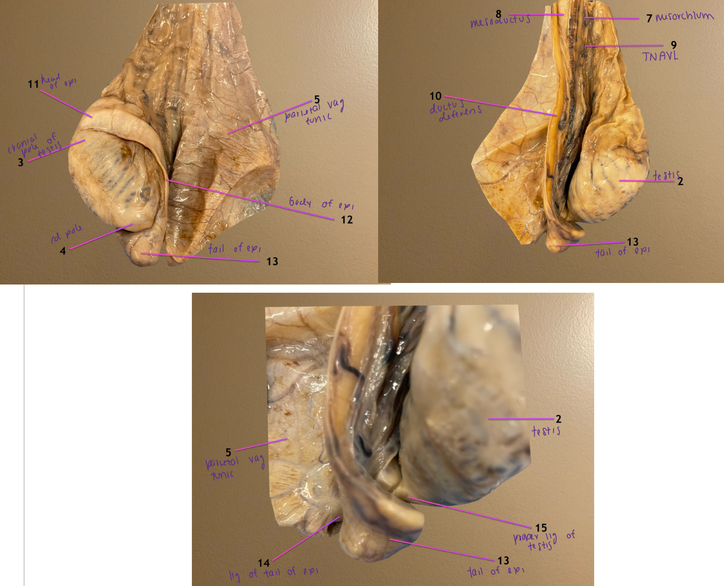
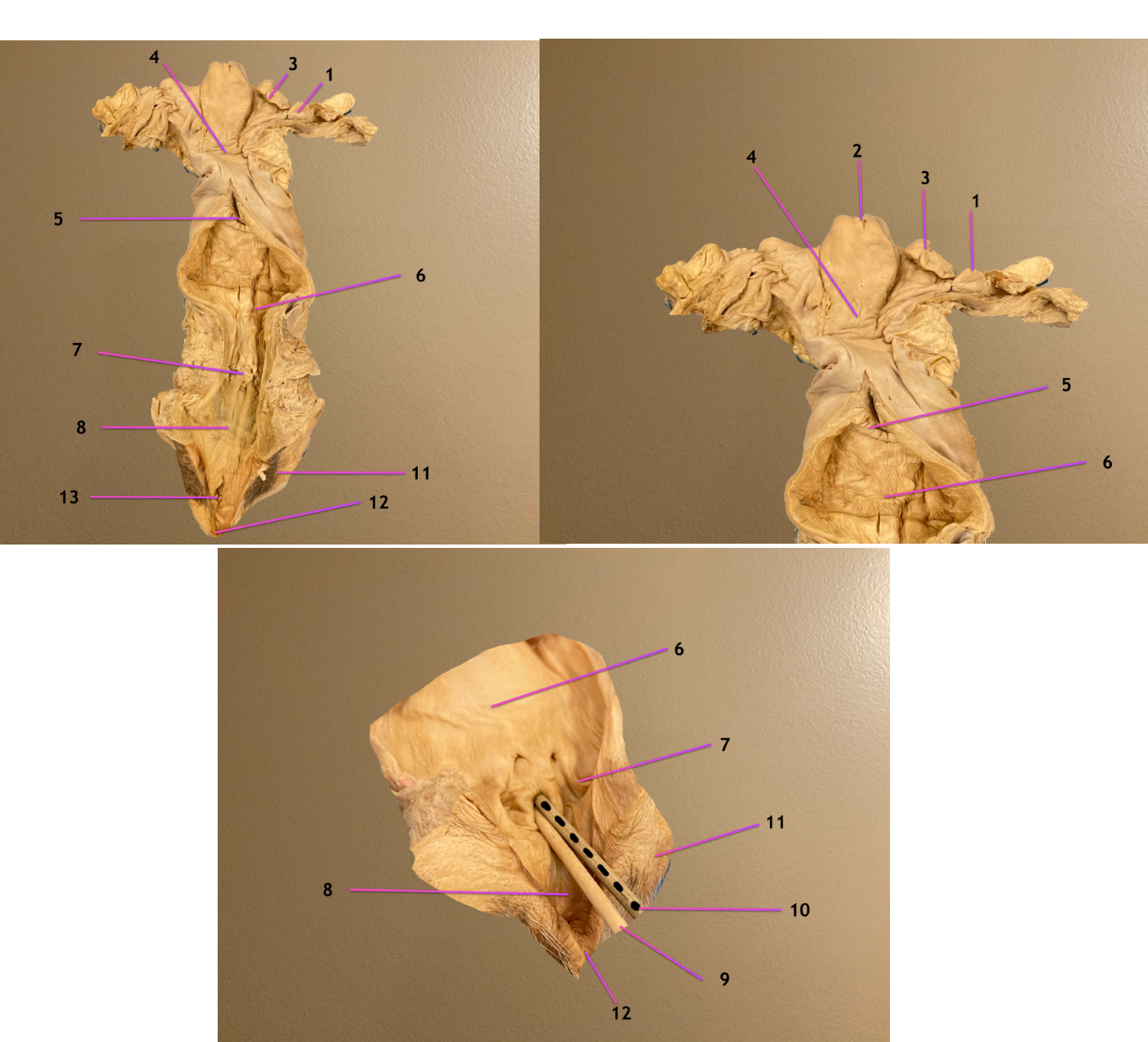
pic 1/2: parietal vaginal tunic opened
pic 3: same but zoomed in to see 14 & 15
.
label 2-5
testis
cranial pole of testis
caudal pole of testis
parietal vaginal tunic


pic 1/2: parietal vaginal tunic opened
pic 3: same but zoomed in to see 14 & 15
.
name the CT that covers the testicle
.
label CT 7, 8
label 9
visceral vaginal tunic
.
mesorchium: portion of 6 that suspends 9
mesoductus: portion of 6 that suspends ductus deferens
testicular vessels, nerve, lymphatics
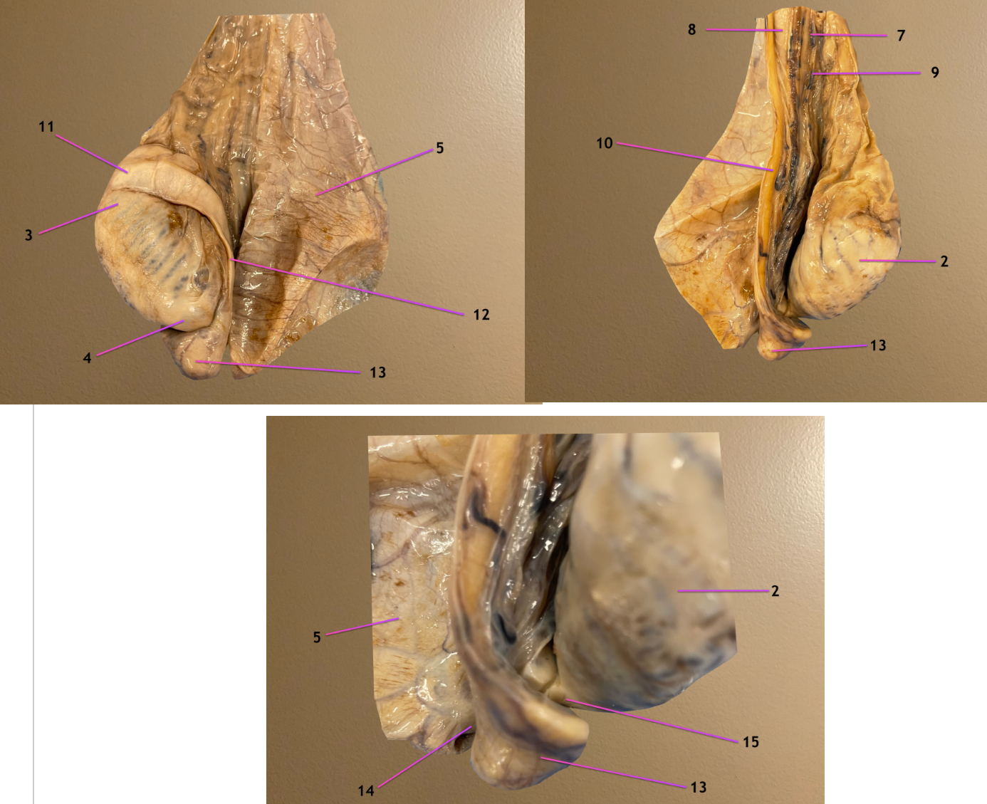
pic 1/2: parietal vaginal tunic opened
pic 3: same but zoomed in to see 14 & 15
.
label 10-13
ductus deferens
head of epididymis: attached to cranial pole of testis
body of epididymis
tail of epididymis: transitions into 10


pic 1/2: parietal vaginal tunic opened
pic 3: same but zoomed in to see 14 & 15
.
label 14, 15
ligament of tail of epididymis: connects tail of epi to parietal vag tunic
proper ligament of testis: directly connects testis to epididymis

view of penis partially extended
.
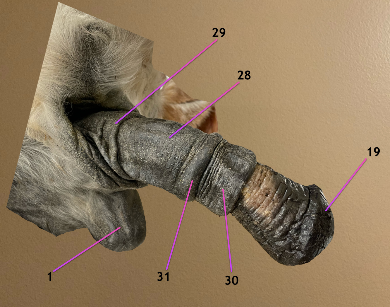
label 1, 28-31
scrotum
outer prepuce fold
preputial orifice: opening of outer fold
inner preputial fold
preputial ring: opening of inner fold
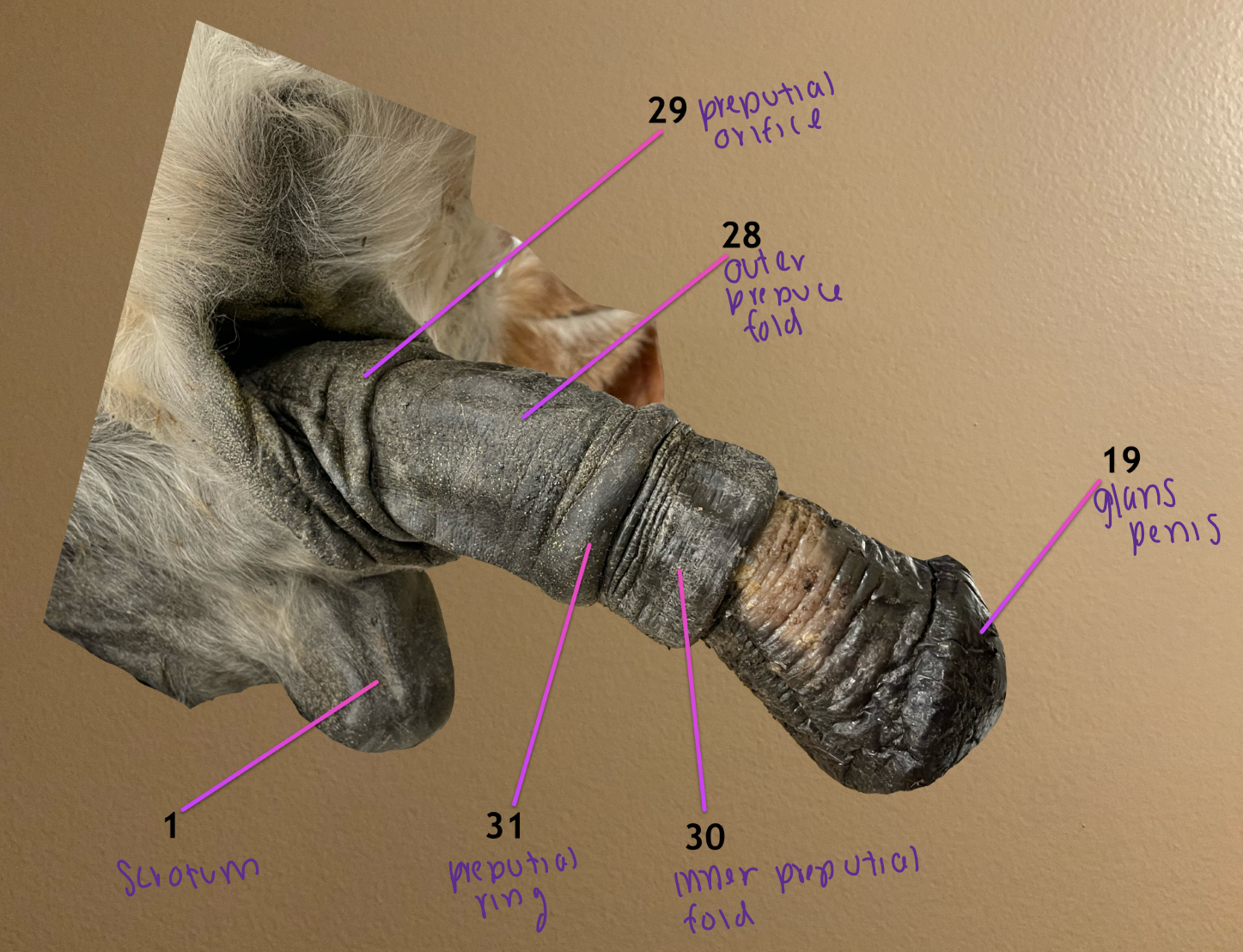
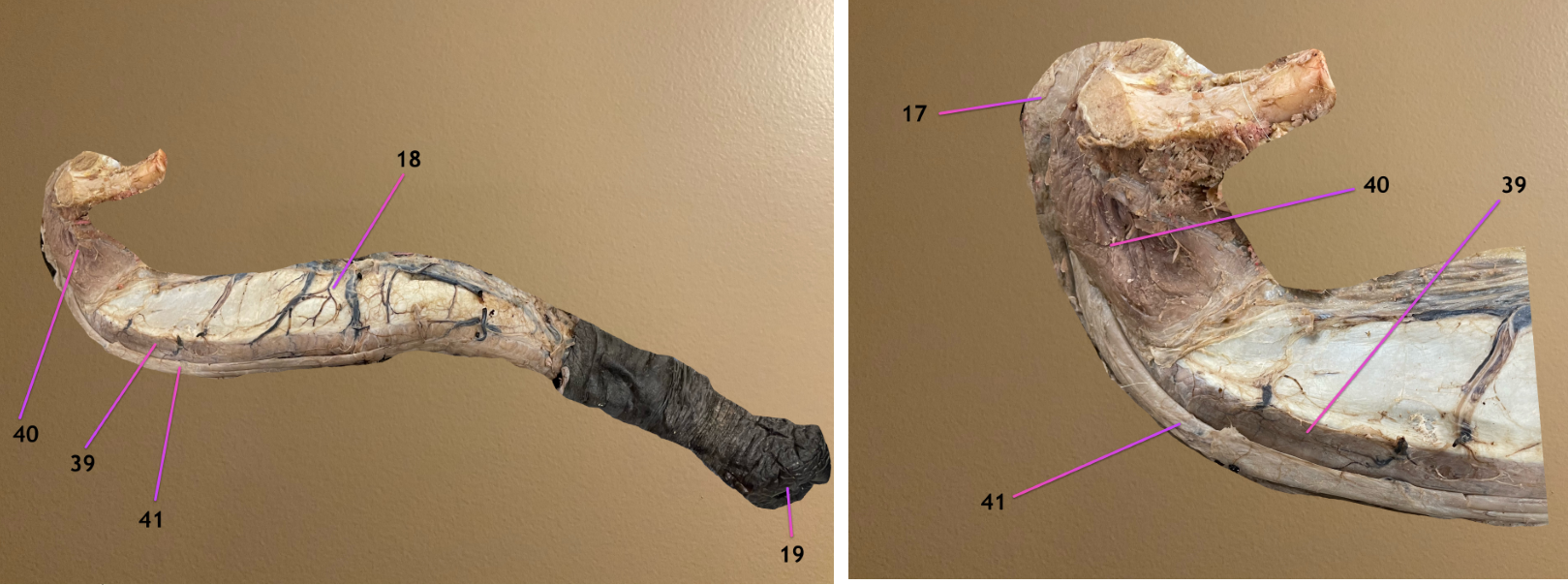
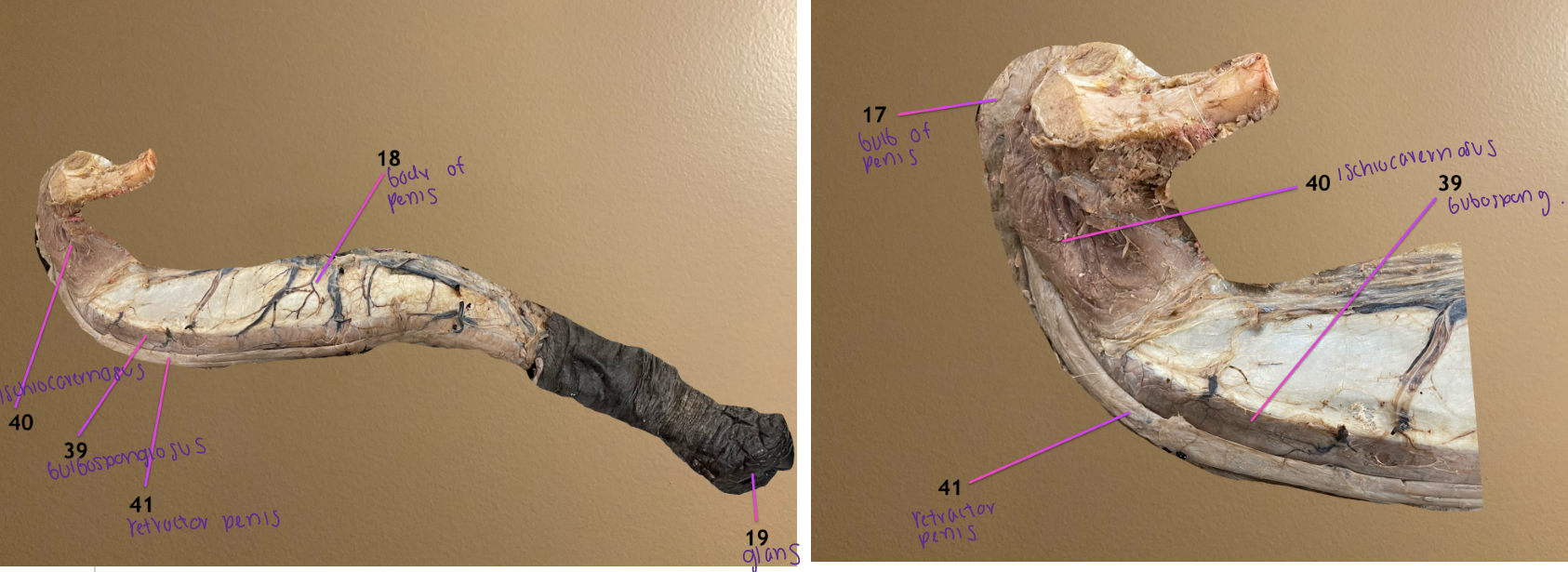
pic 1: lateral view of penis
pic 2: same but zoomed in on root of penis
.
label 17-19
bulb of penis: sits between the crura & serves as beginning of corpus spongiosum penis
body of penis
glans penis


pic 1: lateral view of penis
pic 2: same but zoomed in on root of penis
.
label 39-41
bulbospongiosus m.: on ventral surf of penis
ischiocavernosus m.: covers left & rt crura
retractor penis m.: on ventral surf of penis
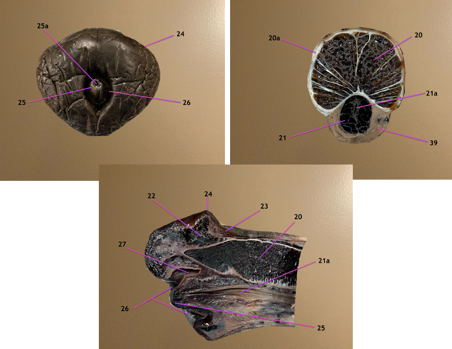
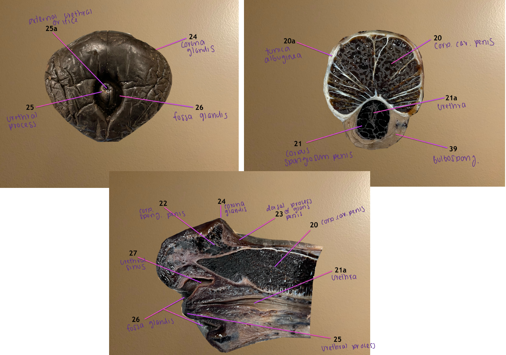
pic 1: cranial view of glans
pic 2: cross section of body of penis
pic 3: longitudinal section of glans
.
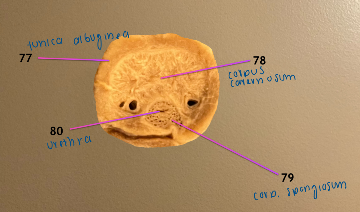
label 20, 20a, 21, 21a
corpus cavernosum penis
20a. tunica albuginea
corpus spongiosum penis: surrounds urethra
21a. urethra

pic 1: cranial view of glans
pic 2: cross section of body of penis
pic 3: longitudinal section of glans
.
label 22-25
corpus spongiosum glandis: cavernous tissue of the glans
dorsal process of glans penis: extension of 22
corona glandis: ridge of tissue surrounding glans
urethral process: protrudes thru fossa glandis


pic 1: cranial view of glans
pic 2: cross section of body of penis
pic 3: longitudinal section of glans
.
label the opening 25a
label 26, 27
25a. external urethral orifice: exit for urine passing thru urethra
fossa glandis: opening the urethral process protrudes thru
urethral sinus: diverticulum that’s a dorsal extension of 26


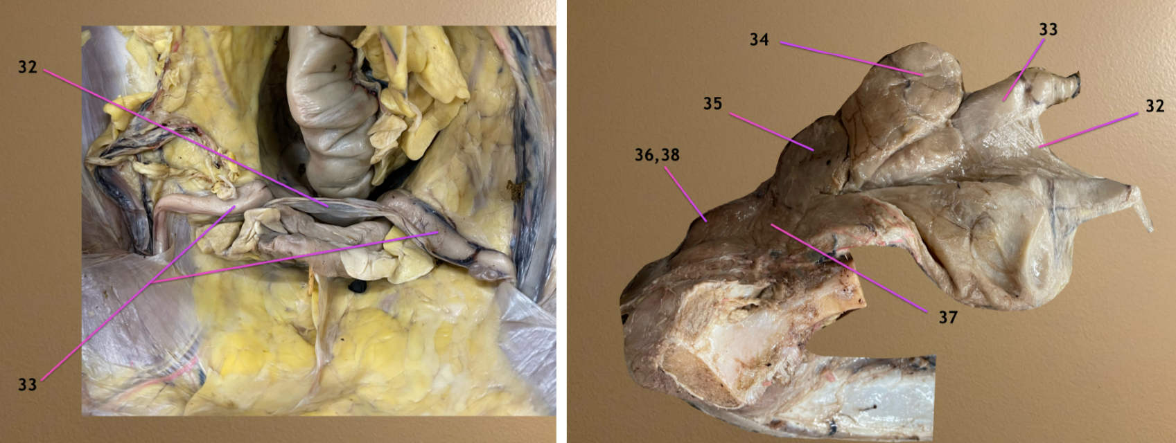
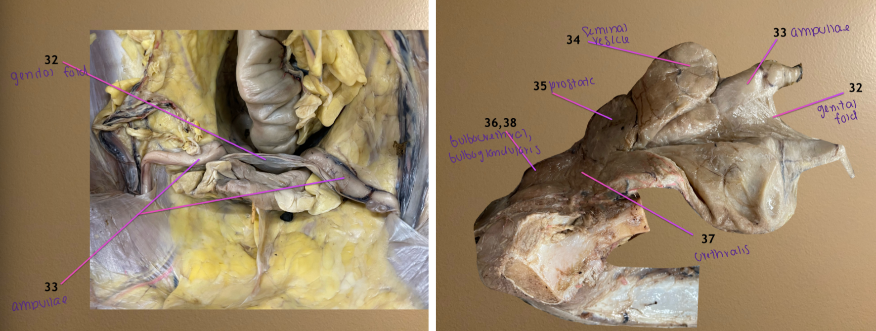
left pic: looking into pelvic cavity, cranial to cd view
rt pic: looking at ischial arch, cranial to rt
.
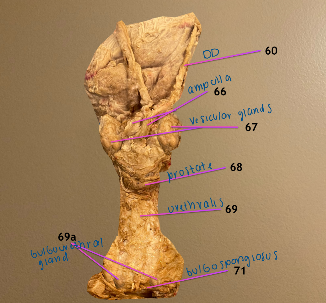
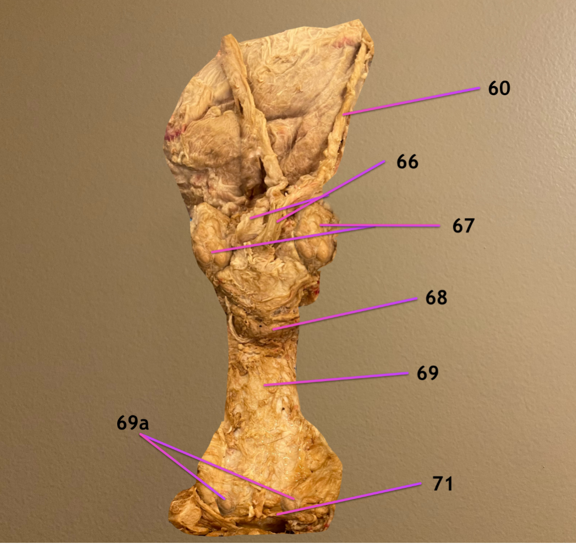
label 32-35
genital fold: contains 33 as they enter pelvic inlet
ampullae: dilated terminal end of DD
seminal vesicle: lateral to 32
prostate gland
.
order of glands you feel during rectal palp (cd to cran): bulbourethral, prostate, seminal vesicle/ampullae

left pic: looking into pelvic cavity, cranial to cd view
rt pic: looking at ischial arch, cranial to rt
.
label 36-38
38 is covering 36
bulbourethral gland
urethralis: covers pelvic portion of urethra
bulboglandularis: covers 36
.
order of glands you feel during rectal palp (cd to cran): bulbourethral, prostate, seminal vesicle/ampullae
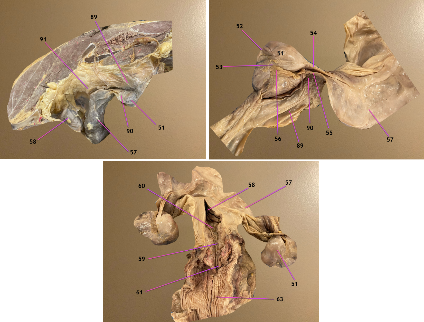
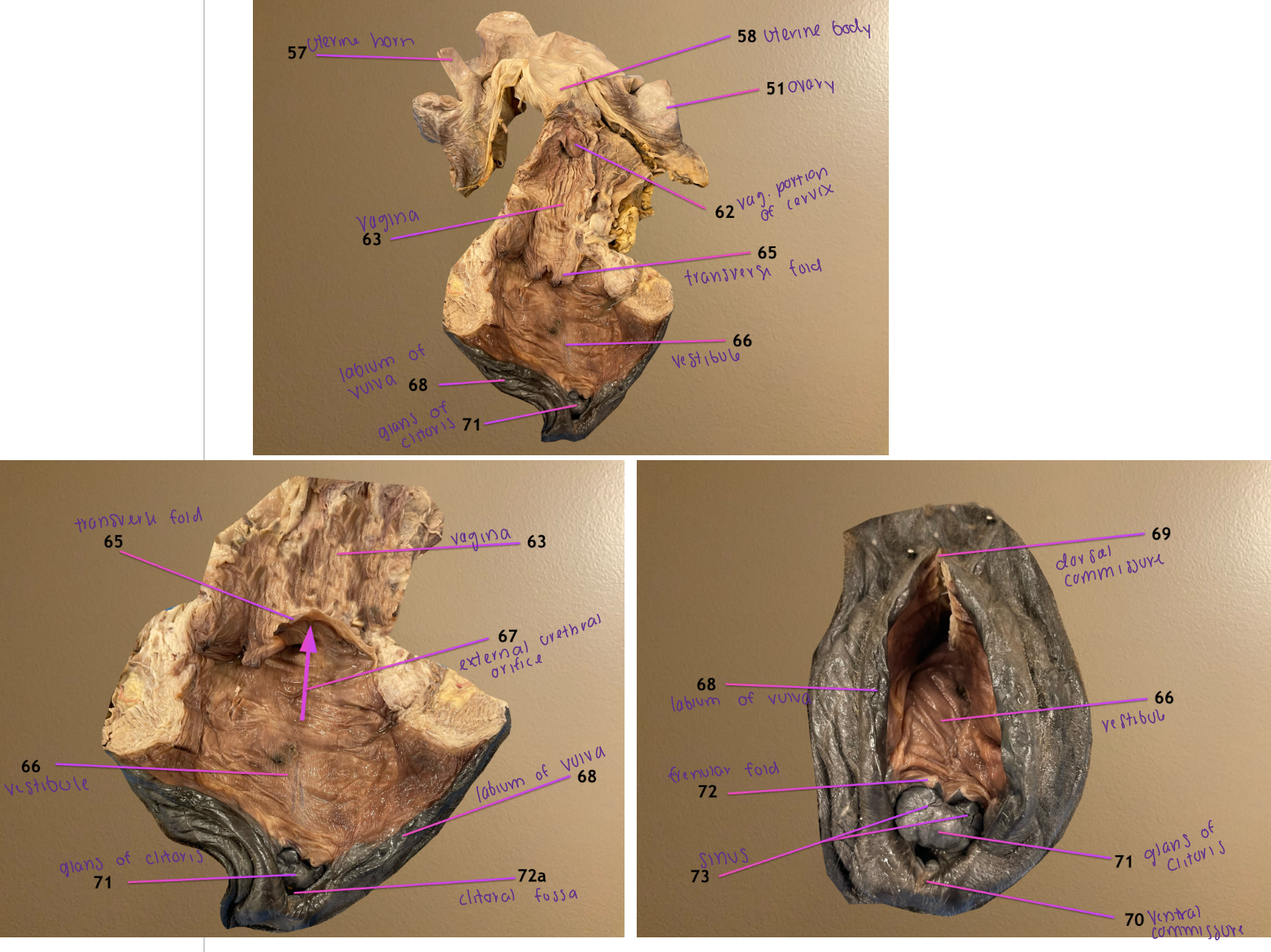
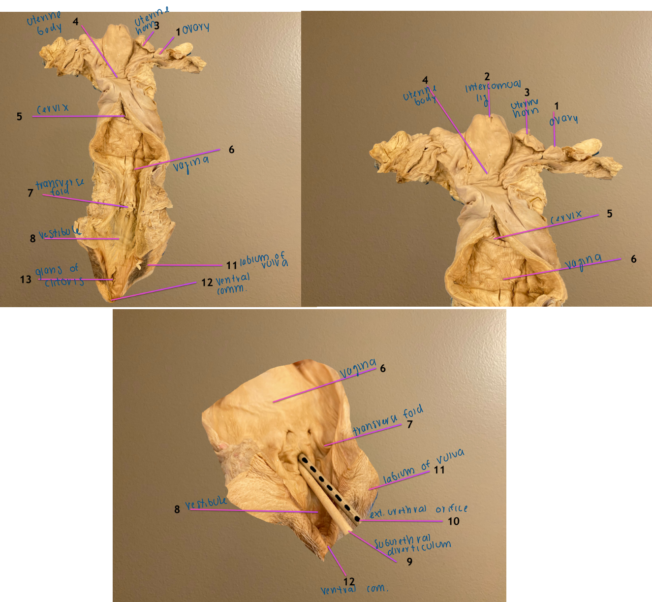
pic 1: female repro tract in situ
pic 3: dorsal view of repro tract
.
label 51-54
ovary
ovarian hilus: convex surface of ovary where BV’s enter
ovulation fossa: concave surf of ovary where ova is released into uterine tube
proper ligament of ovary: connects ovary to uterine horn

pic 1: female repro tract in situ
pic 3: dorsal view of repro tract
.
label 55-58
uterine tube
fimbriae: sweep the egg from ovary to uterine tube
uterine horn
uterine body


pic 1: female repro tract in situ
pic 3: dorsal view of repro tract
.
label 59
label the openings 60, 61
cervix
internal uterine orifice: entrance of cervix
external uterine orifice: exit of cervix


pic 1: female repro tract in situ
pic 3: dorsal view of repro tract
.
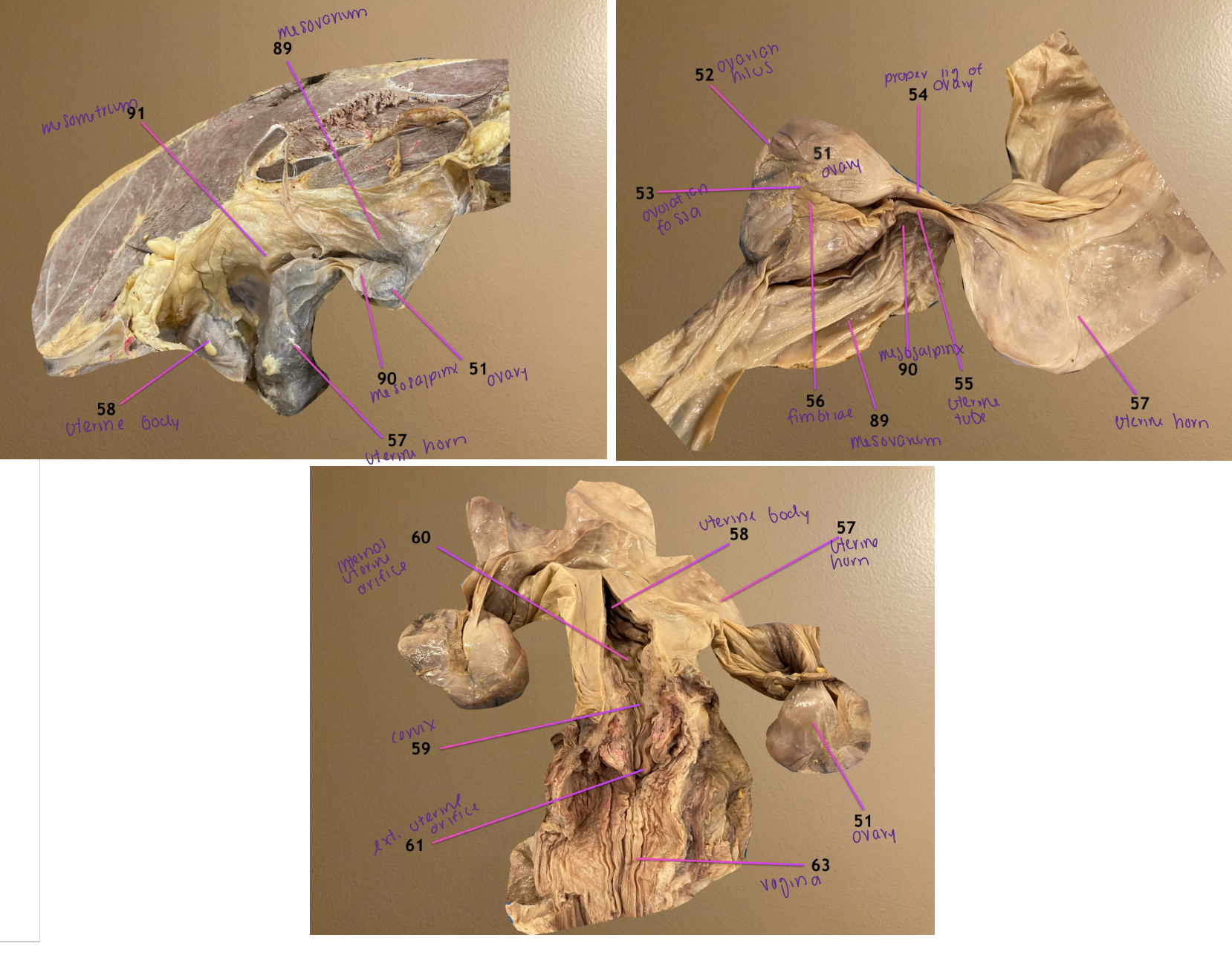
label the CT 89-91
mesovarium: attaches to ovary
mesosalpinx: suspends uterine tube
mesometrium: attaches to uterine horn & body

pic 1: dorsal view of female repro tract
pic 2: same but zoomed in
pic 3: cd to cran view of repro tract
.
label 62, 63, 65, 66
vaginal portion of cervix
vagina
transverse fold: landmark separating 63 & 66
vestibule


pic 1: dorsal view of female repro tract
pic 2: same but zoomed in
pic 3: cd to cran view of repro tract
.
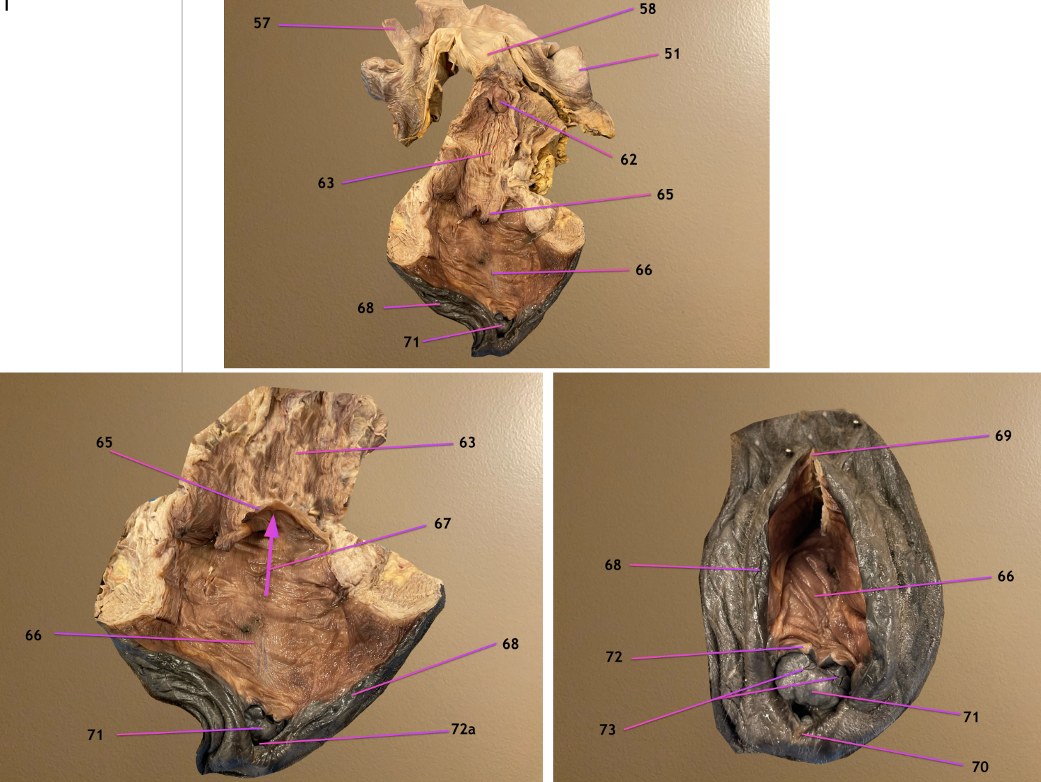
label 67-70
external urethral orifice
labium of vulva
dorsal commissure
ventral commissure


pic 1: dorsal view of female repro tract
pic 2: same but zoomed in
pic 3: cd to cran view of repro tract
.
label 71, 72, 72a, 73
glans of the clitoris
frenular fold
72a. clitoral fossa
sinus
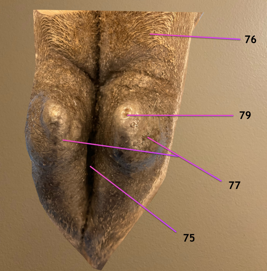
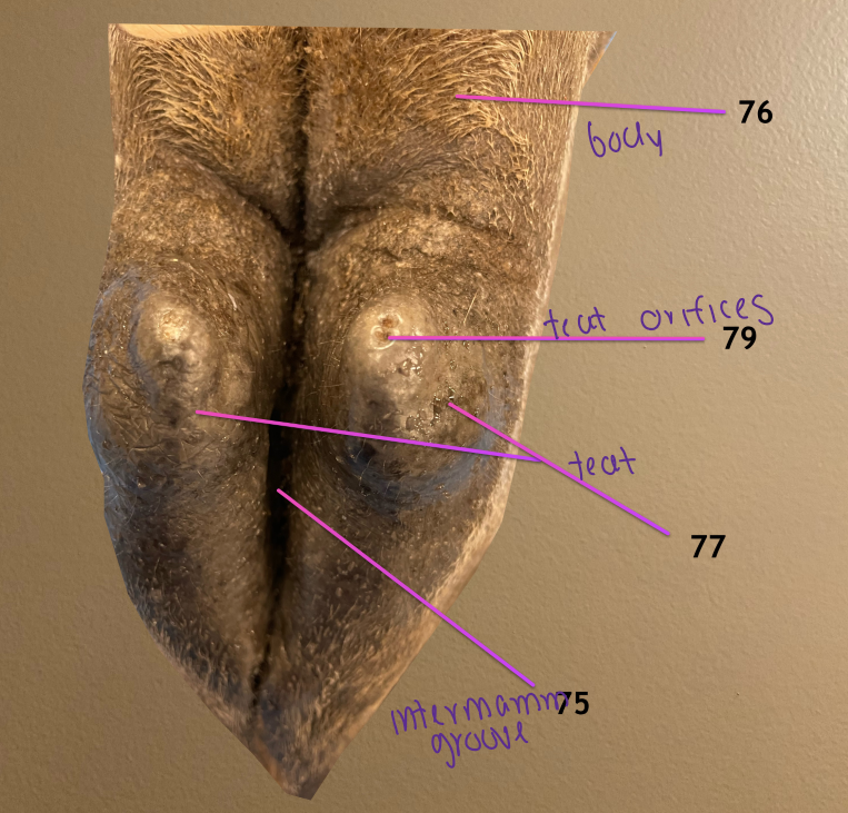
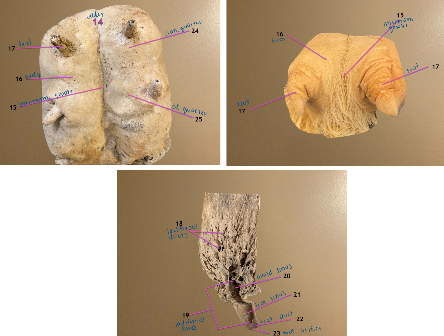
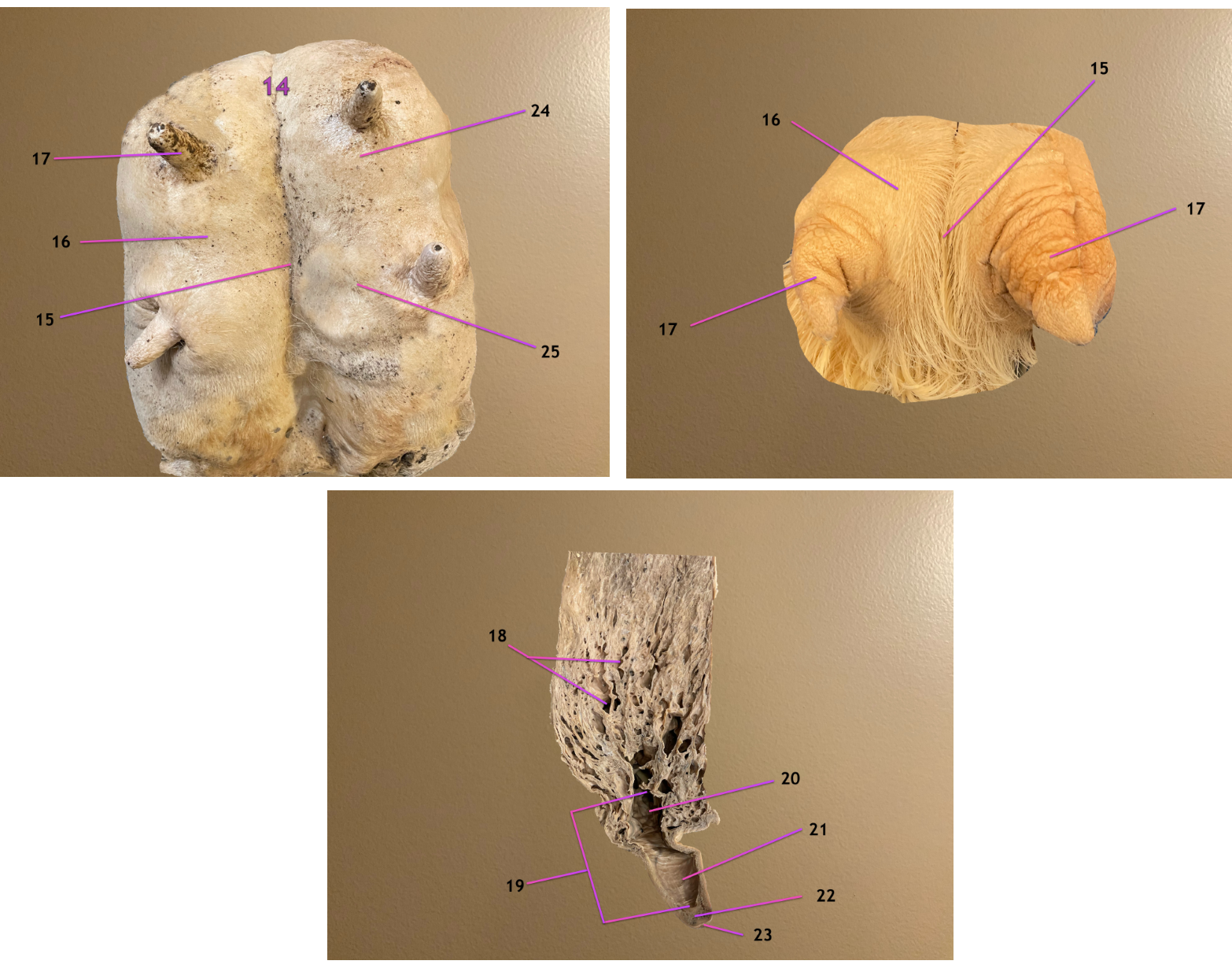
view of mammary gland
.
label 75-77, 79
intermammary groove: divides 76 into left & rt halves
body of mammary gland
teat
teat orifices: there’s 2 of these on each teat
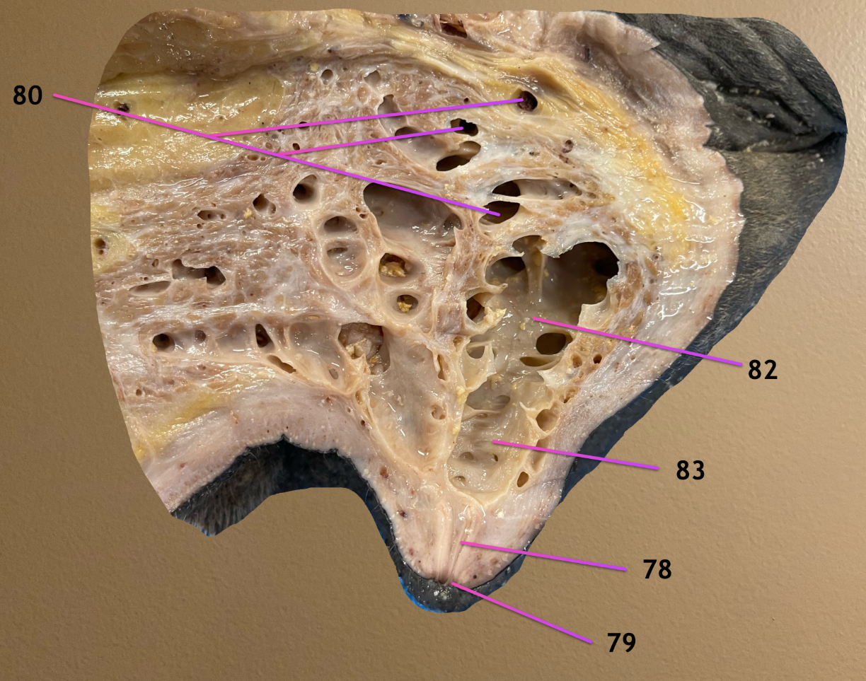
sagittal section of mammary gland
.
label 78, 80, 82, 83
teat duct: milk passes thru this
lactiferous ducts: milk travels thru these to reach lactiferous sinus where milk is stored
gland sinus: part of lactif sinus
teat sinus: same ^
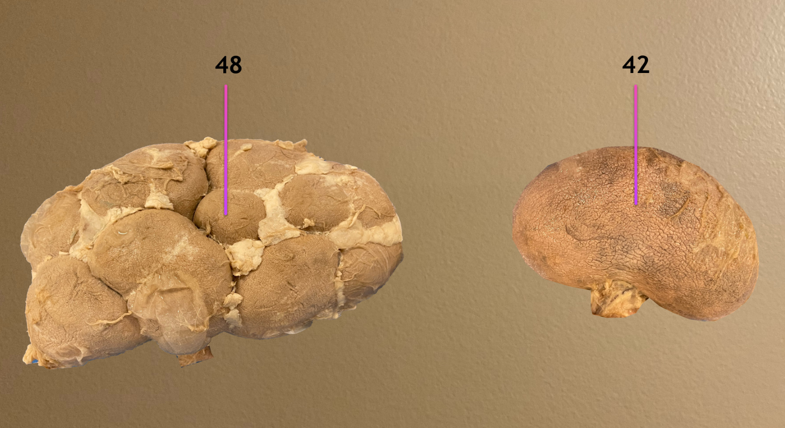
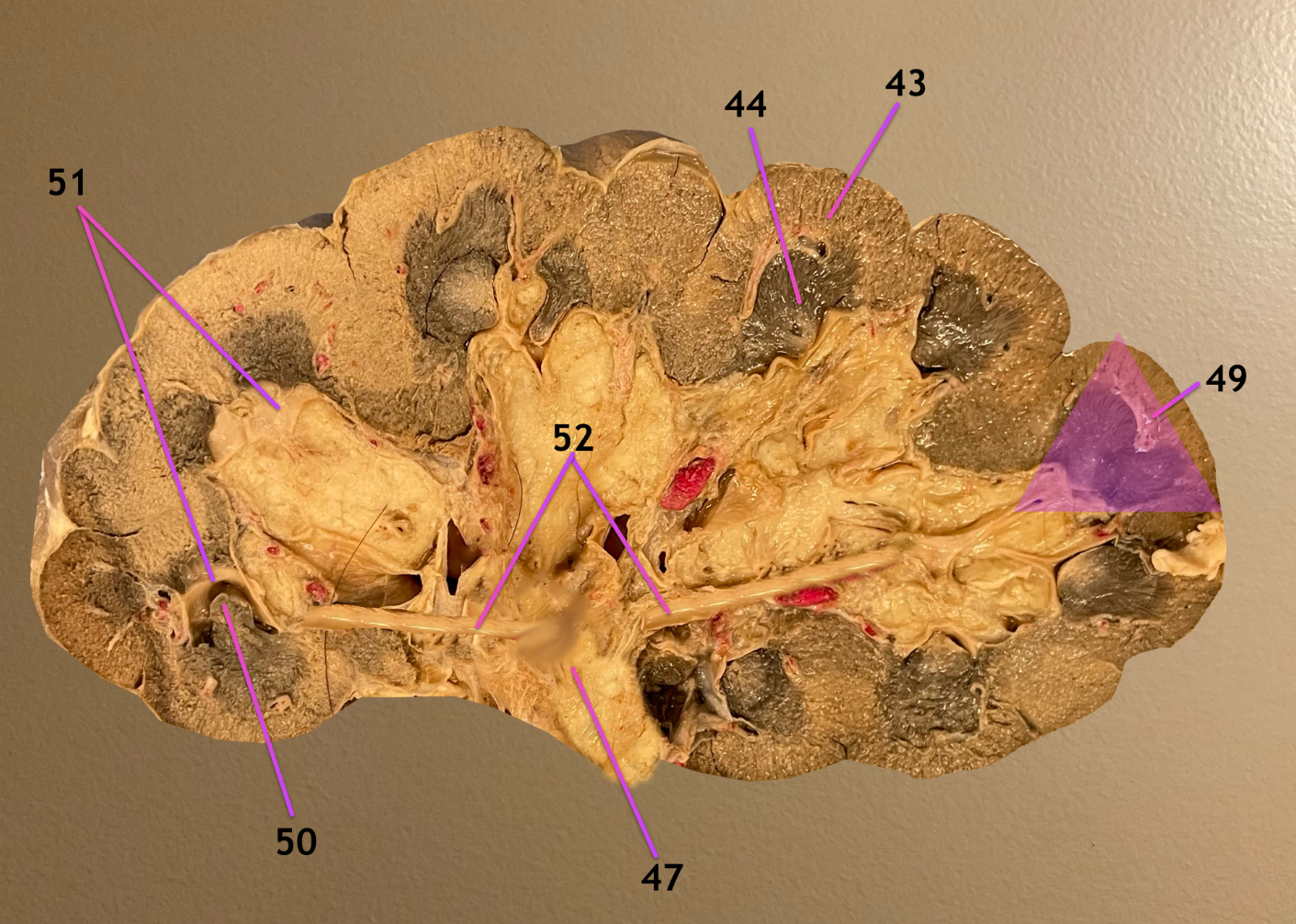
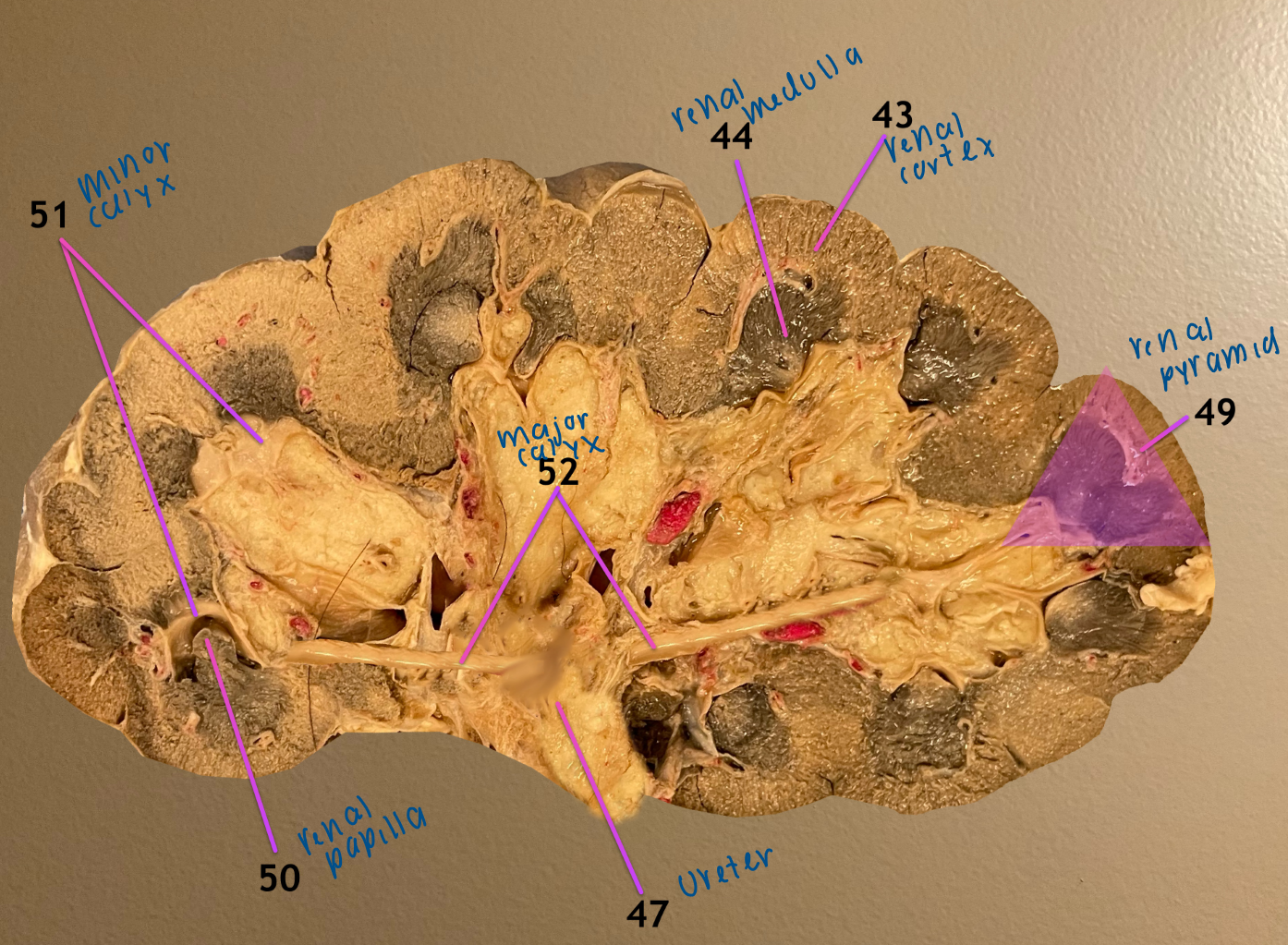
label the types of kidney
unilobar kidney: small ruminant
multilobar kidney: in cows
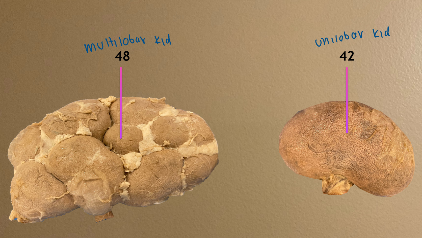
longitudinal section of kidney
.
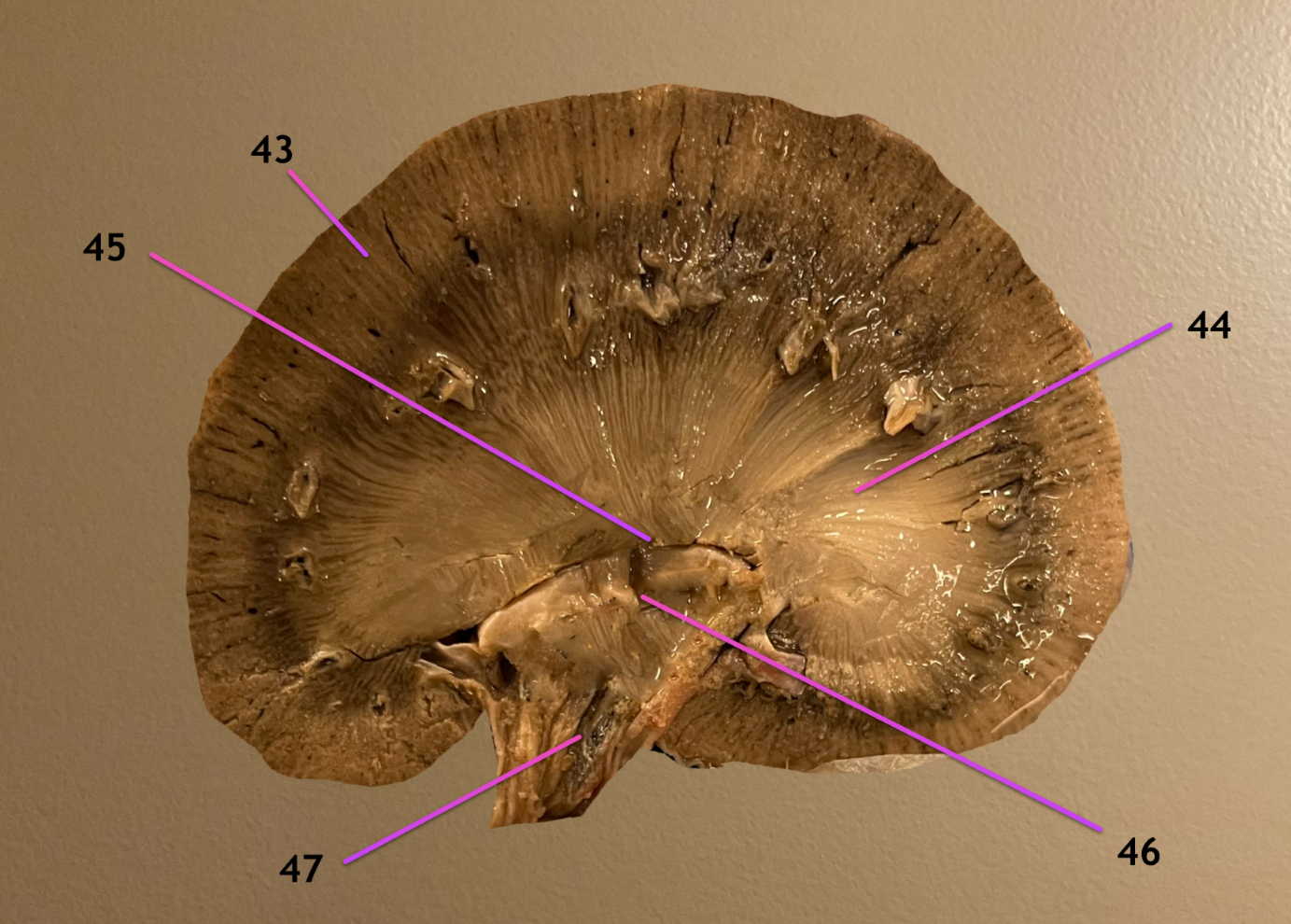
label the topographic region 43, 44
label 47
renal cortex
renal medulla
.
ureter
longitudinal section of kidney
.
label the topographic regions 43, 44
.
label 47, 49-52
include both names of 52
renal cortex
renal medulla
.
ureter
renal pyramid
renal papilla
minor calyx
major calyx / principal branch of the ureter
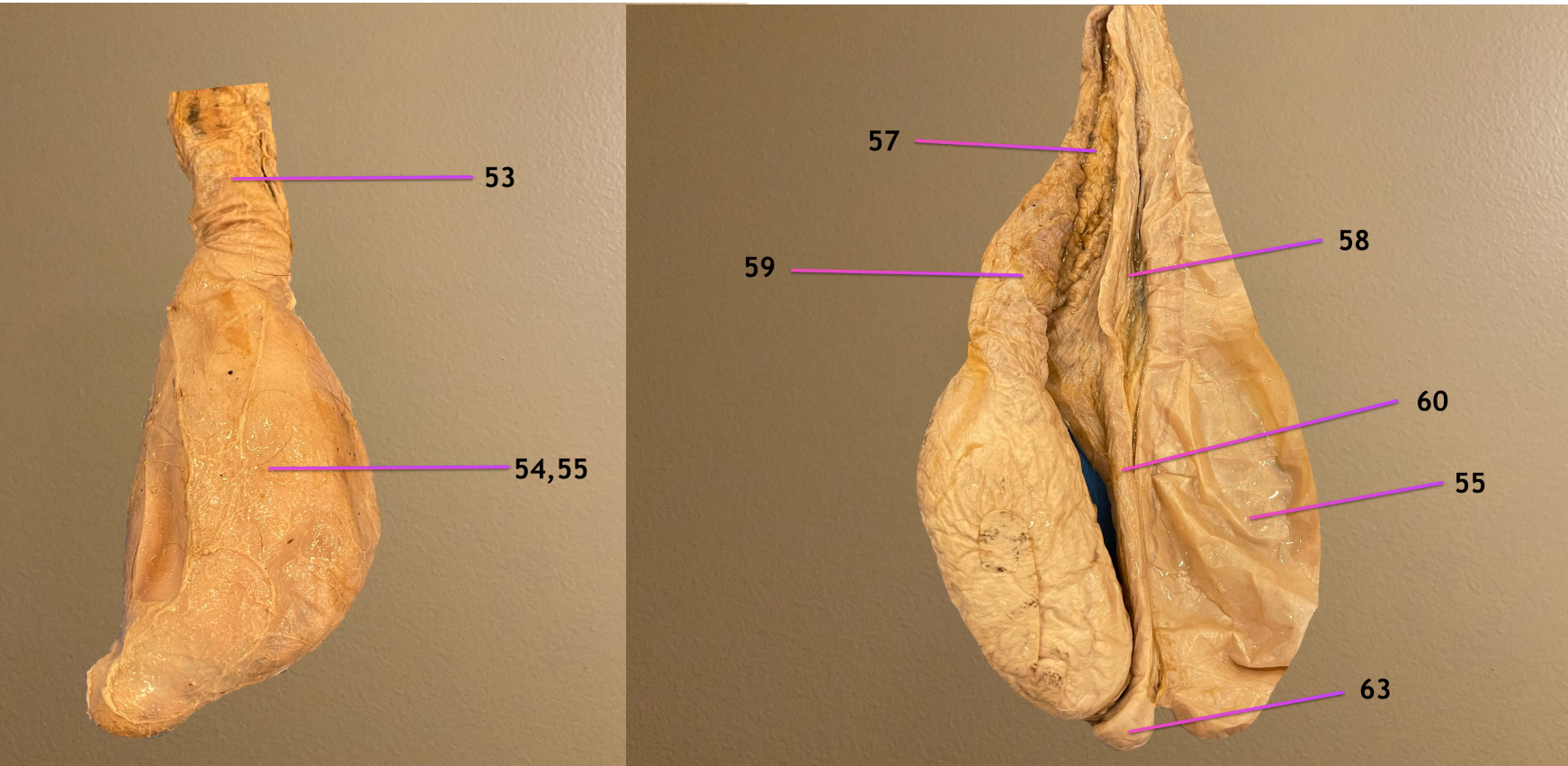
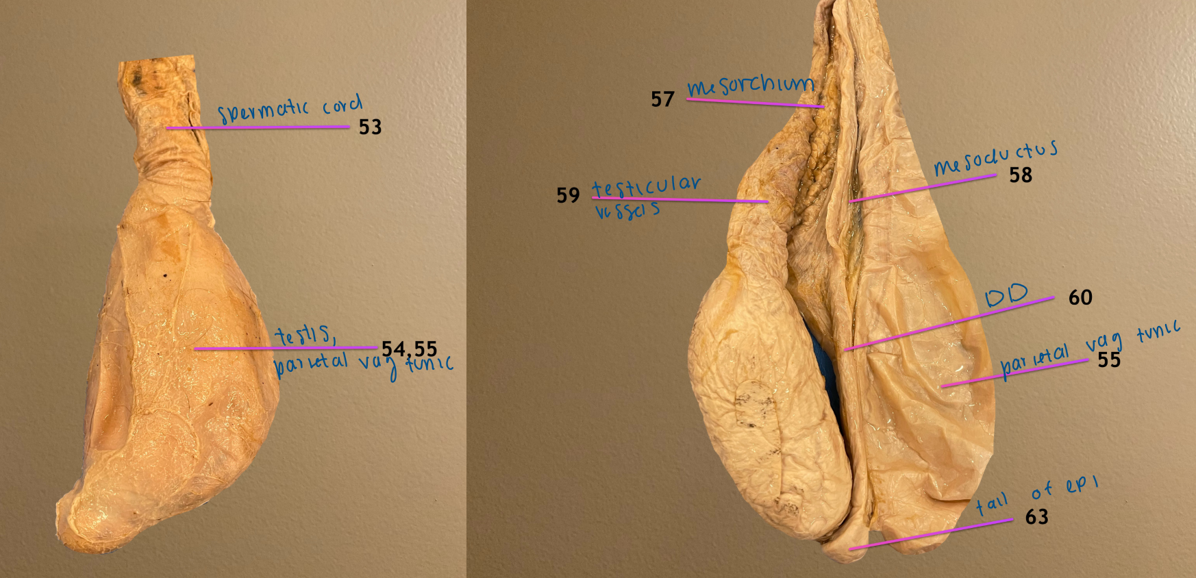
view of testicle
.
label 53, 59, 60
.
label the CT 57, 58
spermatic cord
testicular vessels
ductus deferens
.
mesorchium
mesoductus
.
note: this is bull testicle

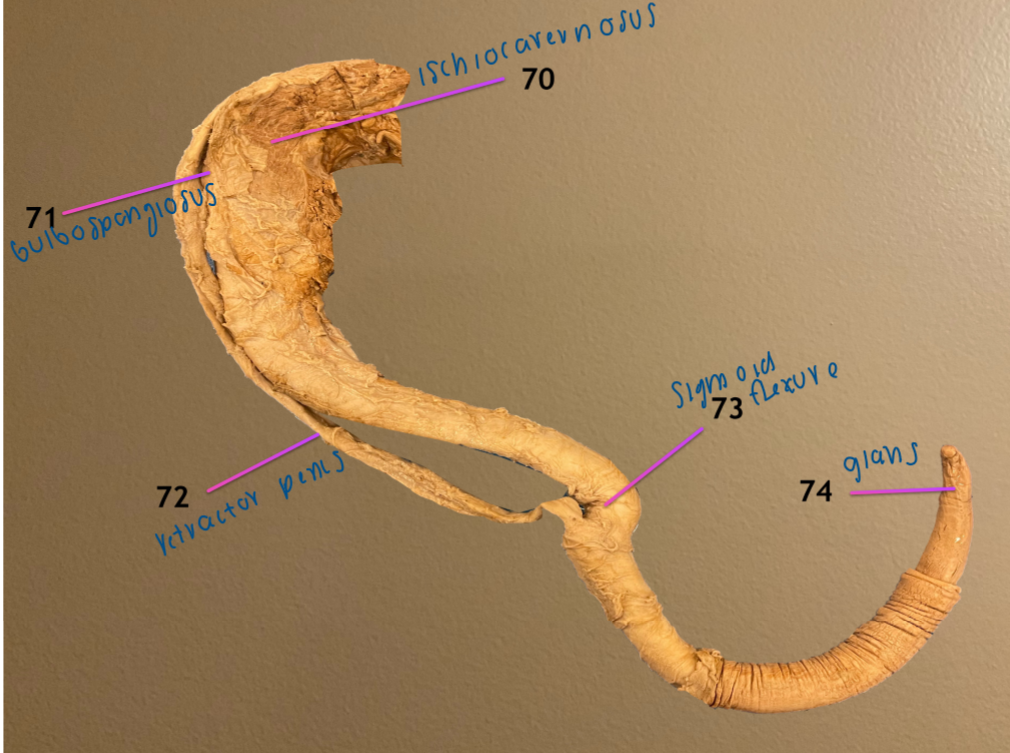
view of penis
.
label 70-73
ischiocavernosus m.
bulbospongiosus m.
retractor penis m.
sigmoid flexure
.
note: this is a bull penis

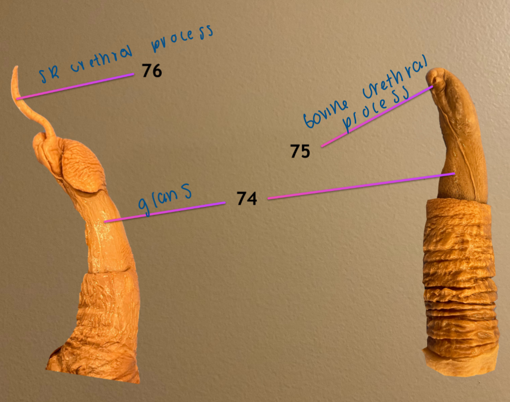
view of glans penis
.
label & note the species: 75, 76
bovine urethral process
small ruminant urethral process
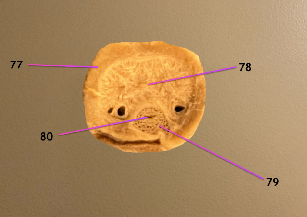
cross section of body of penis
.
label 77-80
tunica albuginea: dense CT surrounding body
corpus cavernosum penis: filled w/ CT in ruminants
corpus spongiosum penis
urethra
.
note: this is a bulls penis

view of bull accessory glands
.
label 60, 66, 67
ductus deferens
ampulla: enlarged end of 60
vesicular glands
.
note: these are labeled from cran to cd
view of bull accessory glands
.
label 68 69, 69a
prostate
urethralis m.: covers pelvic part of urethra
69a. bulbourethral gland: covered by bulbospongiosus m.
.
note: these are labeled cran to cd

pic 1/2: dorsal view of cow repro tract
pic 3: close up of vaginovestibular junction
.
label 1-4
ovary
intercornual ligament: used to stabilize the uterus during rectal palp
uterine horn
uterine body


pic 1/2: dorsal view of cow repro tract
pic 3: close up of vaginovestibular junction
.
label the opening 9, 10
label 13
suburethral diverticulum: ventral to 10
external urethral orifice: dorsal to 9
glans of the clitoris

pic 1: ventral view of cow mammary gland, cranial to top
pic 2: same but of SR
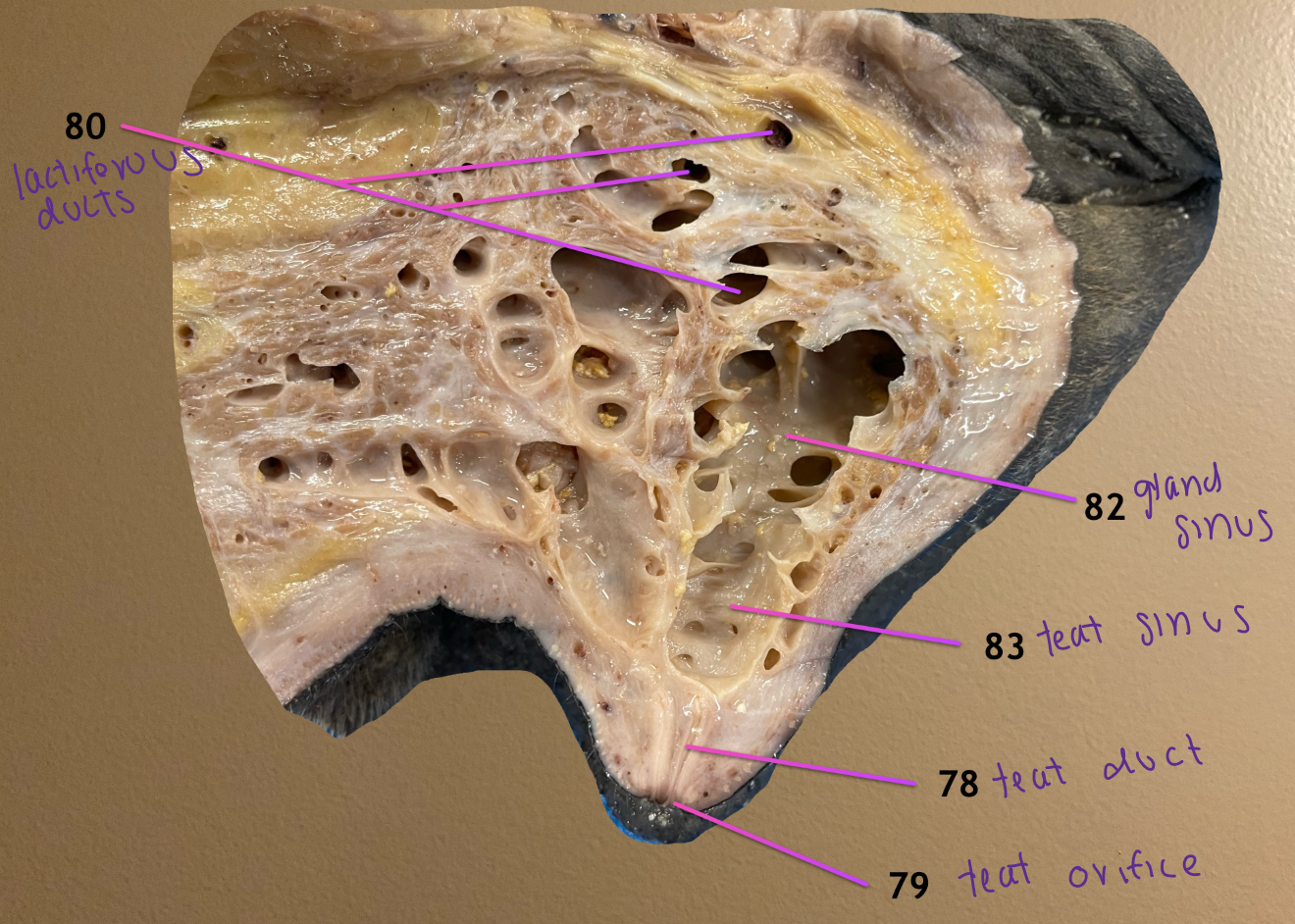
pic 3: inside a quarter
.
label the entire structure 14
label 18-21
udder
lactiferous ducts
lactiferous sinus
gland sinus
teat sinus
pic 1: ventral view of cow mammary gland, cranial to top
pic 2: same but of SR
pic 3: inside a quarter
.
label 22-25
teat/papillary duct
teat/papillary orifice
cranial quarter
caudal quarter

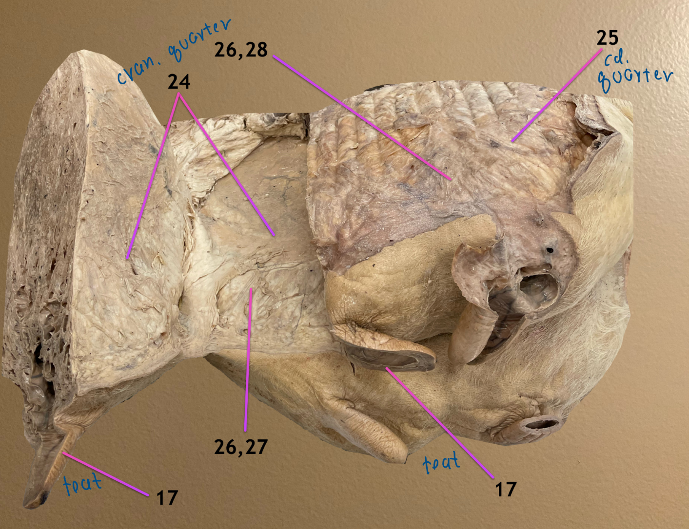
cranial quarter cut & reflected to expose midline of udder
.
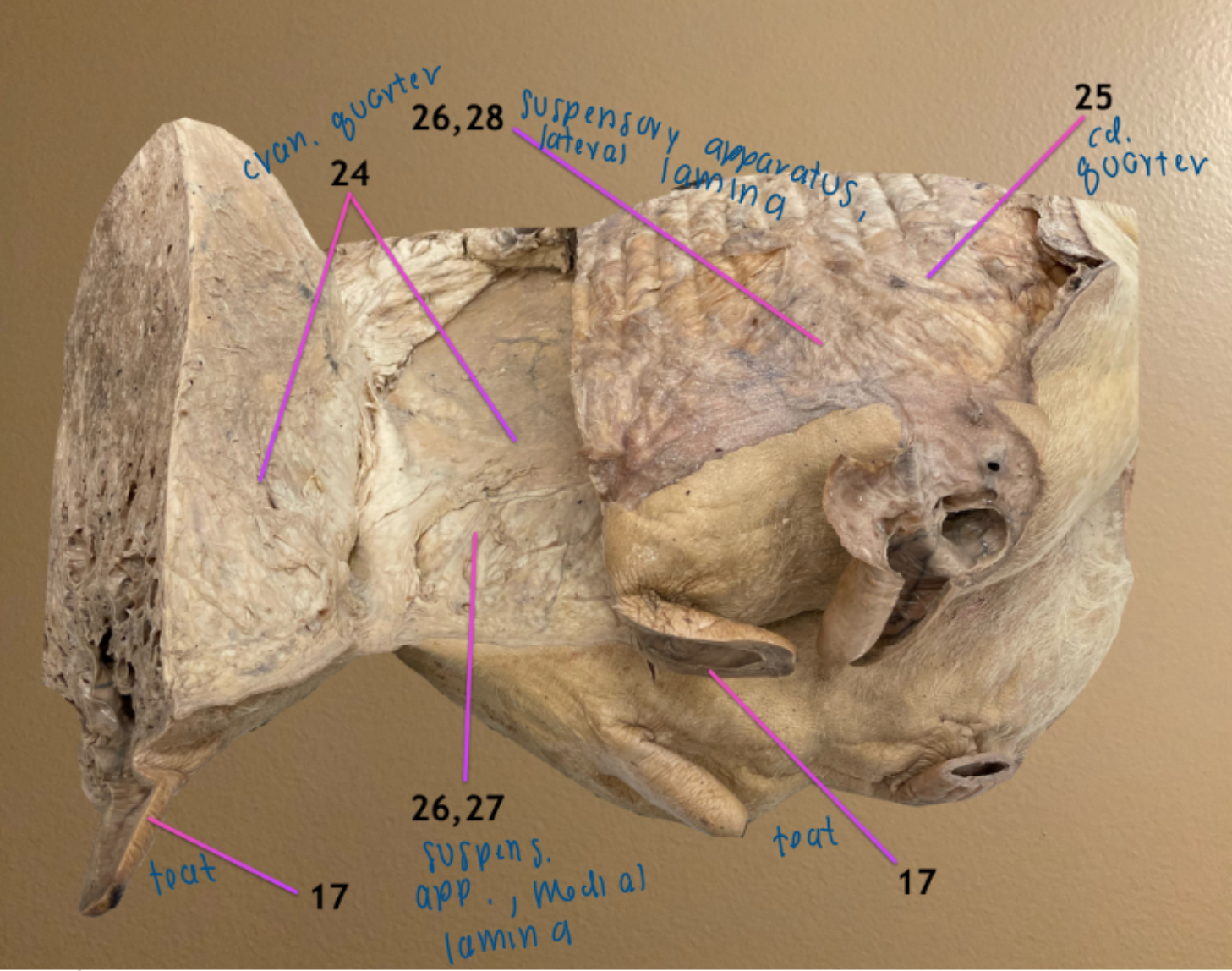
label the entire structure 26
label the CT 27, 28
suspensory apparatus
medial lamina of the suspensory apparatus: meets at midline
lateral lamina of the suspensory apparatus: covers lateral surf of mamm gland, deep to skin
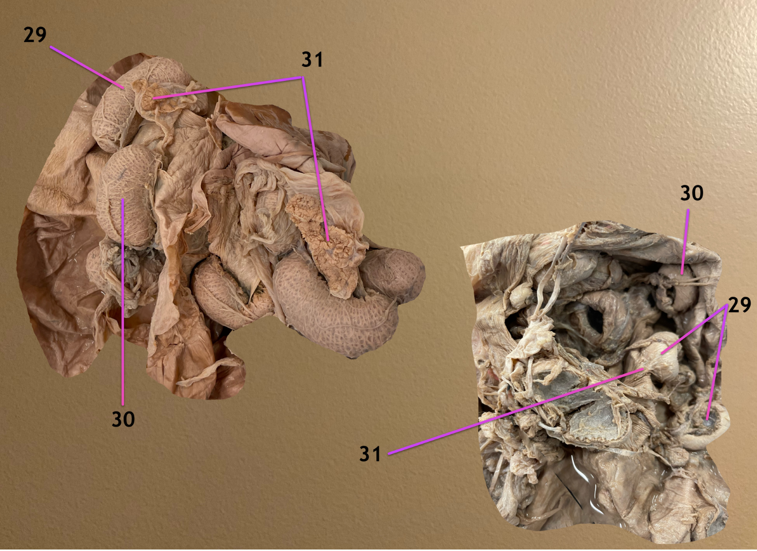
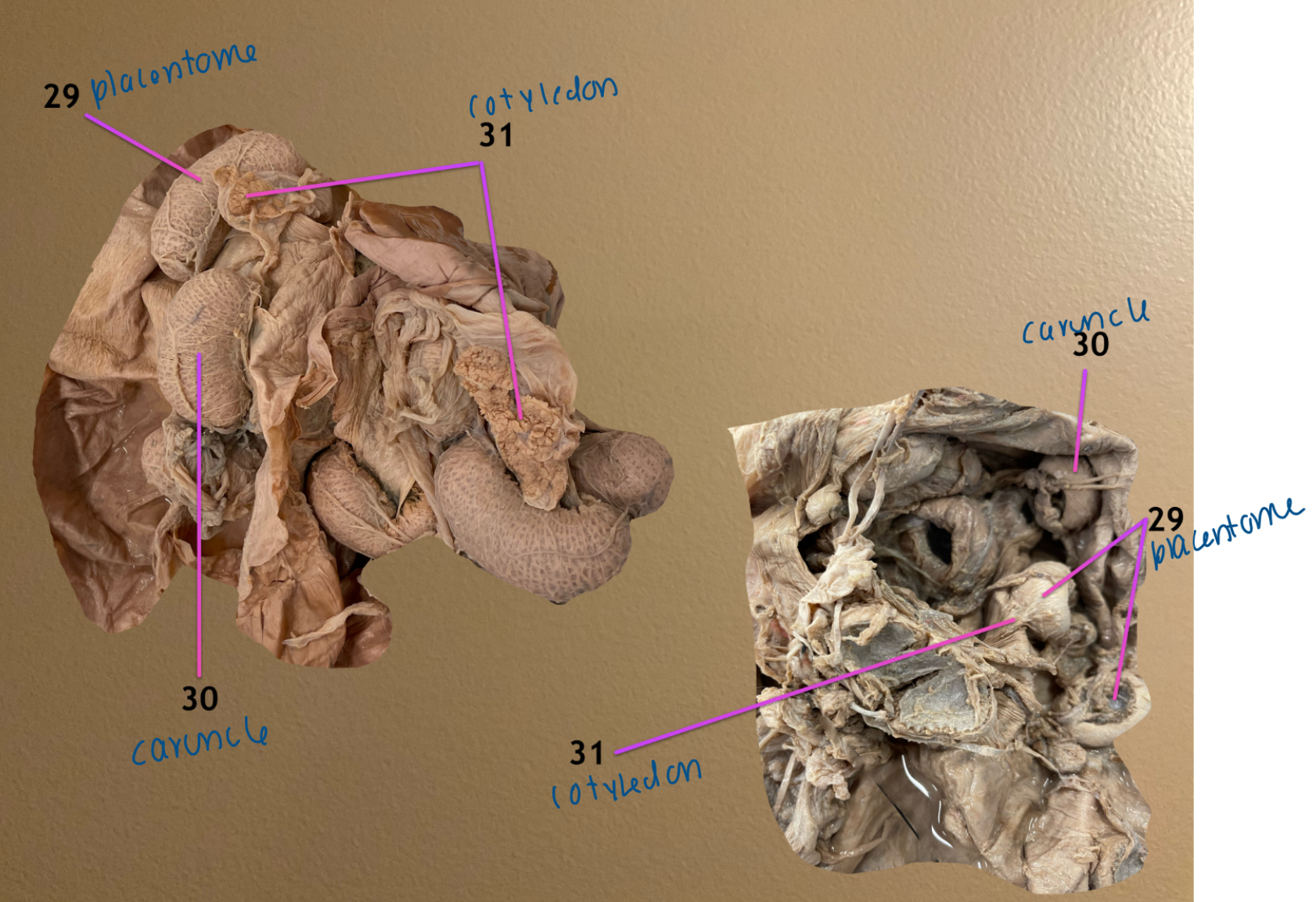
view of placentas
.
label 29-31
.
which species do the left & rt belong to
placentome
caruncle
cotyledon
.
left: cow
rt: SR