RNFL Analysis, Ganglion Cell Analysis
1/29
There's no tags or description
Looks like no tags are added yet.
Name | Mastery | Learn | Test | Matching | Spaced | Call with Kai |
|---|
No analytics yet
Send a link to your students to track their progress
30 Terms
retinal ganglion cells, retinal nerve fiber layer (RNFL)
POAG is a progressive, chronic optic neuropathy which is characterized by a loss of _____ and thereby their axons resulting in a thinning of the ____
before
Damage to the retinal nerve fiber layer occurs (before or after) observable optic nerve head and visual field changes
6, 60
Structural loss of the nerve fiber layer precedes functional loss by as much as ___ years in ___% of patients
30-50%
percentage of retinal nerve fiber layer loss that may occur before a detectable change in visual field testing appears
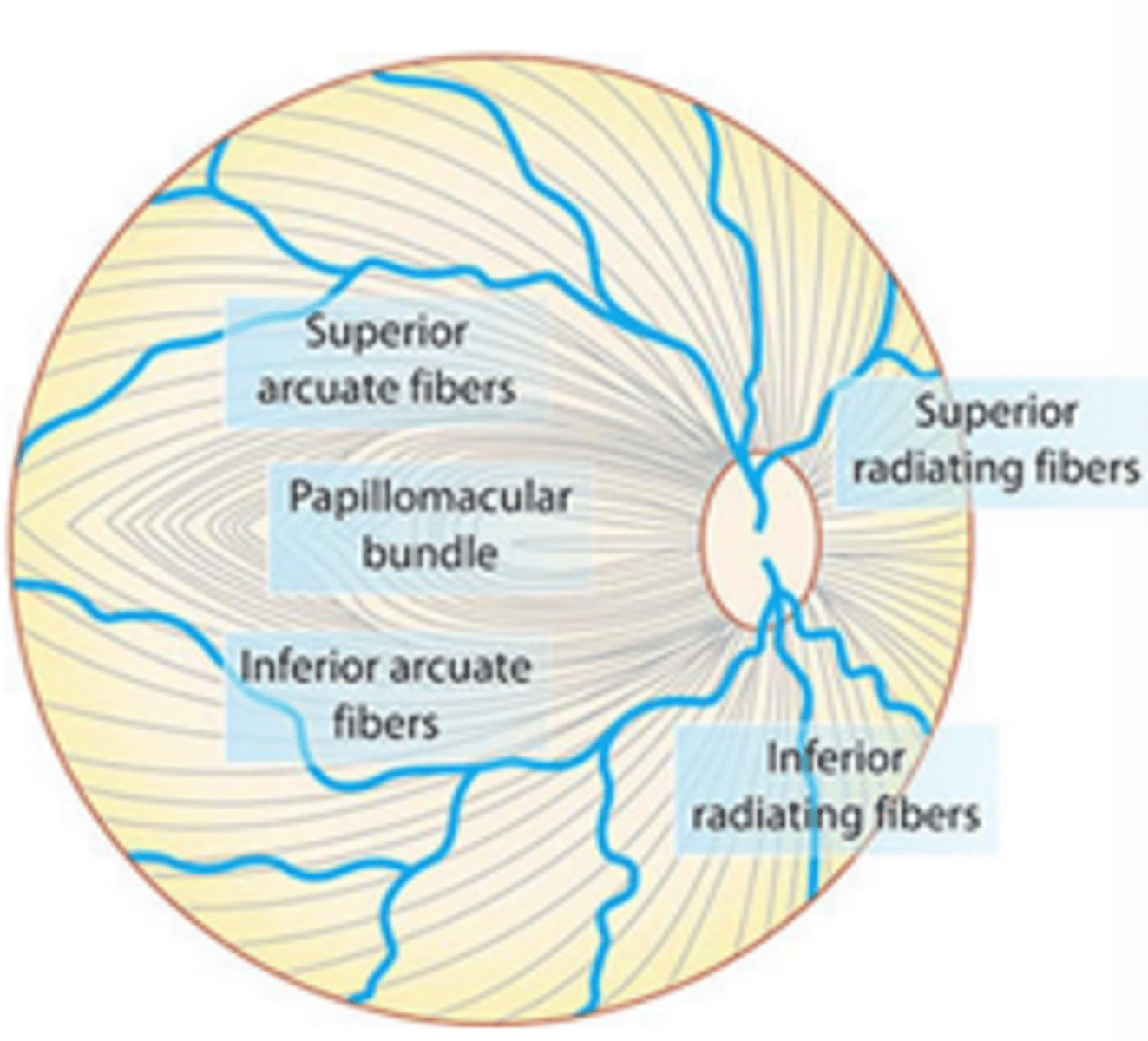
superior and inferior
Axons originating from the temporal retina insert into the ____ rim. This is the area most susceptible to damage caused by glaucoma due to larger laminar pores of the lamina cribrosa as well as the fact that it is a watershed area for vascular supply.

nasal
Axons originating from the nasal retina insert into the ____ rim

temporal
Axons originating from the macula (papillomacular bundle) insert into the ____ rim. This is the area least susceptible to damage caused by glaucoma

peripheral
Fibers of the ____ retina are more susceptible to damage caused by glaucoma due to their peripheral arrangement in the optic nerve causing more mechanical strain from the lamina cribrosa
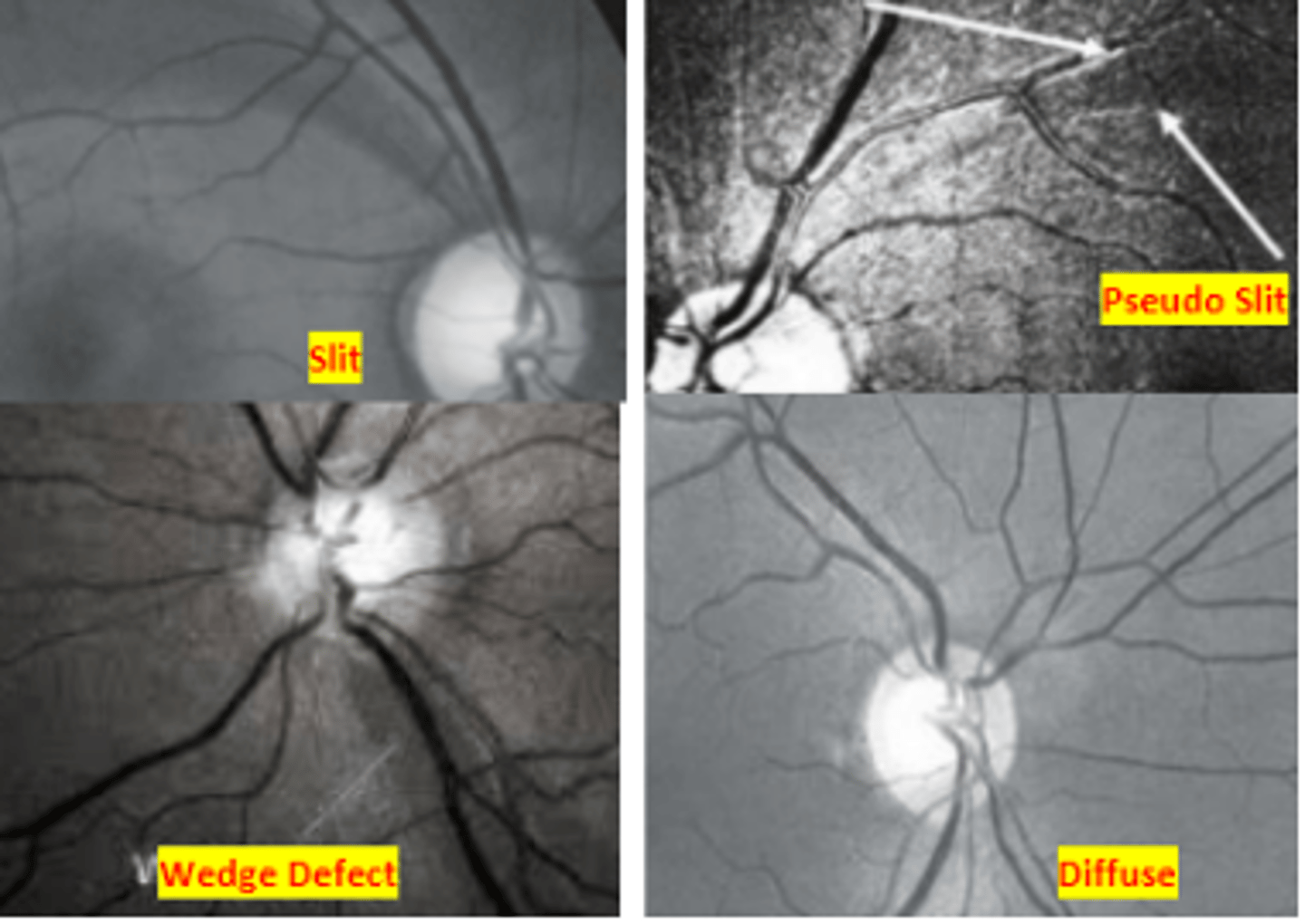
red free filter
The use of a ____ used on direct or slit lamp ophthalmoscopy and fundus photography can be useful in increasing contrast in order to better view the nerve fiber layer. Requires clear media to acquire a bright enough image. Nerve fiber layer loss can be slit, wedged, or diffuse (less easy to identify, but most common).
10
Pseudo slit defects seen of the nerve fiber layer that do not extend all the way to the optic nerve head occur in ____% normal eyes
80, 94
RNFL photography has a sensitivity of ___% and a specificity of ____%. This reveals the importance of RNFL assessment as an indicator for early glaucomatous damage
Optical coherence tomography (OCT)
retinal imaging technique that creates a cross sectional image analogous to an ultrasound, but utilizing light reflectivity. Is used for both the detection and analysis of progression of glaucoma and other retinal diseases.
floor effect
OCT is most usually when the patient is a glaucoma suspect or has early to moderate disease. Its usefulness in late disease is limited due to the ____ where the RNFL thickness becomes so thin that it is no longer detectable via OCT
Red disease
occurs when the OCT is flagging information for a patient who does not actually have any disease
Green disease
occurs when the OCT is not flagging information for a patient who does have disease. I.e.) the presence of an epiretinal membrane in a patient having NFL thinning due to glaucoma . This may result in normal OCT testing in the presence of RNFL thinning
inferior, 84, 90
The ____ quadrant on OCT is very specific to glaucoma and is the least affected by age. It has a ___% sensitivity and a ___% specificity for the detection of glaucoma
92.9 microns
mean RNFL thickness
>80 microns
normal average RNFL thickness
70-79 microns
average RNFL thickness that is normal, but suspicious for glaucoma
60-69 microns
average RNFL thickness indicative glaucoma seen in less than 5% of normal eyes
0.2-0.5 microns
normal RNFL loss per year
4 microns
acceptable amount of inter visit variability of RNFL thickness that can be attributed to measurement error. Real change is considered twice this
Inferior
OCT RNFL thickness quadrant that is best used to discriminate healthy from glaucomatous eyes due to the fact that it is least affected by age decay and highly affected by glaucoma.
>119 microns
normal inferior quadrant RNFL thickness
92.5-119 microns
inferior quadrant RNFL thickness that is suspicious for glaucoma (56% normal, 44% glaucoma)
<92.5 microns
inferior quadrants RNFL thickness indicative of glaucoma (100%)
ganglion cell dropout, paracentral, inferior temporal, macular vulnerability zone
NFL loss as measured by OCT is an indirect method of measuring ____ which preceding NFL loss. This has been shown to have better diagnostic ability for _____ VF loss when compared to RNFL changes and is seen as ____ GCC reduction known as the ____. Other retinal conditions affecting ganglion cell count must also be ruled out before diagnosing glaucoma
nerve fiber layer, ganglion cell layer, and inner plexiform layer
The ganglion cell complex as measured by OCT includes these three retinal layers
68-74.8 microns
average GCC thickness in normal eyes
OCT A, inferotemporal
____ testing shows loss of vessels in the peripapillary region of patients having glaucoma, especially of the ____ sector. However, this testing has a lack of normative database, high vascular variability between patients, and it is unclear if systemic HTN meds will affect these measurements