Auburn Microbiology Lab Final!
1/159
There's no tags or description
Looks like no tags are added yet.
Name | Mastery | Learn | Test | Matching | Spaced | Call with Kai |
|---|
No analytics yet
Send a link to your students to track their progress
160 Terms
BSL 1
microorganisms not known to cause disease in healthy adults; protective equipment not required
BSL 2
indigenous microorganisms that can lead to diseases of varying severity in healthy adults; protective equipment required
BSL 3
indigenous or exotic microorganisms that cause serious or potentially lethal disease through respiratory transmission; immunizations may be required
BSL 4
microorganisms that are dangerous and exotic with high risk of aerosol transmitted infections; rarely are there treatments or vaccines; diseases may be fatal
Brightfield microscope
simplest form of microscopy where light is either passed through, or reflected off, a specimen. Illumination not altered by devices that change properties of light; requires use of stains to visualize cells
Phase contrast microscope
converts the differences in optical density of cells into shades of brightness; allows visualization of morphology, external structures, and some internal structures; stain not required
Fluorescent Microscope
uses high intensity illumination to excite fluorescent molecules which absorb photons, are excited to a higher level, as they relax back to ground-state vibrational energy is lost and emission spectrum shifted to longer wavelengths; fluorescence emirates from sample
Dark field Microscope
Contrast is created by a bright specimen on a dark background. It is ideal for revealing morphology and external structures, but does not provide a great deal of information about internal structure. Stains are not required.
base/arm
frame of microscope
light source
LED
stage
horizontal area supporting the slide; has stage adjustment knobs
lens system
oculars, objectives, condenser
oculars
10X magnification
objectives
attached to nosepiece; magnification of 10X, 40X ( high dry), 100X (oil immersion)
total magnification
ocular power x objective power
condenser
collects and directs light from the light source to the slide; located under the stage
diaphragm
controls the amount of light that reaches the slide
focusing knobs
coarse focus knob (outer) & fine focus knob (inner)
fine focus knobs
used after reaching 40X and 100X
coarse focus knobs
only used when using 10X
immersion oil
same refractive index as glass; used with 100X objective only; fills gap between objective and slide to form a continuos lens path which increases image resolution by reducing light refraction
parfocal
ability of a microscope to remain relatively in focus when changing from lower to higher power objective
numerical aperture
mathematical expression that describes how the condenser lens concentrates and focuses the light rays from light source
resolving power
ability of a lens to show two closely spaced objects as distinct and separate
working distance
distance between the bottom of the objective lens and the slide
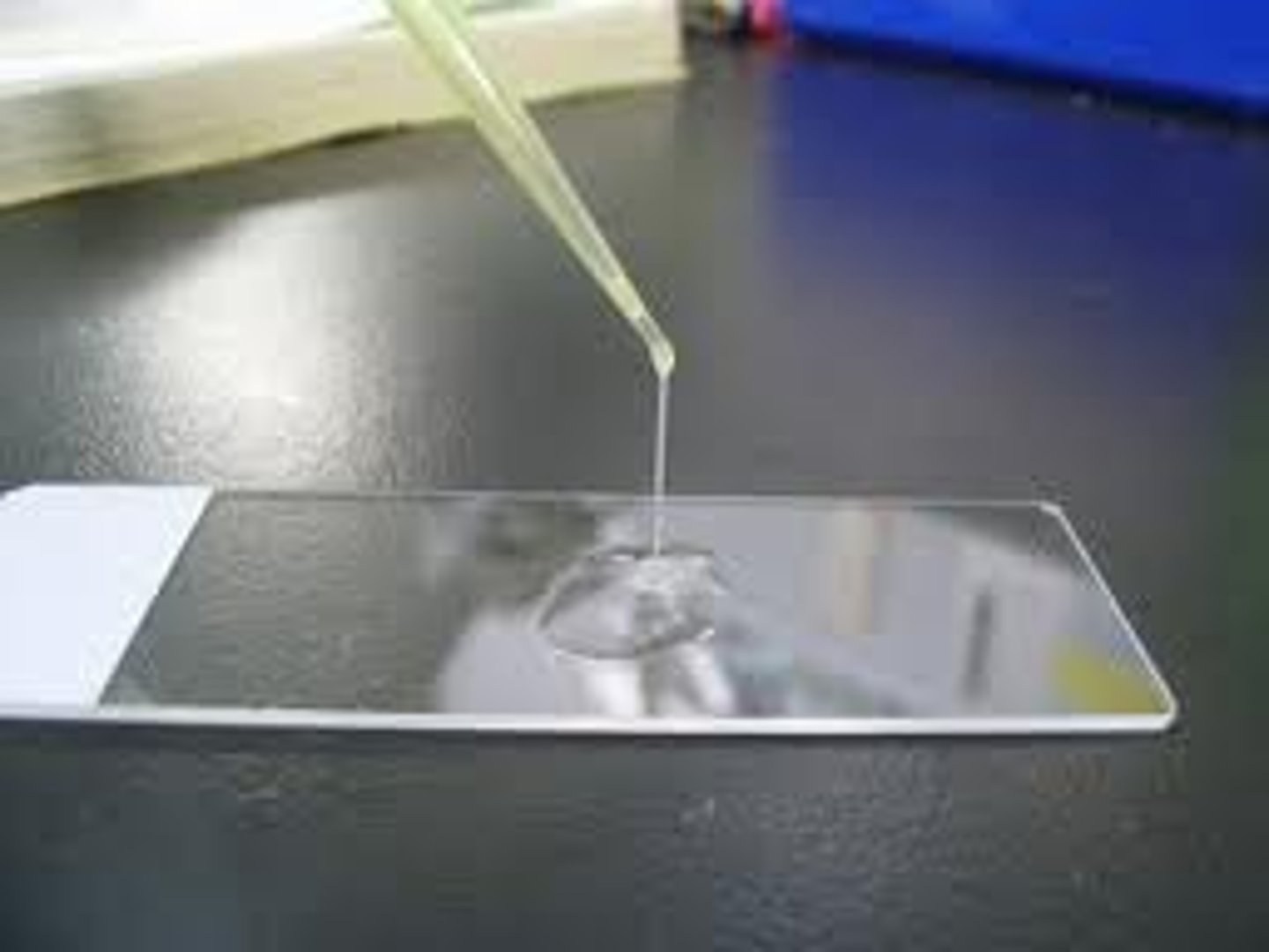
ubiquity of microorganisms
microorganisms are everywhere
simple stain
staining with a single stain; allows visualization of cell morphology, cell arrangement, and internal storage materials
methylene blue
stain used in simple staining
types of cell morphology
cocci, rods, spiral/curved
cocci
spherical morphology; singly, pairs, tetrads, chains, clusters
rods (bacilli)
rod shaped morphology; singly or chains
spiral/curved
corkscrew rods morphology
Hans Christian Gram
responsible for Gram stain
Gram positive
have thick peptidoglycan layer in cell wall, retain crystal violet, purple
Gram negative
thin layer of peptidoglycan in cell wall; do not retain crystal violet; pinkish (safranin)
Gram stain steps
crystal violet, Gram's iodine, ethanol, safranin
crystal violet
primary stain (60 sec)
Gram's iodine
mordant (60 sec)
ethanol
decolorizer (10 sec)
safranin
counter stain (60 sec)
endospore
allow microorganisms to survive environmental conditions that are not favorable to growth
endospore staining
spores- malachite green (green)
vegetative cells- safranin (red)
standard plate count (SPC)
most common method of determining the number of live bacteria in a sample
spread plate technique
used with serial dilutions, one of the most common standard plate count methods
colony forming units (CFU)
SPC reports; between 30-300 considered statistically valid
serial dilutions
lab procedure in which an amount of one substance is added to a sterile solvent to reduce the concentration of original substance
#cells counted x #dilutions x reciprocal of dilution factor
= # cells in original sample
complex medium
exact composition and amounts of amino acids, vitamins, and growth factors are not exactly known in this medium
defined medium
specific chemical composition is know and the individual components are weighed out exactly to make up the medium
selective medium
allow certain bacteria to grow but will inhibit others from growing
differential medium
cause some bacteria to take on an appearance that distinguishes them from other bacteria
Agar
complex polysaccharide isolated from seaweed
autoclave
heating media to 121C for at least 15 min at 15 psi of steam pressure
fastidious
slow growers requiring specific nutrients and growth conditions
E coli
found in human intestine
anaerobic
can grow without oxygen
aerobic
requires oxygen to grow
pyscrophiles
optimal growth between -5C and 20C; in icy waters
mesophiles
optimal growth between 20C and 50C; most bacteria
thermophiles
optimal growth between 50C and 80C; in soils
hyperthermophiles
optimal growth is above 80C; deep in ocean floor
psychrotrophs
bacteria that can grow at temperatures higher or lower than their optima
Proteus, Pseudomonas, Campylobacter, Leuconostoc
psychrotrophs that can grow at refrigerator temps and cause food spoilage
prodigiosin
red pigment antibiotic tested for treatment of pancreatic cancer; in Serratia marsecens
neutrophiles
grow at or near neutral pH (7)
acidophiles
grow at acidic pH values (<7)
alkaliphiles
grow at alkaline pH values (>7)
UV light
non ionizing short wavelength radiation that falls between 4nm and 400nm in visible spectrum; shorter wavelength=more damaging
obligate (strict) aerobe
must grow in oxygen
MOST Pseudomonas species
microaerophiles
aerobic bacteria that prefers 2-10% oxygen
facultative anaerobes
grows well in aerobic conditions but can also grow anaerobically when oxygen isn't available
E. coli
aerotolerant anaerobes
tolerate oxygen and grow in its presence but do not require oxygen for energy production
Streptococcus pyogenes
obligate (strict) anaerobes
are harmed or killed by oxygen
Clostridium
toxic forms of oxygen
hydrogen peroxide & superoxide
Bacitracin disk (0.02-0.05 IU)
Purpose: Differentiates microorganisms based on susceptibility to bacitracin.
Procedure: Use a swab to make a lawn streak on a TSA or SBA plate. Apply a Bacitracin disk to the center of the lawn streak plate. Gently tap the disk to secure it onto the media.
Incubate for 24 hours at 37°C then read the zone of inhibition.
Interpretation:
Susceptible: Any size zone of inhibition around the disk
Resistant: No zone of inhibition (growth up to the edge of the disk)
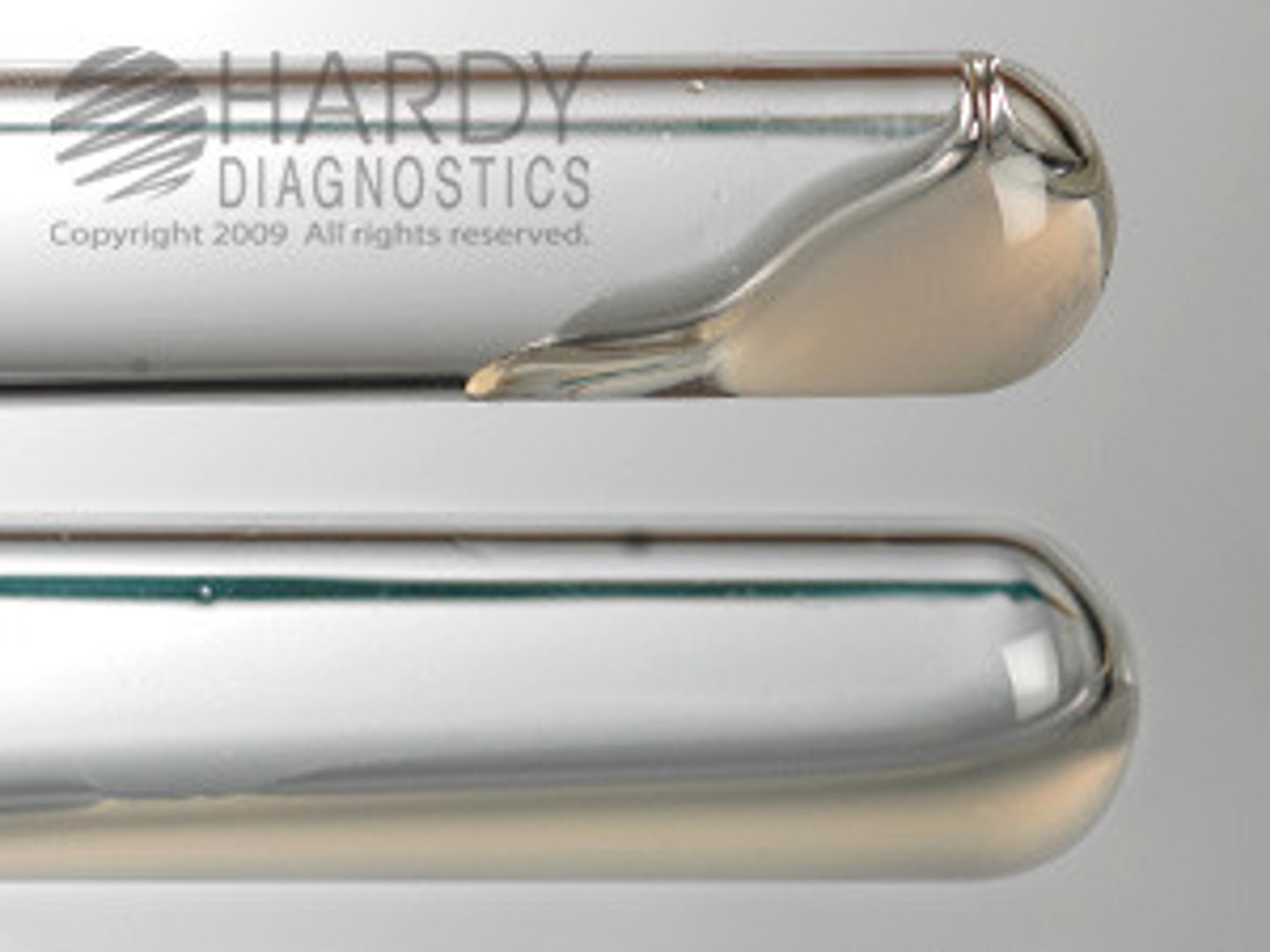
BHI with 6.5% NaCI
Purpose: Differentiates microorganisms based on their ability to grow in 6.5% NaCl.
Procedure: Inoculate a BHI broth containing 6.5% NaCl with a small amount of organism. Incubate for 24-48 hours at 37°C with a loose cap. Too heavy of an inoculum will give a false positive result.
Interpretation:
Positive: Growth (turbidity)
Negative: No growth (clear)
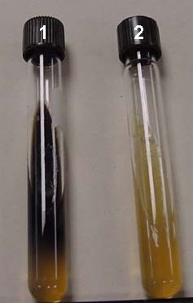
Bile esculin hydrolysis
Purpose: Differentiates microorganisms based on their ability to hydrolyze esculin to esculetin and dextrose. The esculetin reacts with ferric citrate in the medium to form a dark brown-black complex.
Procedure: Using a loop, inoculate the media by streaking in a zig-zag pattern up the slant. Incubate for 24 hours at 37°C with a loose cap.
Interpretation:
Positive: Blackening of the entire slant
Negative: No color change or less than 1⁄2 of the slant is black
catalase
degrades H2O2 to oxygen and water
EMB (eosin methylene blue agar)
Purpose: Selects microorganisms based on their ability to grow in presence of eosin Y and methylene blue. Differentiates microorganisms by their ability to vigorously ferment lactose and produce acid.
Procedure: Using a sterile loop, inoculate an EMB plate and streak for isolation. Incubate the plate at 37°C for 24 hours.
Interpretation:
Positive (metallic green sheen): Growth + / Acid ++. Vigorous lactose fermenter with excessive acid formation (primarily, but not only, E. coli)
Positive (purple colonies): Growth + / Acid +. Moderate to slow lactose fermenter with moderate acid formation (non-E. coli coliforms)
Negative: Growth + / Acid -. Pink or colorless colonies (no lactose fermentation) or no growth
Growth at 5°C (walk-in cooler)
Purpose: Differentiates microorganisms based on their ability to grow at 5°C.
Procedure: Using a sterile loop, inoculate a TSA plate and streak for isolation. Incubate the plate at 5°C for 3-5 days.
Interpretation:
Positive: Growth in quadrants 3 or 4 of plate
Negative: No growth in quadrants 3 or 4 of plate (clear)
Growth at 41°C
Purpose: Differentiates microorganisms based on their ability to grow at 41°C.
Procedure: Using a sterile loop, inoculate a TSA plate and streak for isolation. Incubate the plate at 41°C for ONLY 24 hours.
Interpretation:
Positive: Growth in quadrants 3 or 4 of plate
Negative: No growth in quadrants 3 or 4 of plate (clear)
Growth at 45°C
Purpose: Differentiates microorganisms based on their ability to grow at 45°C.
Procedure: Using a sterile loop, inoculate a TSA or SBA plate and streak for isolation. Incubate the plate at 45°C for ONLY 24 hours.
Interpretation:
Positive: Growth on plate
Negative: No growth (clear)
superoxides
converted to oxygen and H2O2 by superoxide dismutase
beta hemolysis
complete lysis of red blood cells around a colony; clear zone around colonies
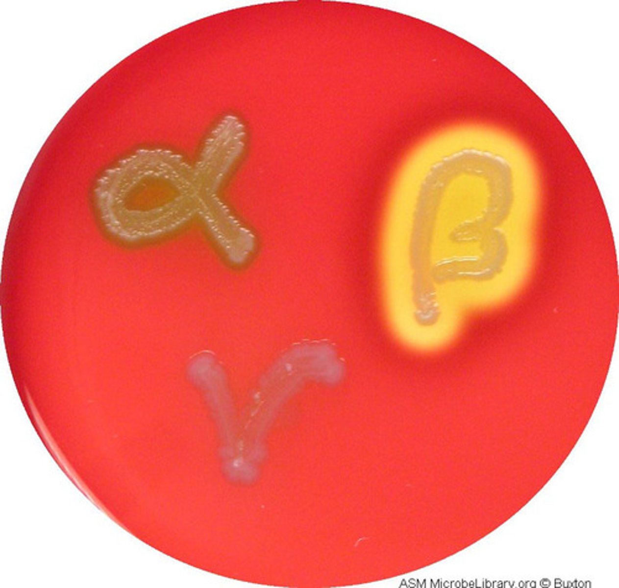
alpha hemolysis
breakdown of red blood cells producing a greenish discoloration around colonies
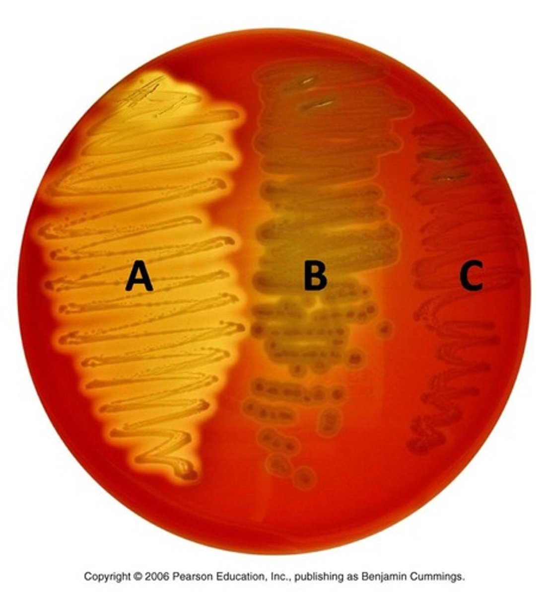
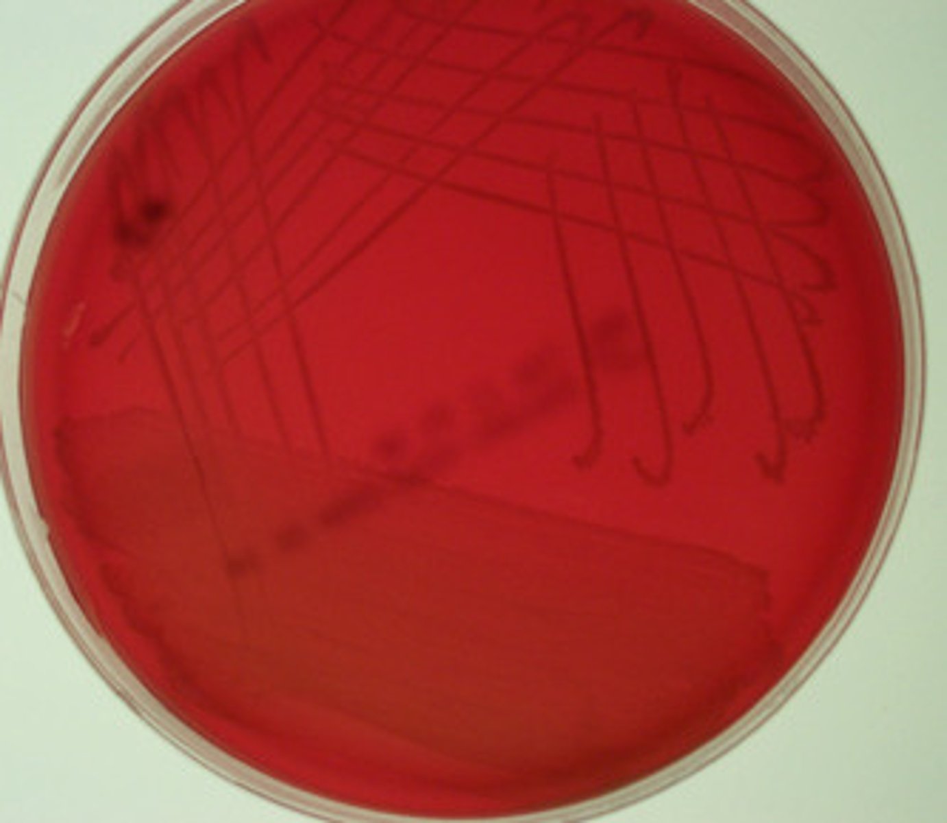
gamma hemolysis
do not exhibit any hemolysis of blood and have no effect on red blood cells in a blood agar plate
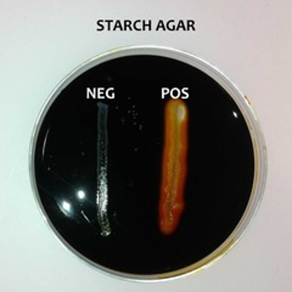
starch hydrolysis
detected by adding gram's iodine to a starch agar plate; media turns black/brown; if starch degraded, media adjacent to growth will be clear after adding iodine
Bile esculin agar
if organism can hydrolyze esculin, media will turn dark brown or black; positive only if over half media turns dark
coagulase
insulating small tube of rabbit blood plasma with loopful of organism; clotting=positive
Positive: Coagulation of the plasma (clot formation)
Negative: No coagulation/clot formation
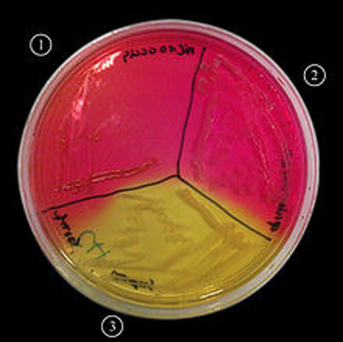
MSA (mannitol salt agar)
ability to grow at 7.5% NaCl and ferment mannitol; Positive: Growth+/Acid+(yellow colonies)
Negative: Growth+/Acid- or Growth-/Acid- (colorless, no yellow, no growth)
phenol red carbohydrate tubes
arabinose, lactose, trehalose, mannitol, sucrose, xylose, etc;
positive: yellow (gas produced & carb fermented)
negative: red (stays red)

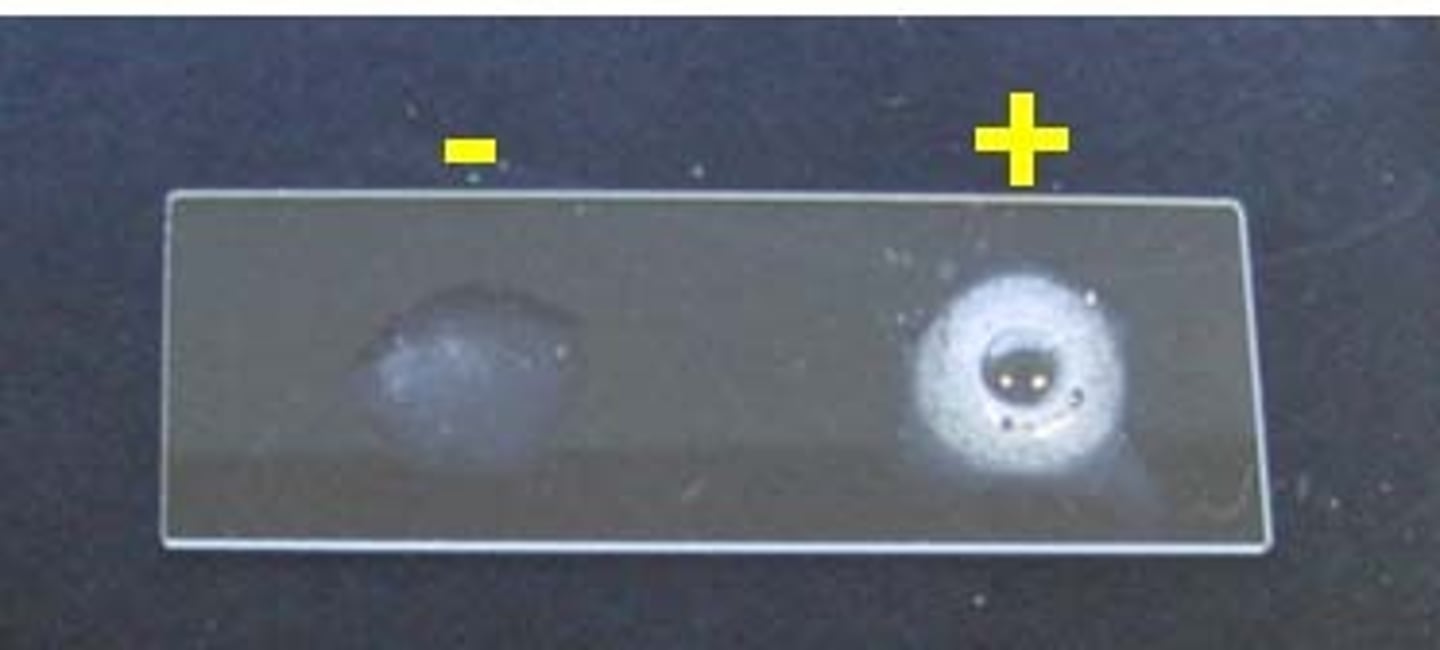
KOH test
confirms gram stain on whether 3% KOH can lyse bacterial cell wall and release DNA
strings present: gram negative
no strings: gram positive
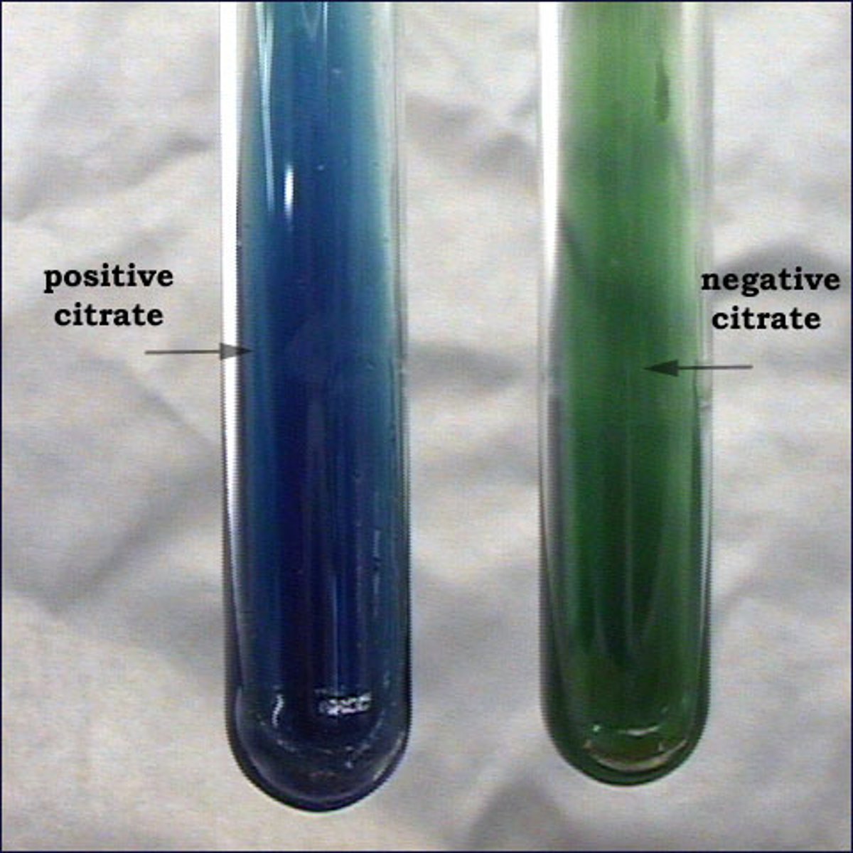
citrate
diff. based on their ability to utilize citrate as a sole carbon source and ammonium salts as sole nitrogen source
positive: growth w/ intense blue color
negative: no growth/no color change (stays green)
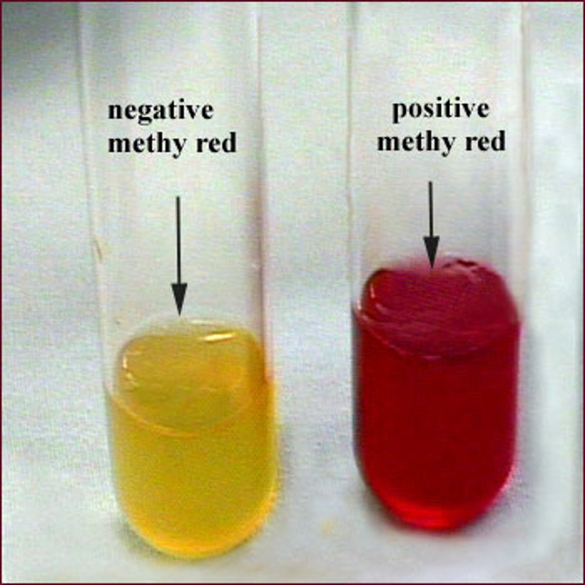
methyl red
diff based on ability to produce and maintain stable acid end products from glucose fermentation
positive: red color (immediate)
negative: yellow color/no color
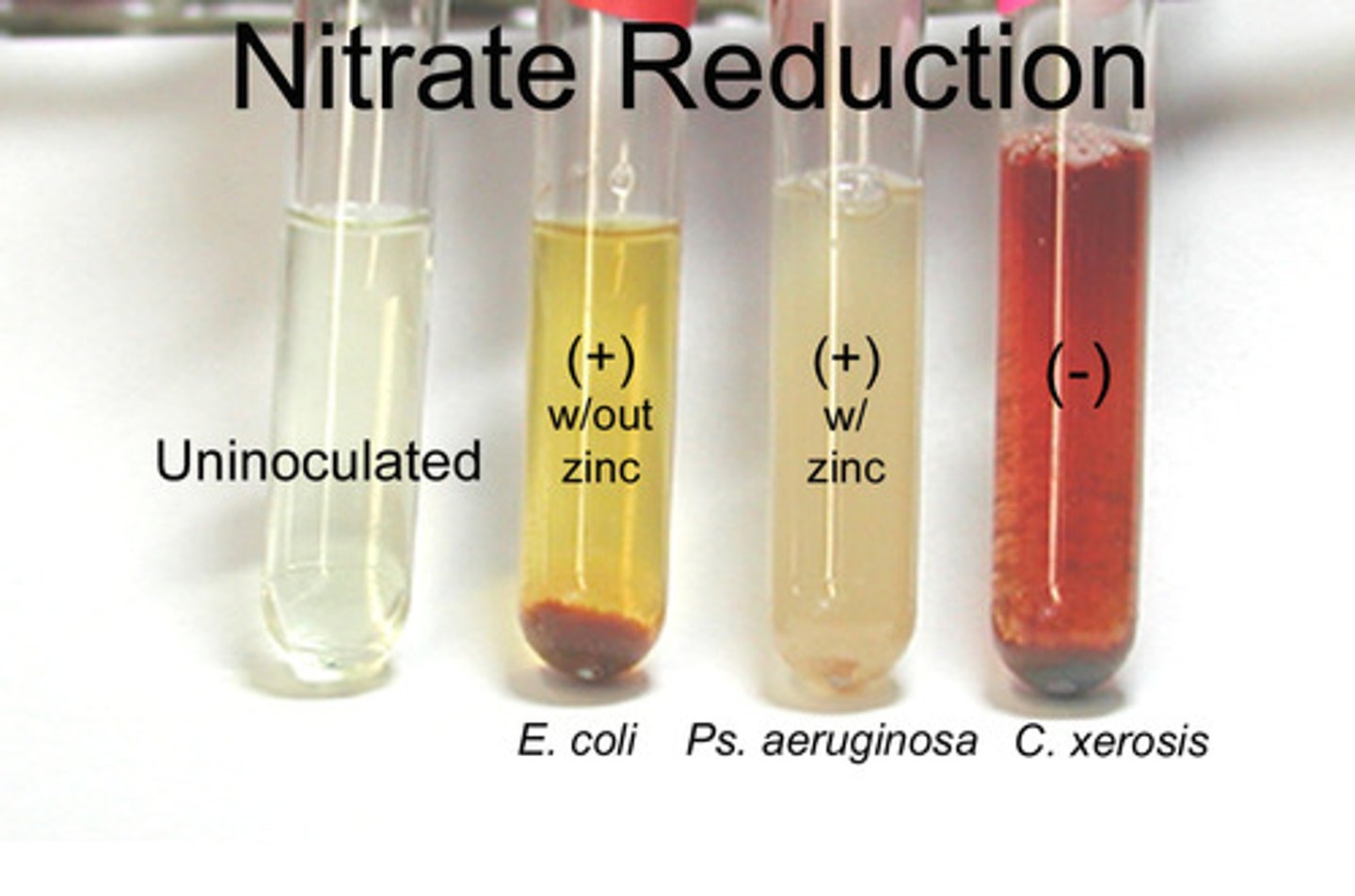
Nitrate reduction
diff based on ability to reduce nitrate to nitrite or ammonia or nitrogenous gas
positive: red color develops
negative: no red color develops; red develops after zinc is added
Novobiocin
Purpose: Differentiates microorganisms based on susceptibility to novobiocin
Procedure: Use a swab make a lawn streak on a TSA or SBA plate. Apply a Novobiocin disk to the center of the lawn streak plate. Gently tap the disk to secure it onto the media. Incubate for 24 hours at 37°C then read the zone of inhibition.
Interpretation:
Susceptible: Zone of inhibition >=17mm
Resistant: Zone of inhibition <= 16mm
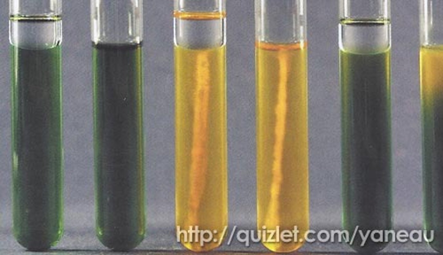
O/F glucose (oxidation/fermentation)
oxidative or fermentative metabolism of carb in tube
oxidative: yellow color in open tube only
fermentative: yellow color in both open tube and closed tube
neither: green or blue color in both open and closed tubes
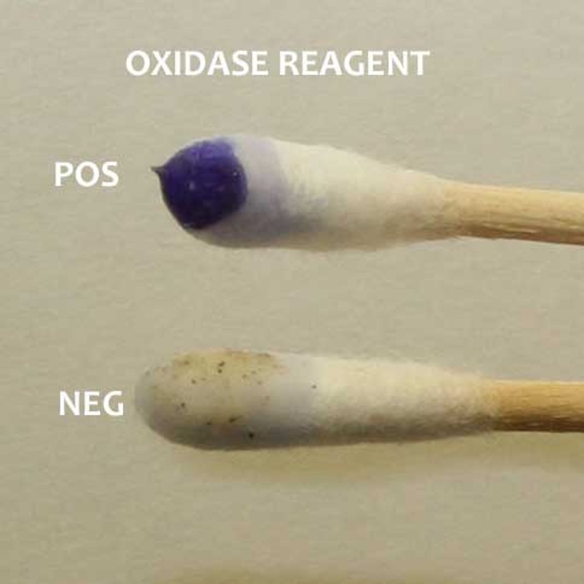
oxidase
presence of enzyme cytochrome C oxidase in bacteria
positive: blue or purple in 10-30 sec
negative: no color change or blue color after 30 sec
phenylalanine
to produce phenylalanine deaminase- enzyme removes amine group from amino acid and releases as free ammonia; produces phenylpryuvic acid
positive: green
negative: yellow
Production of Red Pigment at 25°C
Purpose: Differentiates microorganisms by their ability to produce a red pigment when incubated at 25°C. Procedure: Inoculate a TSA plate and incubate at 25°C for 24 hours. Observe for red pigment (prodigiosin) production.
Positive: Red pigment
Negative: No pigment production