Anatomy Lecture - Popliteal Fossa and Leg Region
1/24
There's no tags or description
Looks like no tags are added yet.
Name | Mastery | Learn | Test | Matching | Spaced | Call with Kai |
|---|
No analytics yet
Send a link to your students to track their progress
25 Terms
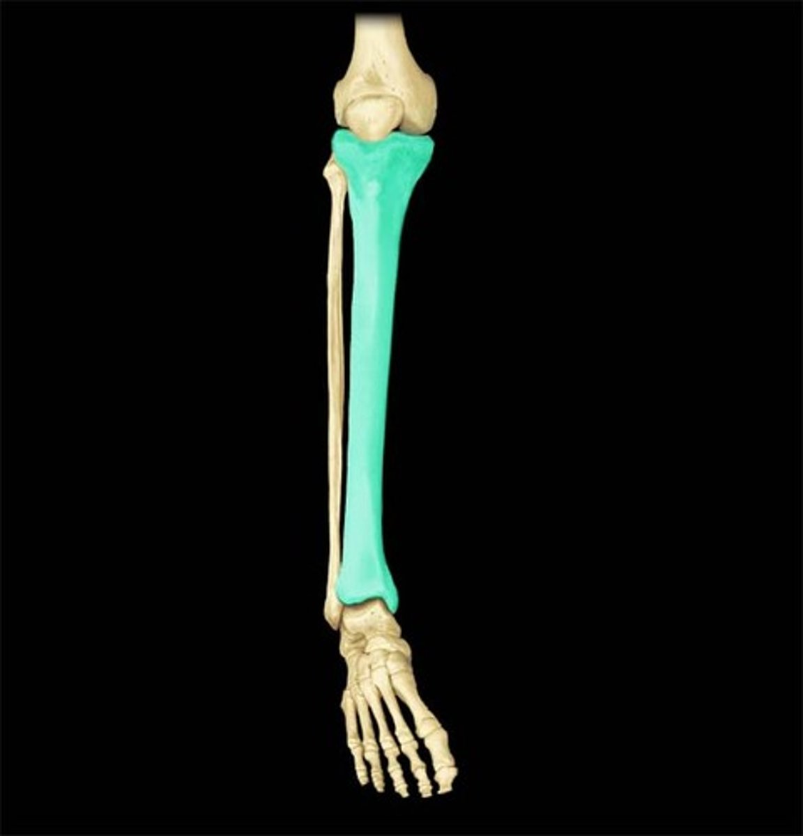
Tibia
- shaft is vertical
- shin bone
- medial side of leg
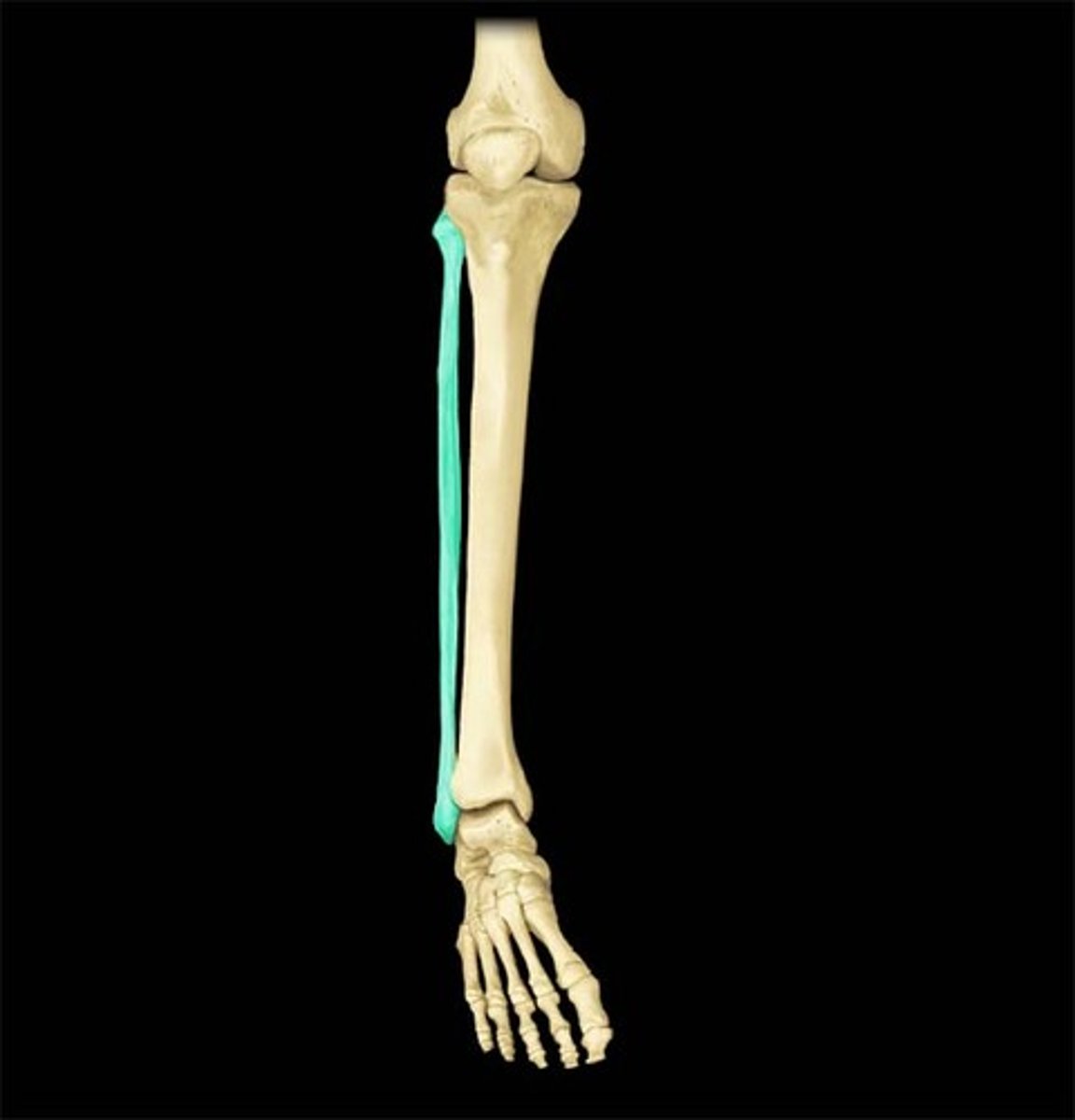
Fibula
- lateral to tibia
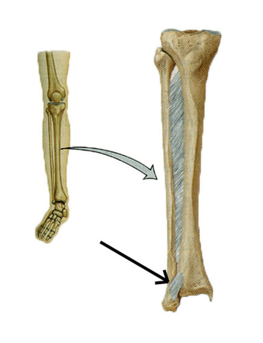
Tibiofibular syndesmosis
- joint between the tibia and fibular
Talus
- no muscular or tendinous attachments
- surface is covered in articular cartilage
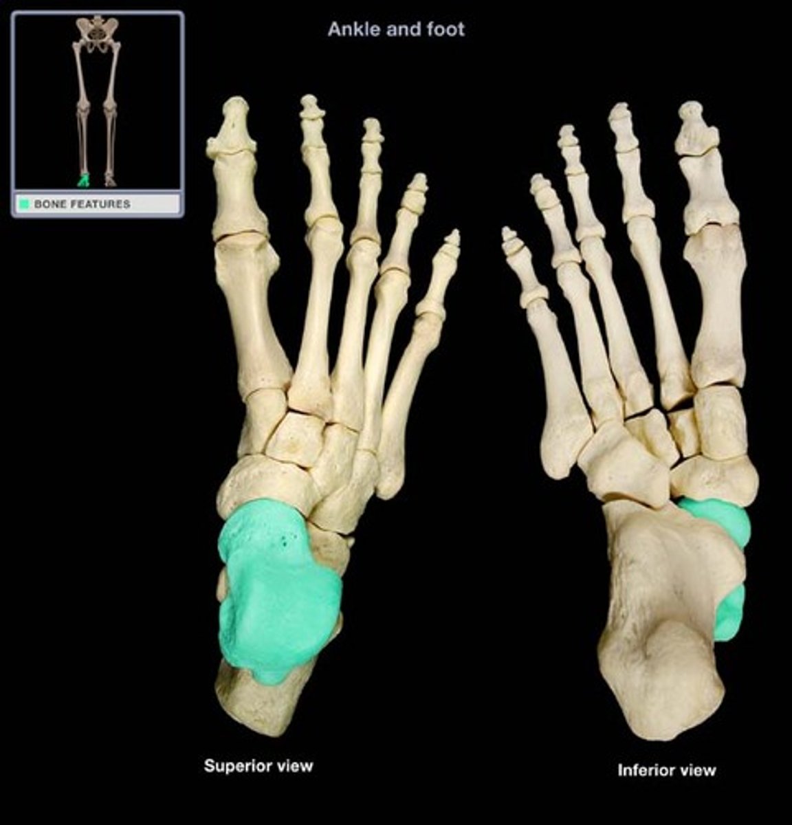
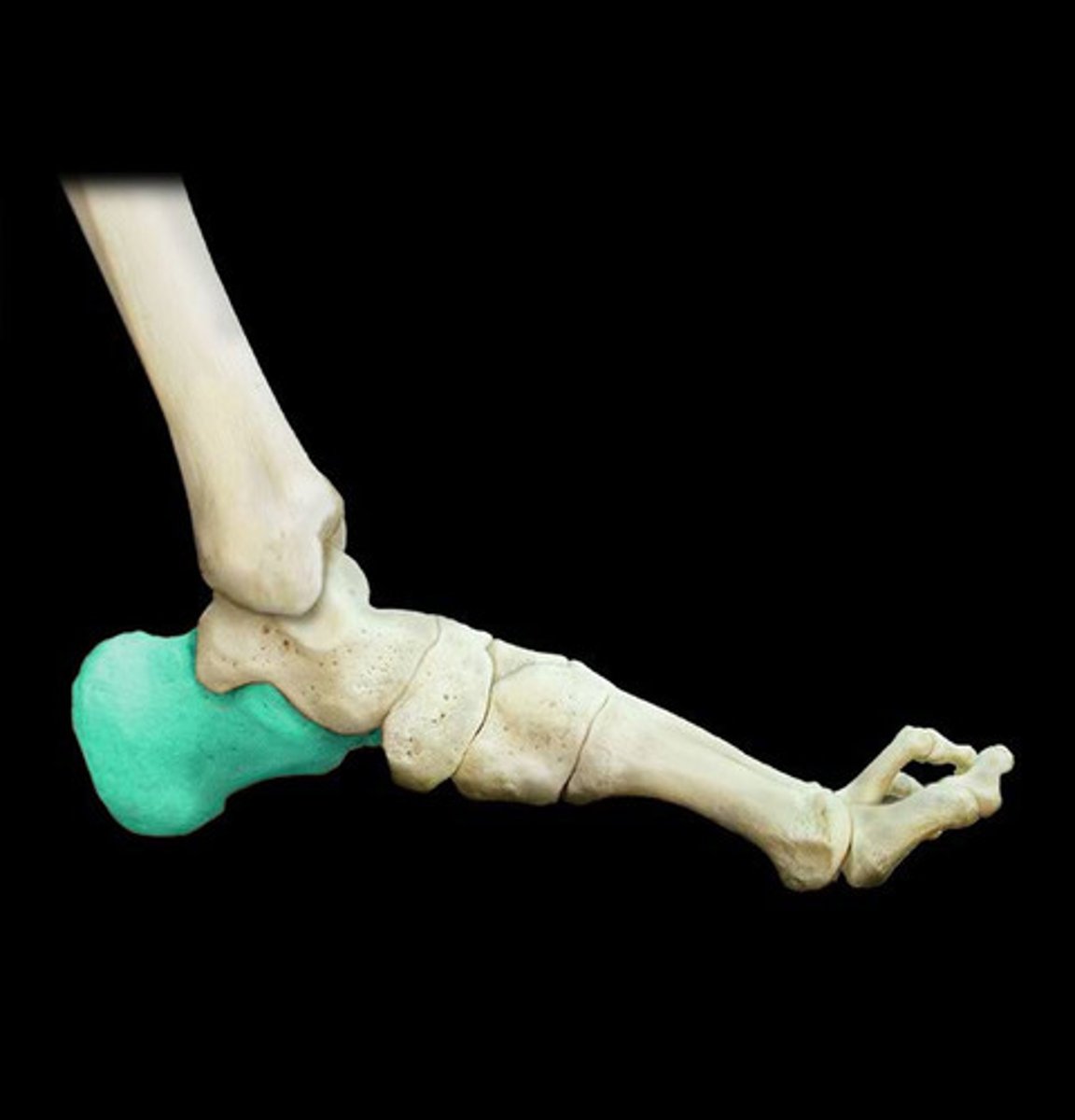
Calcaneus
- "heel bone"
- transmits the majority of body weight from talus to calcanues
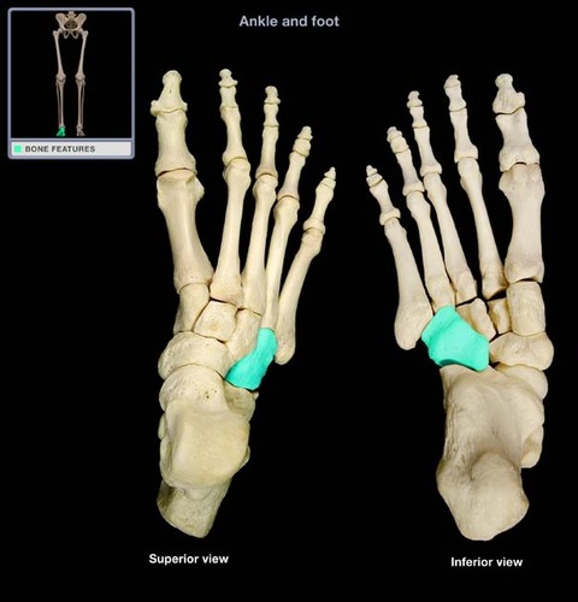
Cuboid
- most lateral bone
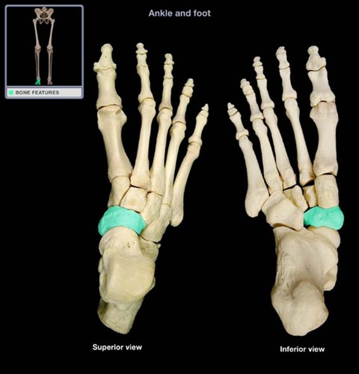
Navicular
- navicular tuberosity
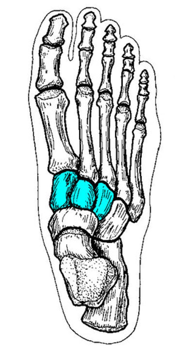
Cuneiforms
medial, intermediate, lateral
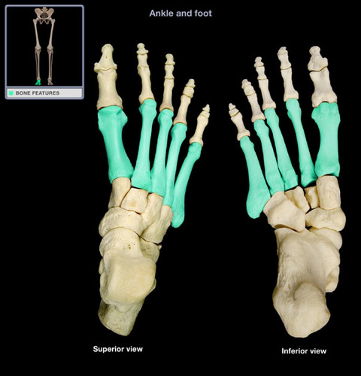
Metatarsals
- foot bones
- labeled medial to lateral
- all have a base, shaft, and distal head
Popliteal Fossa
- posterior to knee
- diamond shape
- fat filled
- contains neurovascular structures
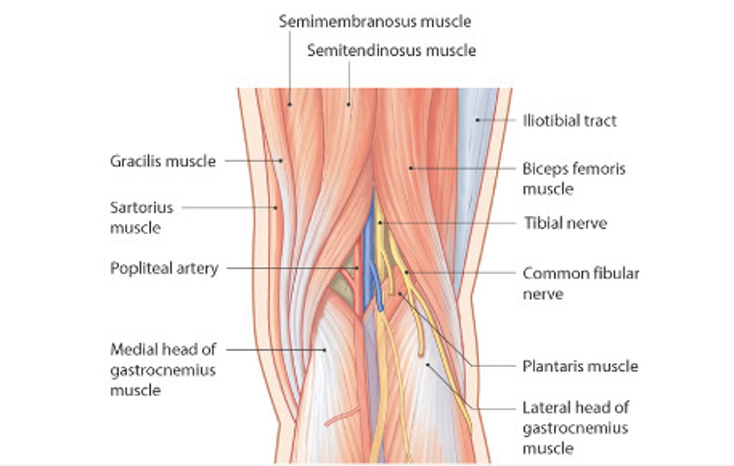
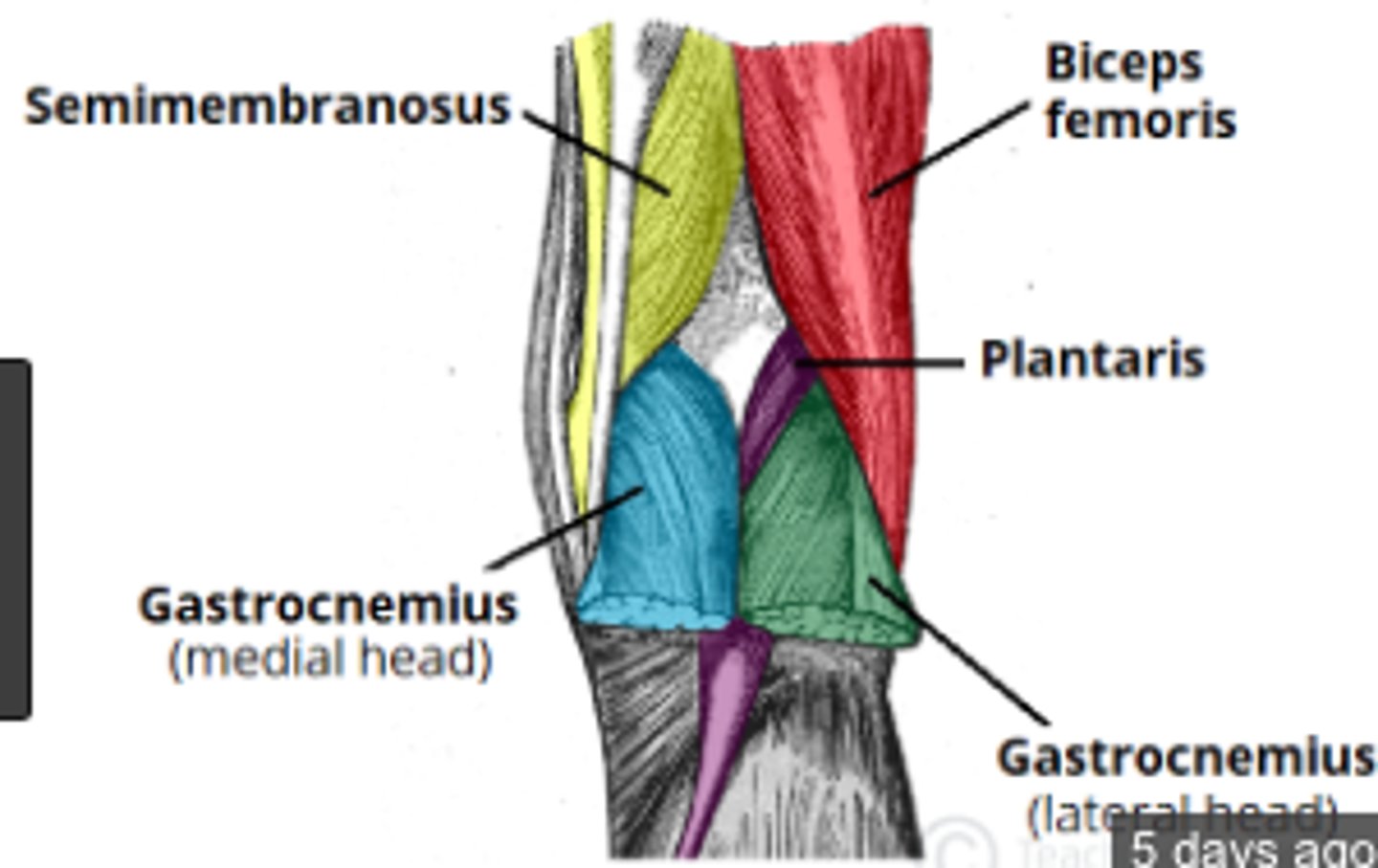
Borders of Popliteal Fossa
-Superolateral Border: Biceps femoris
-Superomedial Border: Semimembranosus, Semitendinosus (lateral to semimembranosus)
-Inferolateral Border: Lateral head of Gastrocnemius
-Inferomedial Border: Medial head of Gastrocnemius
Posterior Border: Skin, fascia
Anterior Border: Popliteal surface of femur, oblique popliteal ligament, popliteal fascia over popliteus, floor of fossa
Popliteal Fossa Nerves
- sciatic nerve splits into tibial and common fibular
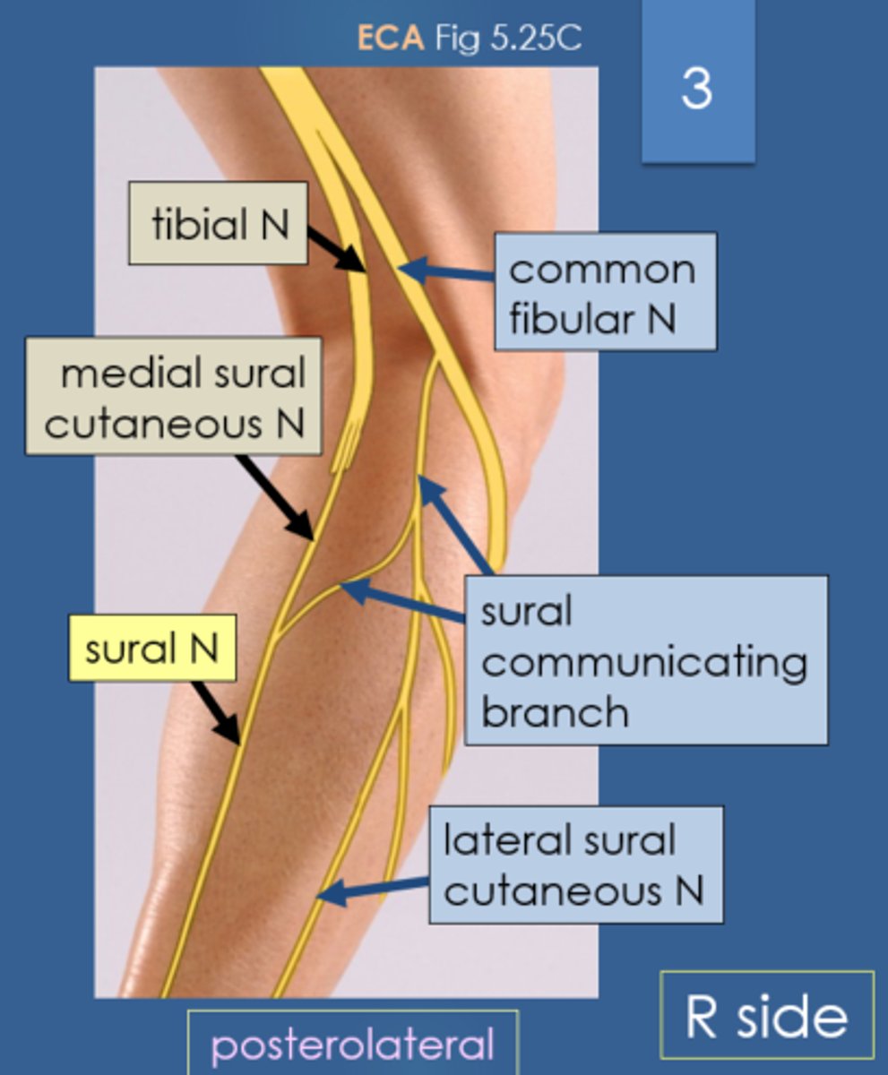
Tibial Nerve
- L4-S3
- Medial
- gives branch to medial sural cutaneous
Common Fibular Nerve
- L4-S2
- Lateral
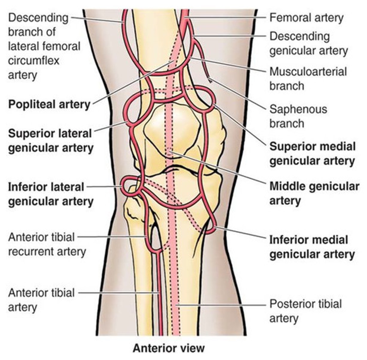
Popliteal Artery
- begins when femoral AA. passes through adductor hiatus
- ends when division starts of anterior and posterior tibial arteries
- gives rise to genicular arteries
genicular arteries
Branches of the Popliteal Artery providing the blood supply to the area about the knee
Three Compartments of the Leg
- anterior
- lateral
- posterior
Anterior Compartment of Leg
- dorsi flexor or extensor compartment
- Retinaculum (fascia wraps tendons tight to bone)
- innervated by deep fibular nerve
Lateral Compartment of Leg
- Evertor compartment
- smallest compartment
Posterior Compartment of Leg
- Plantar Flexion
- Largest
- separated by transverse intermuscular septum
Superior extensor retinaculum
- from fibula to tibia proximal
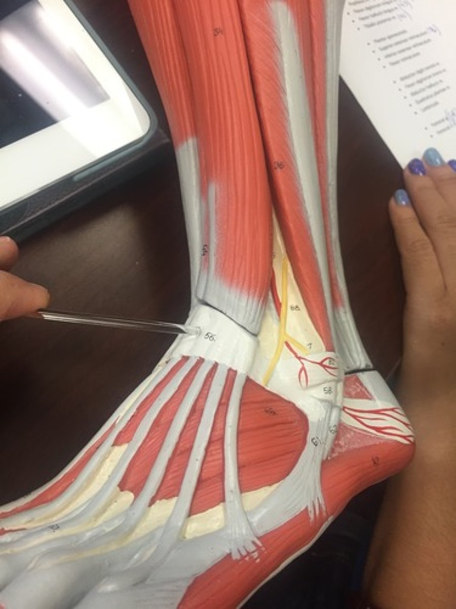
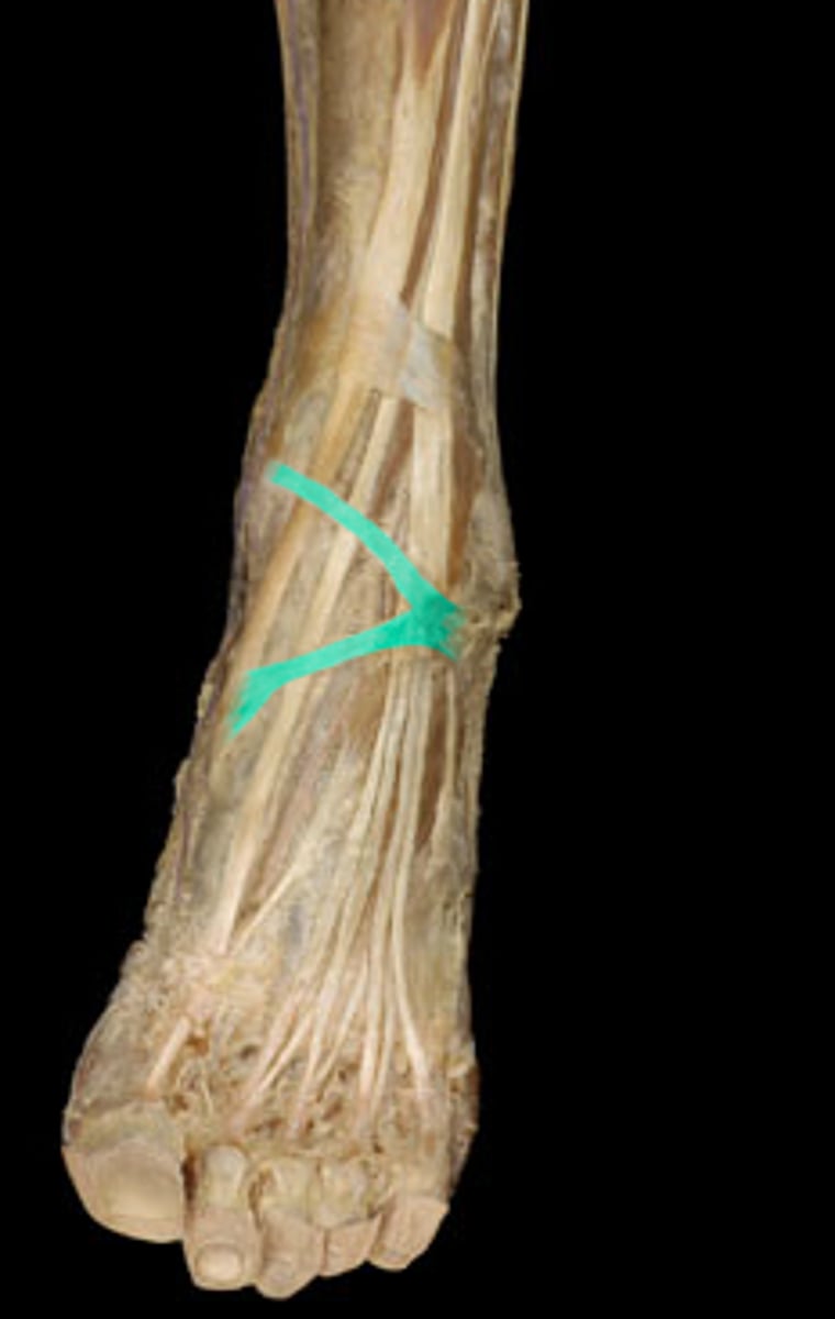
Inferior extensor retinaculum
- Y shaped
- lateral calcaneus to medial malleolus
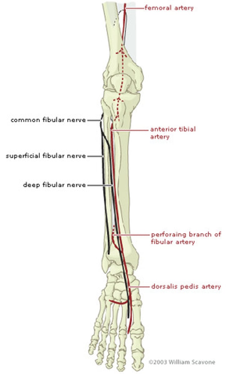
Anterior Tibial AA.
- branch of popliteal AA.
- Becomes dorsalis pedis
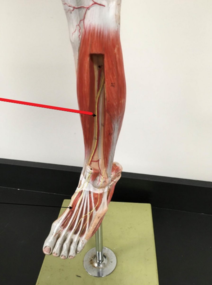
Dorsalis pedis
top of foot
Deep Fibular Nerve
- terminal branch of the common fibular NN.
- accompanied by the tibial AA.