BMS2011
1/95
There's no tags or description
Looks like no tags are added yet.
Name | Mastery | Learn | Test | Matching | Spaced | Call with Kai |
|---|
No analytics yet
Send a link to your students to track their progress
96 Terms
-
-
-
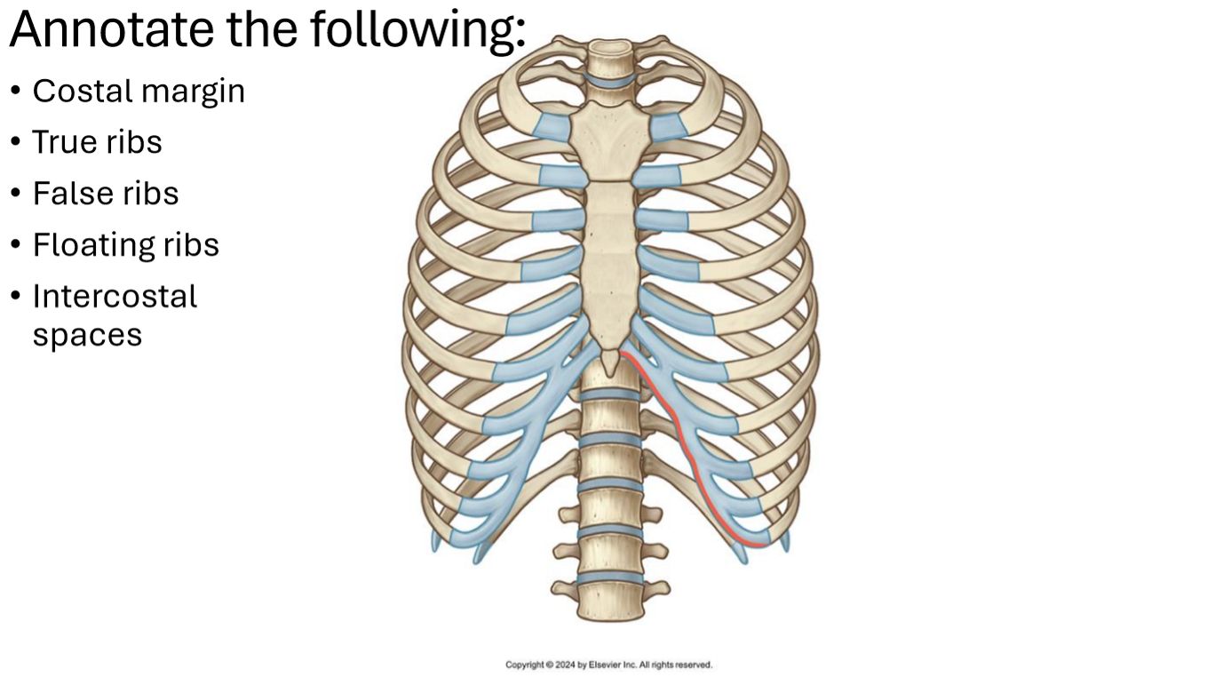
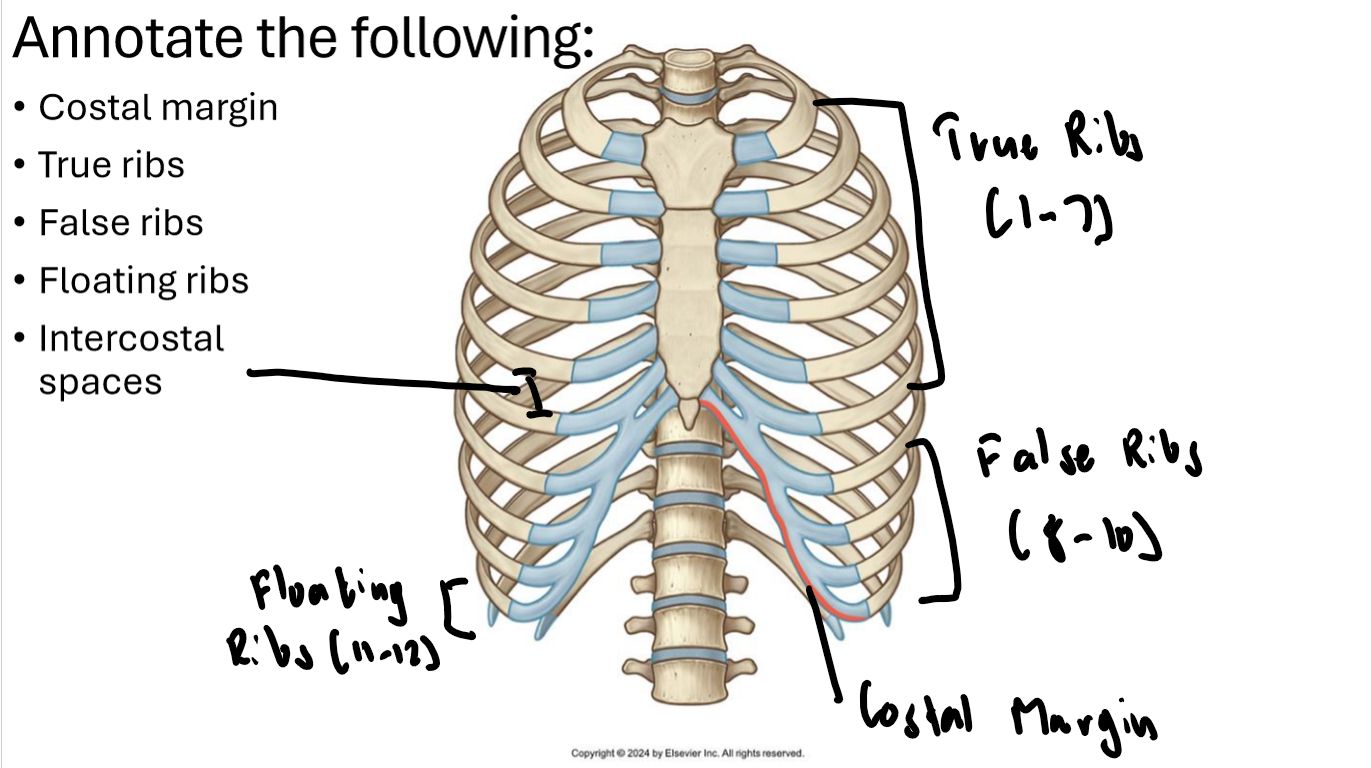
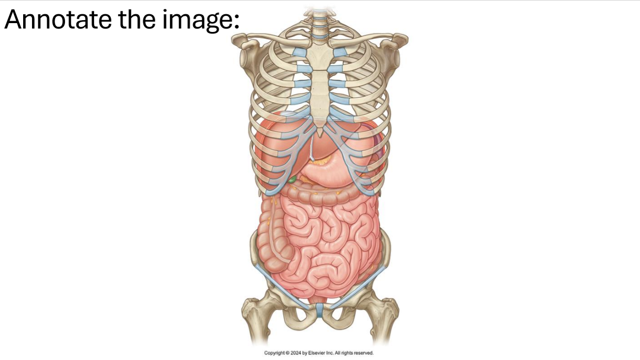
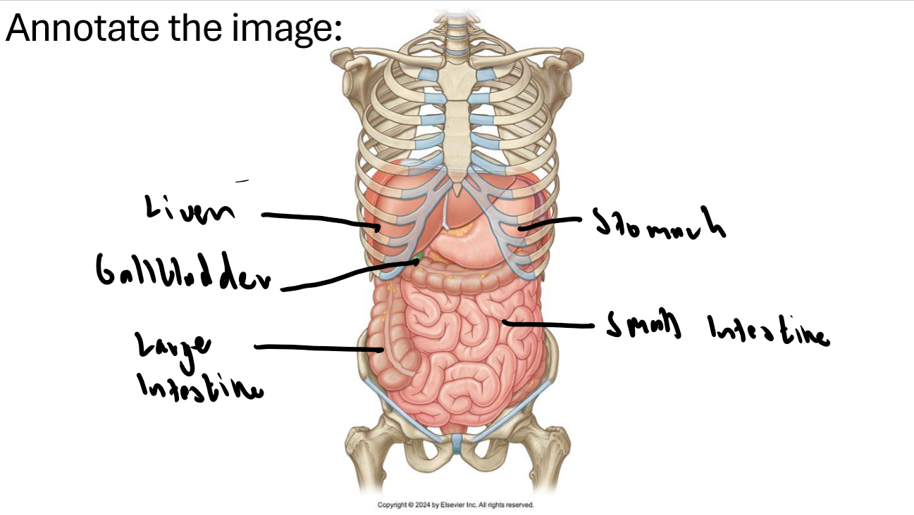
Define what a true rib, false rib and floating rib is? Where are these types of ribs located?
True Rib: Rib that connects directly to sternum (Ribs 1-7)
False Rib: Rib that indirectly connects to sternum via 7th rib cartilage (Ribs 8-10)
Floating Ribs: Ribs that do not connect to the sternum (Ribs 11 and 12)
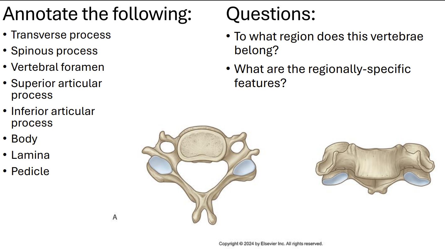
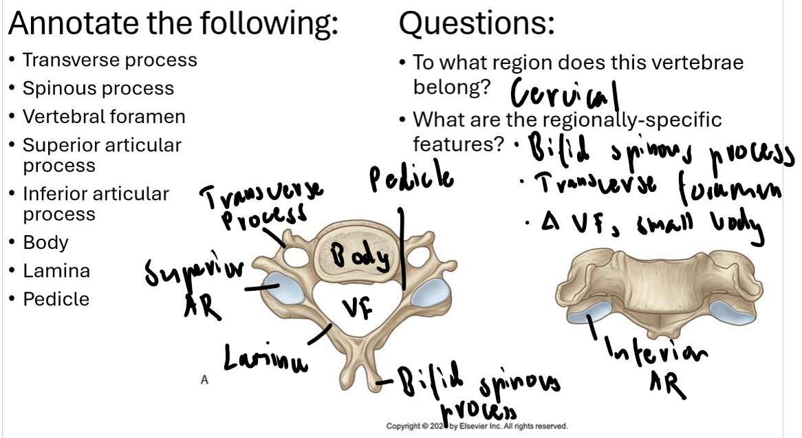
What are the features of a cervical vertebrae?
Bifid spinous process
Transverse formation
Triangular vertebrae foramen
Small body
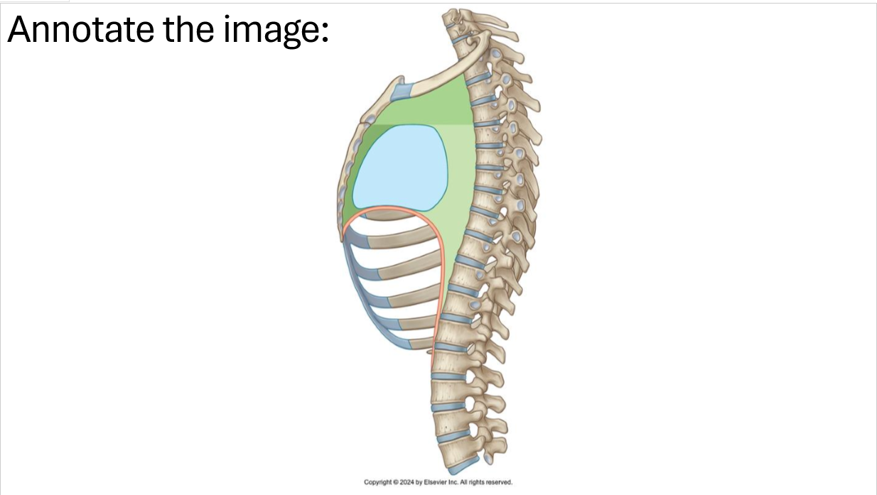
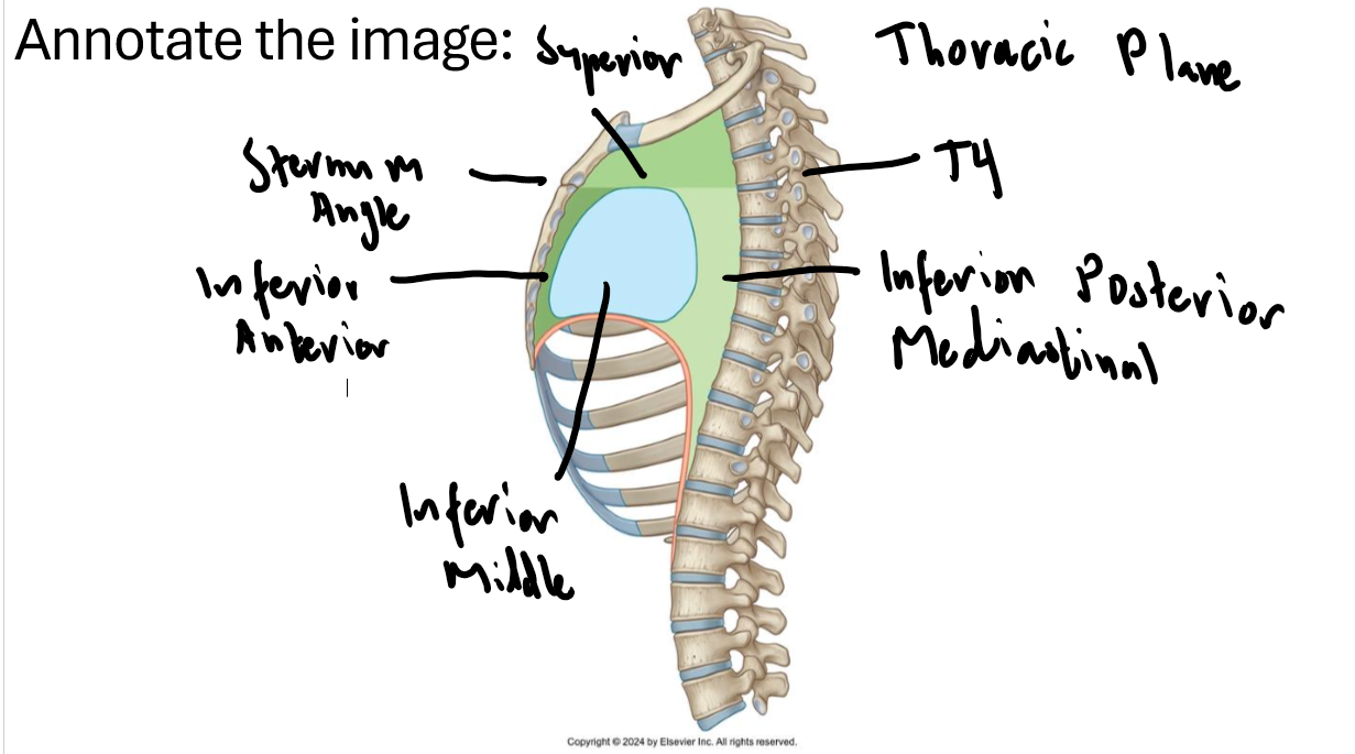
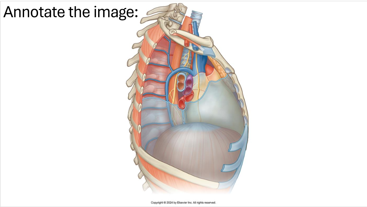
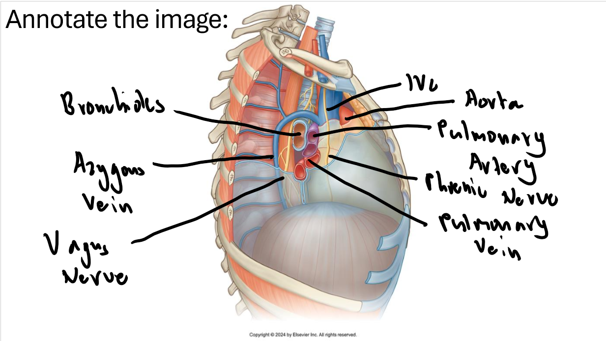
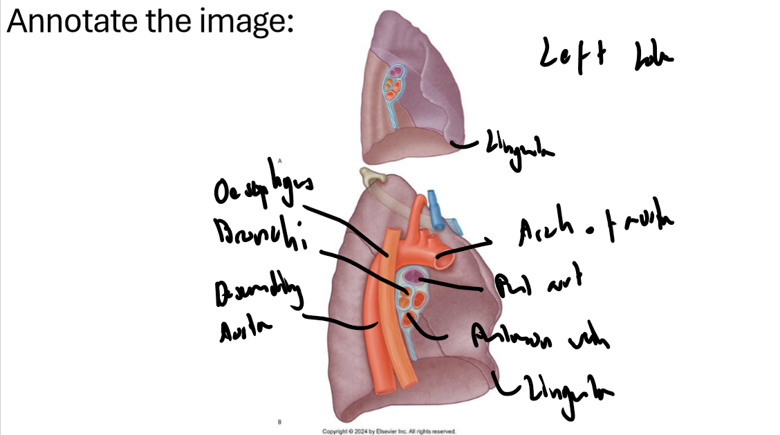
What are the contents of each mediastinal divsion?
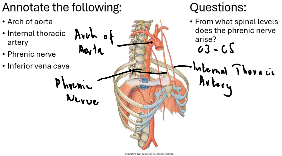
Superior: Arch of Aorta, Trachea, SVC, arteries + veins, Oesophagus, Vagus nerve, brachiocephalic vein
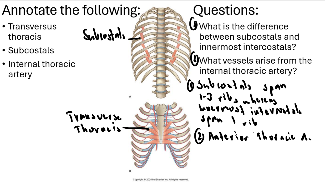
Inferior Anterior: Thymus, internal thoracic artery
Inferior Middle: Heart, phrenic nerve, pericardium
Inferior Posterior: Vagus nerve, oesophagus, descending aorta, R + L bronchioles, azygous veins, thoracic ducts
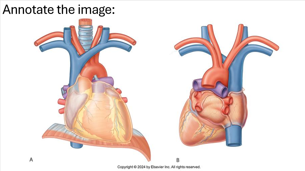
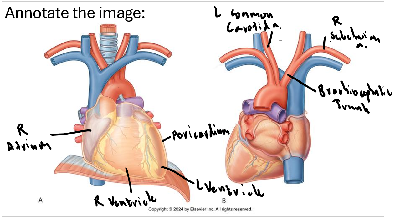
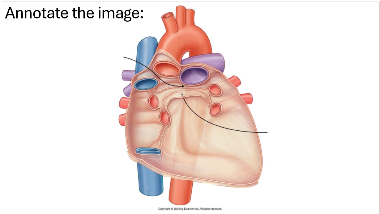
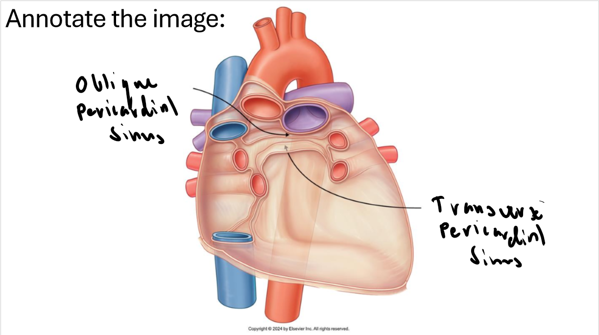
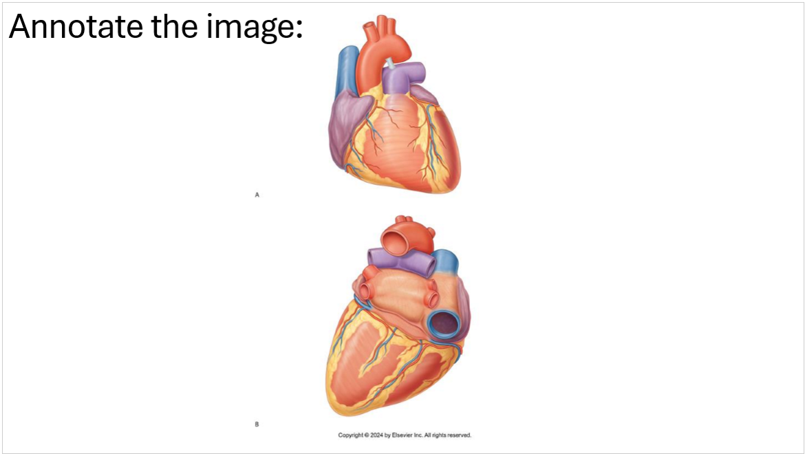
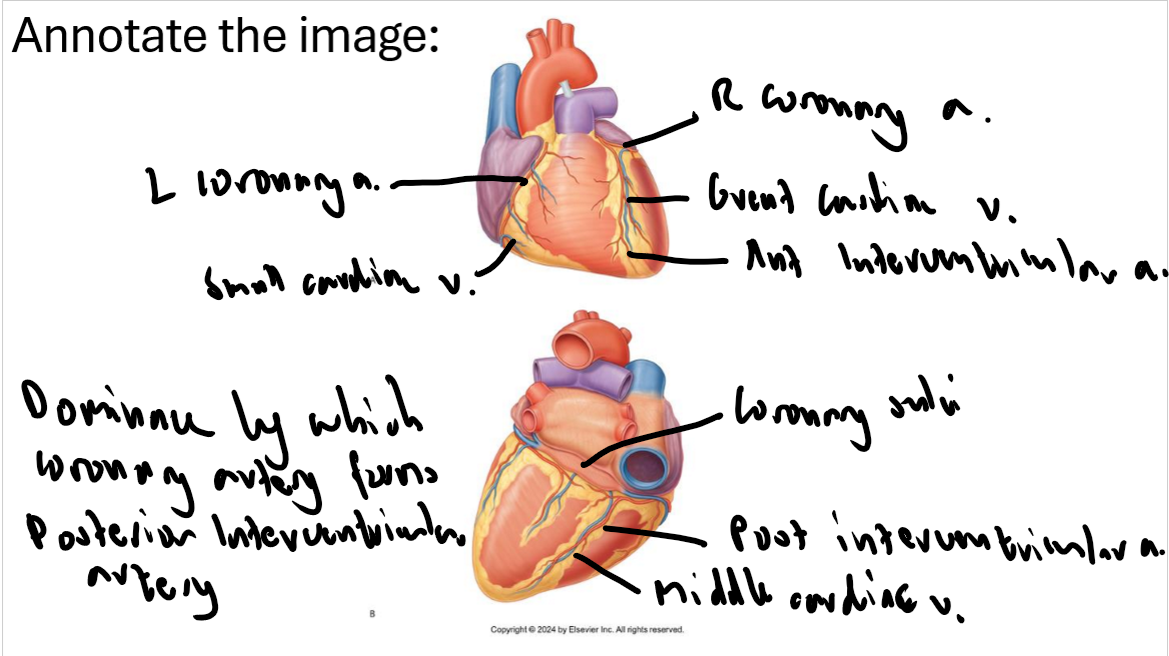
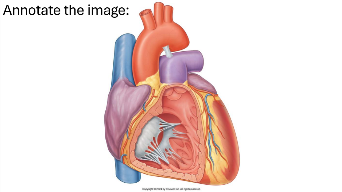
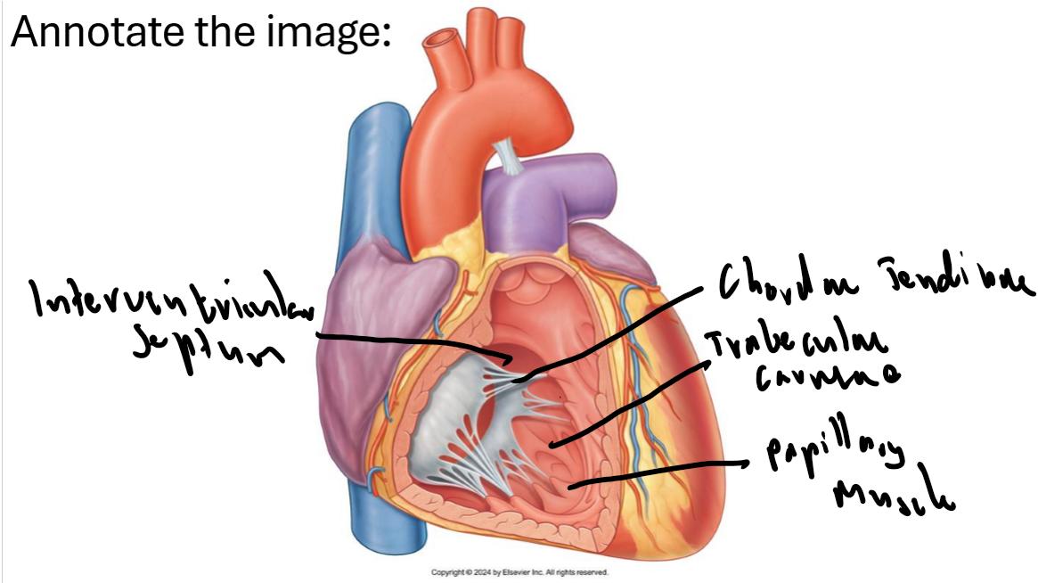
What is the pericardium?
Tough, fluid-filled, double-layered sac that encloses the heart and the roots of the major blood vessel
The heart is covered by a serous membrane with visceral and parietal layers like a balloon, visceral touches the heart and parietal is the outer layer
Between these layers is a fluid filled cavity called the pericardium space
Parietal layer is also attached to a fibrous layer which attaches to the diaphragm and roots of the great vessels
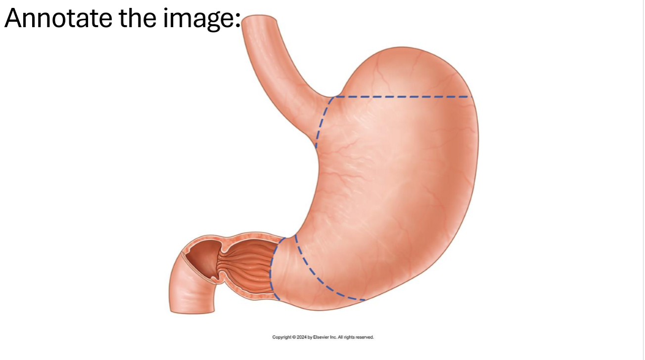
What are the folds inside the stomach called?
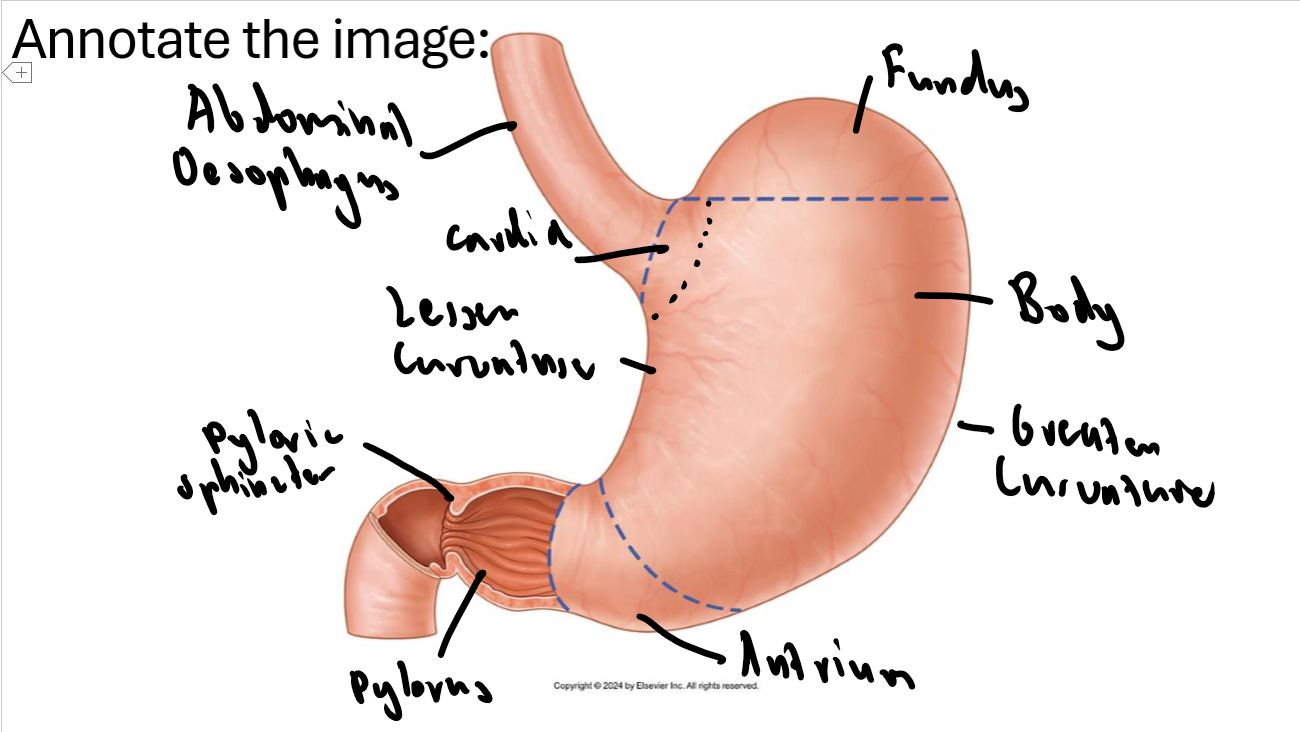
Gastric folds or gastric rugae
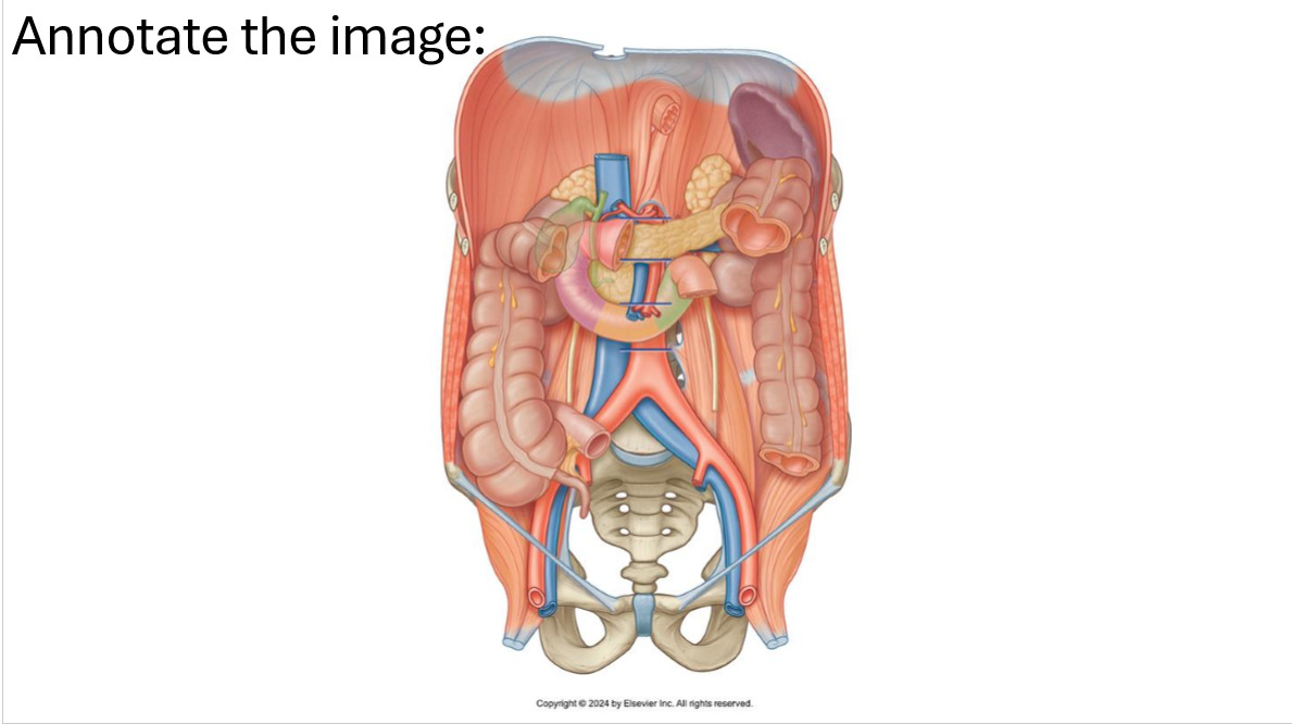
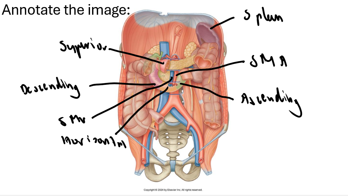
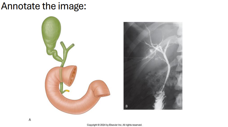
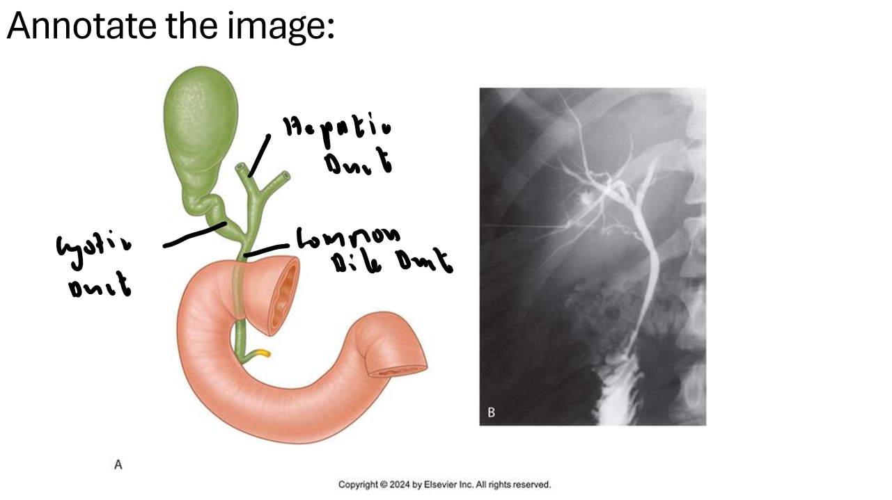
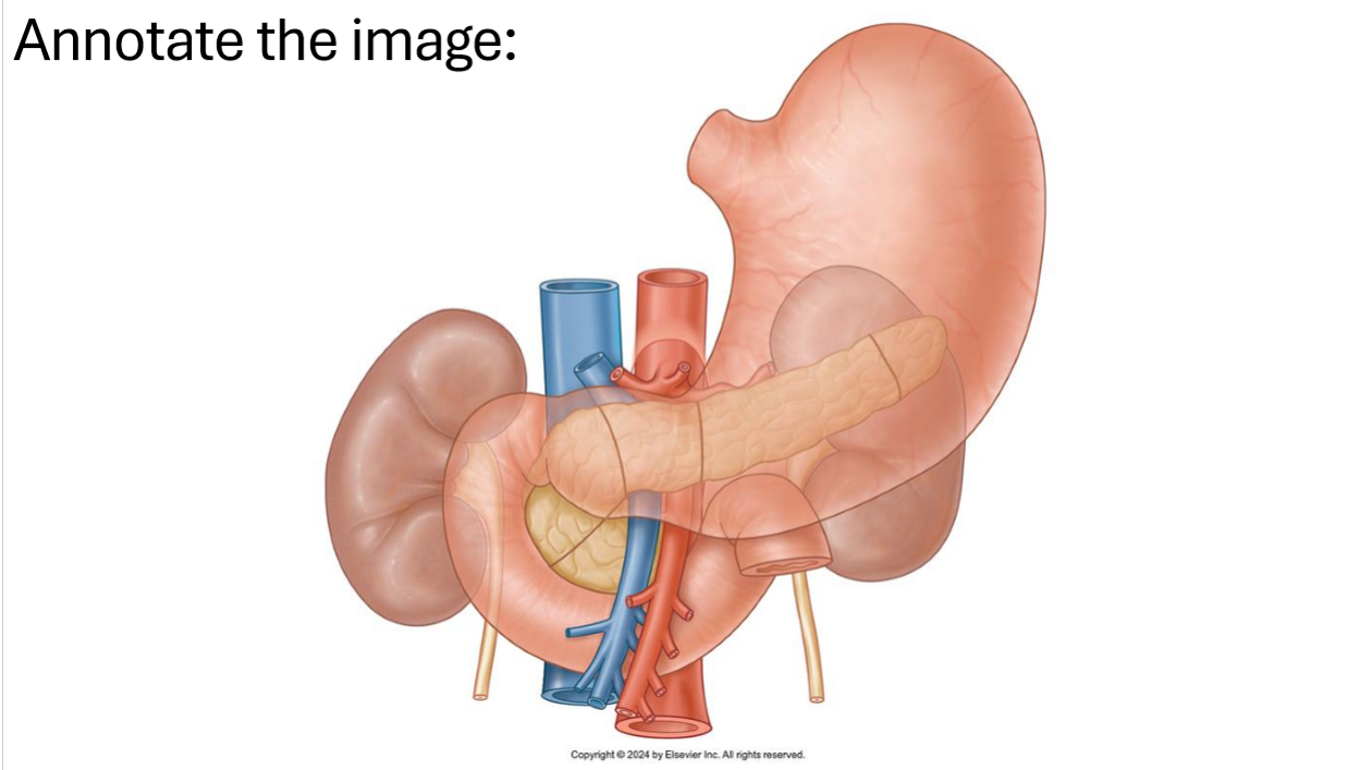
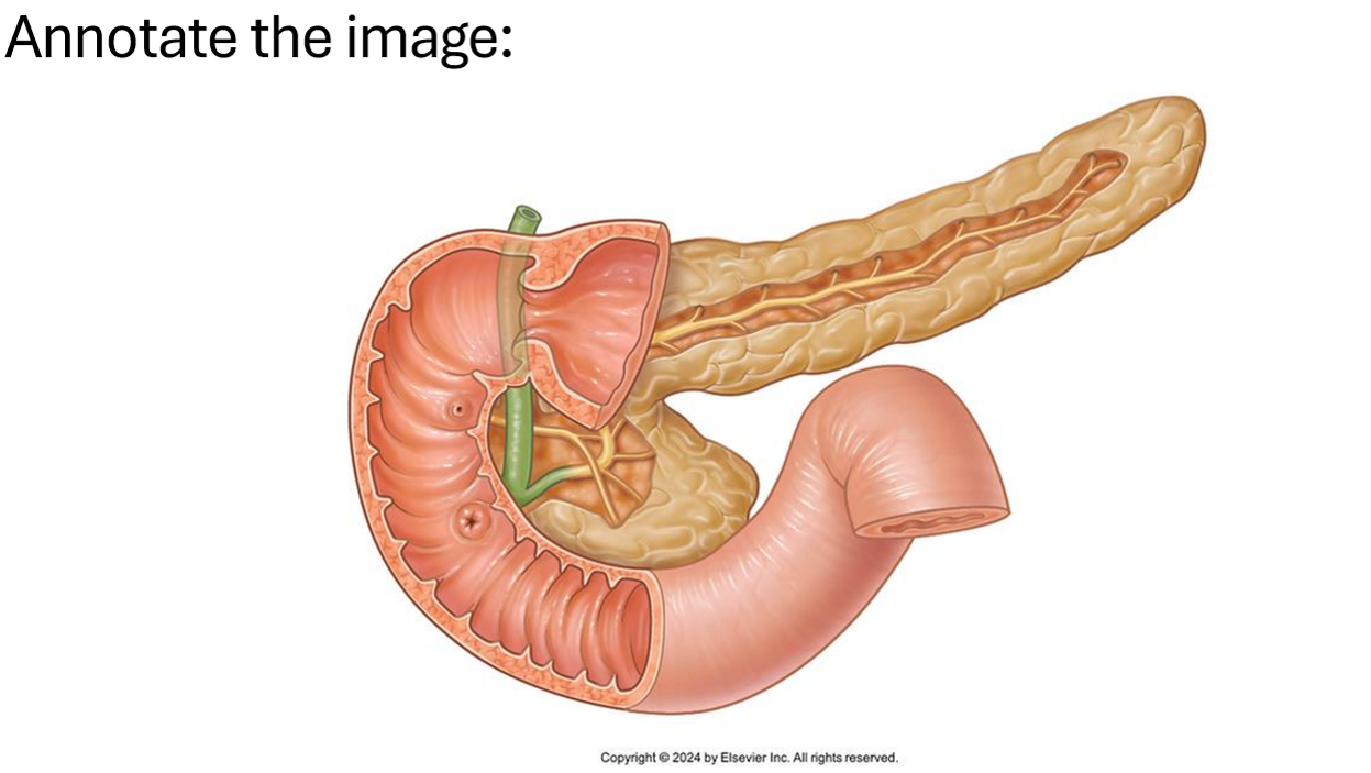
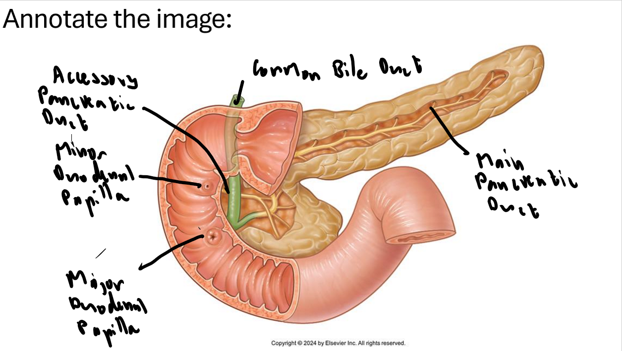
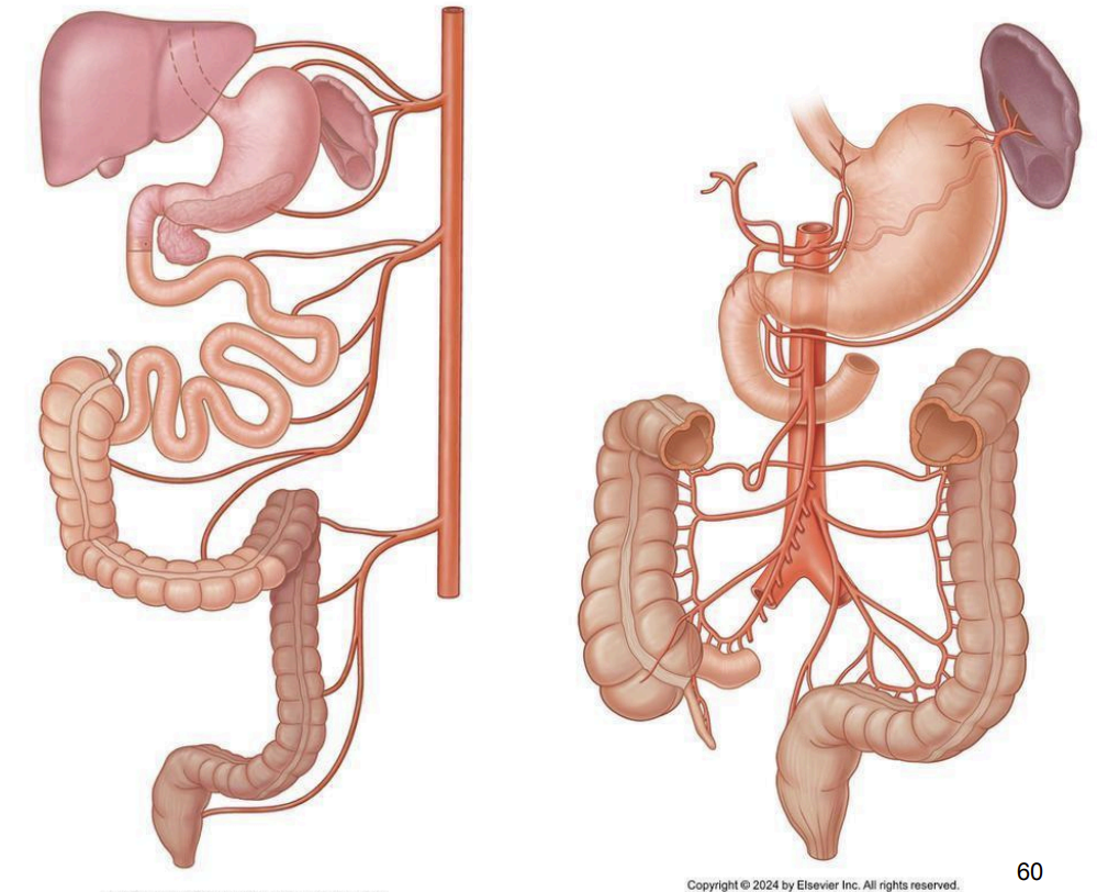
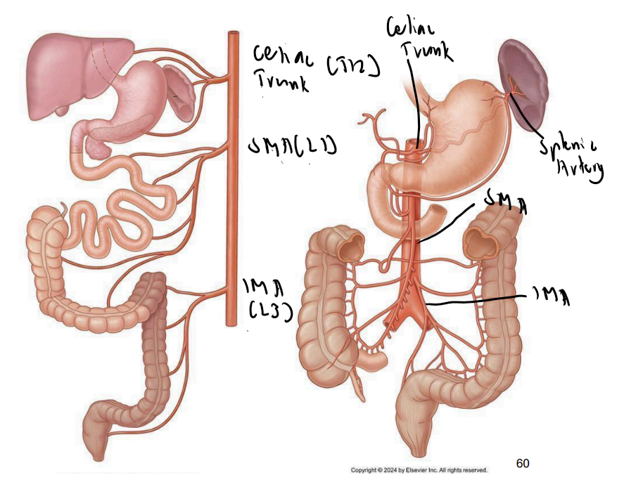
Name parts of the duodenum and important arteries.
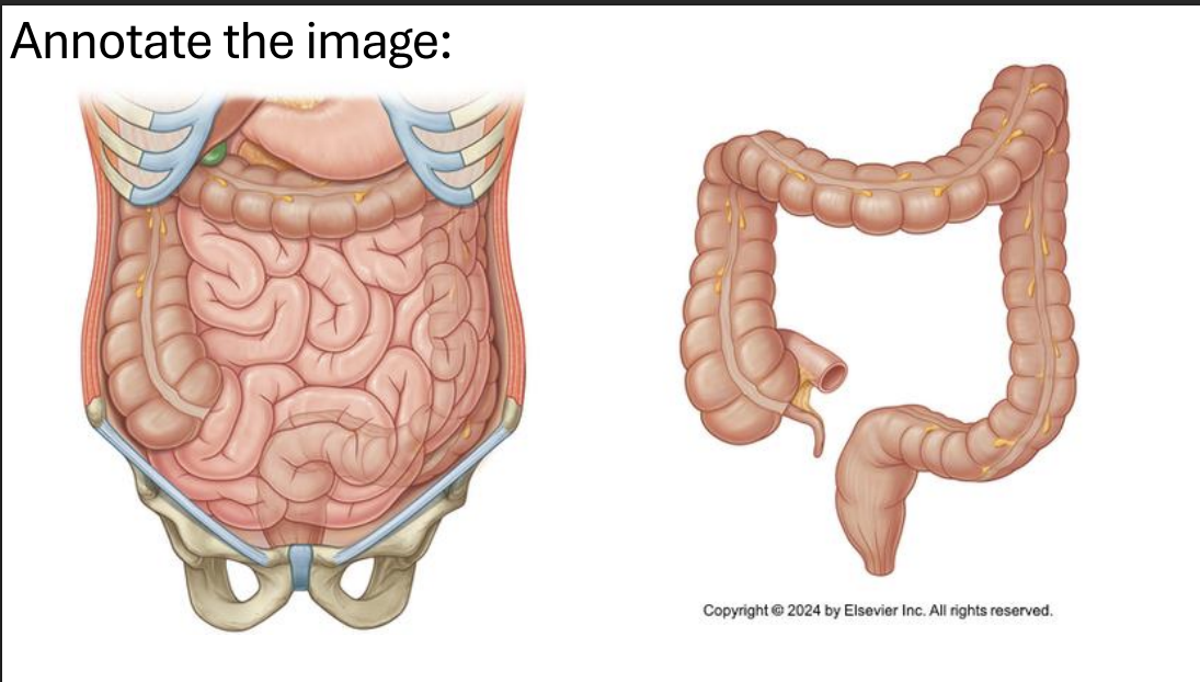
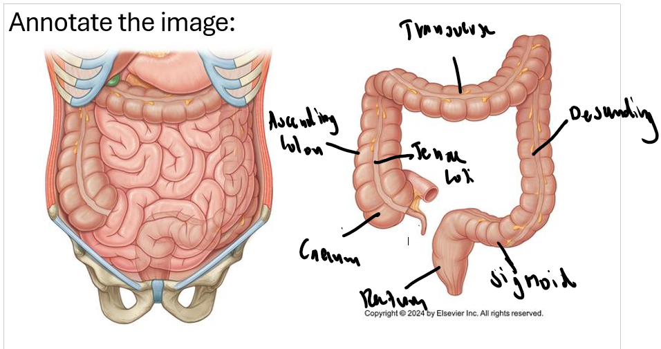
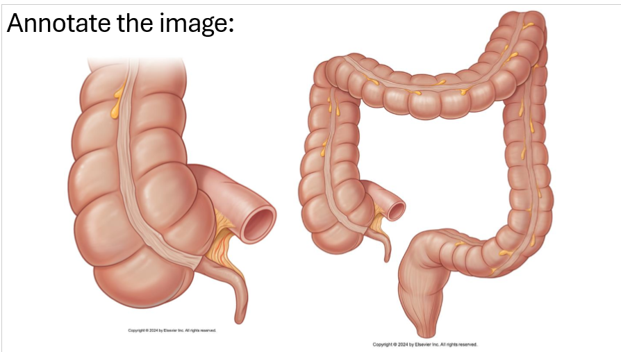
What are features of the large intestine?
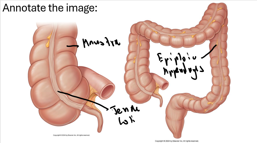
Haustra: Pousches
Taenia Coli: Longitudinal muscle layer
Epiploic Appendages: pouches of fat
Inside (evenly distributed semilunar folds
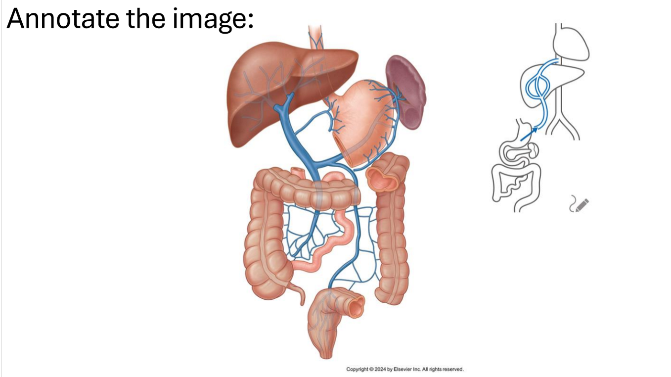
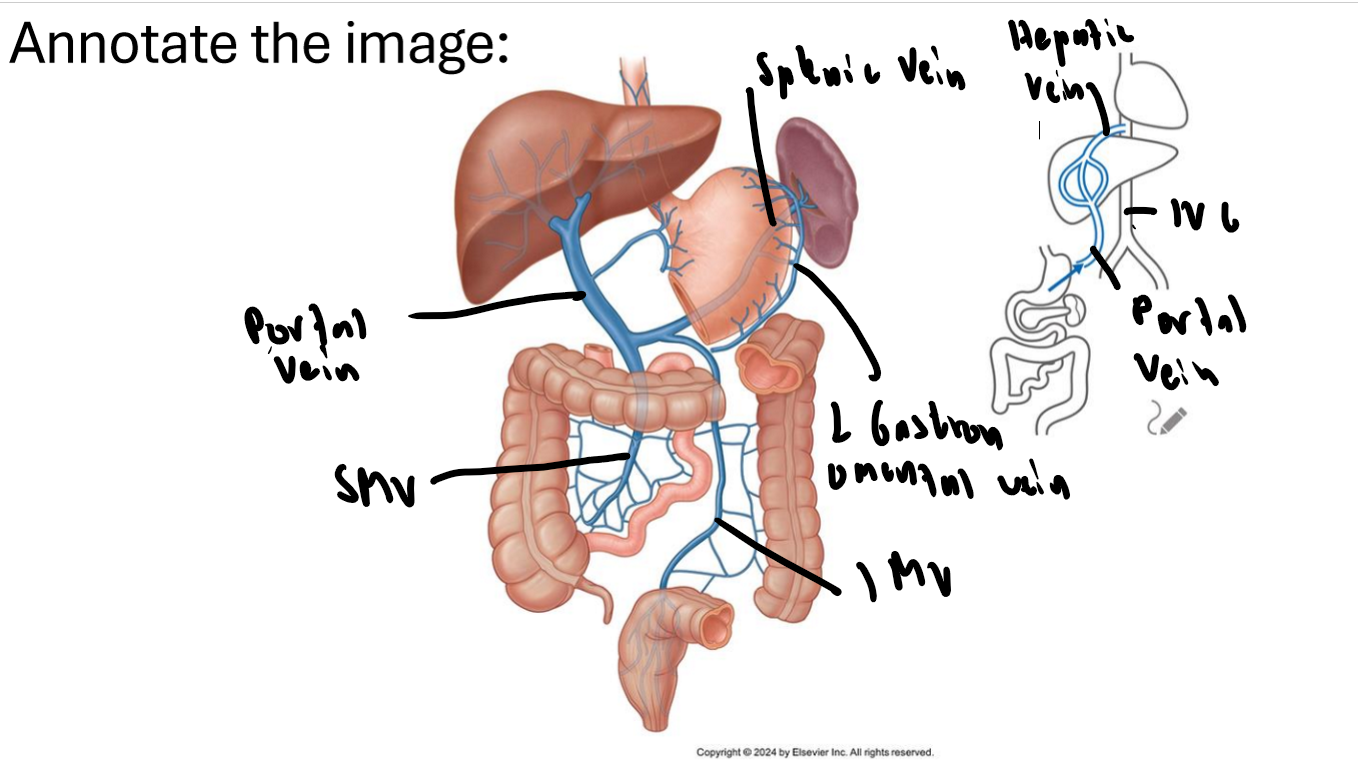
What’s the difference between the portal hepatis and portal triad (what are the contents)?
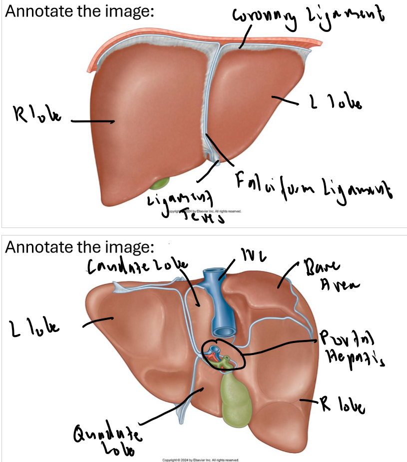
Portal hepatis is the opening and portal triad is the structures that pass through it, these include: Common bile duct, hepatic portal vein and proper hepatic artery

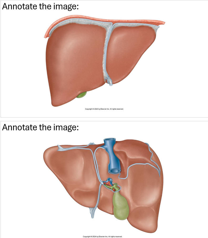
What are the names of the ligaments and where are located/covering?
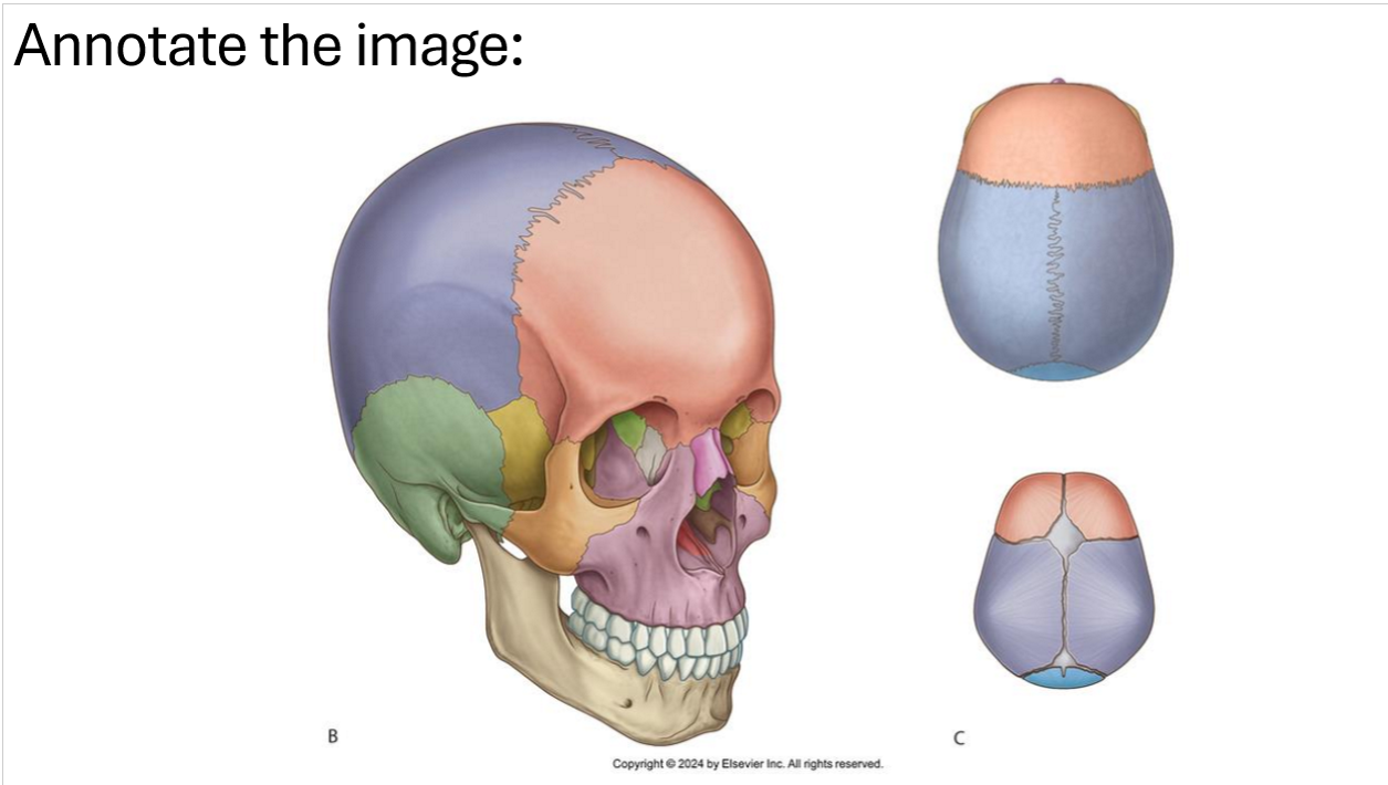
Name the neurocranium bones and sutures that separate them
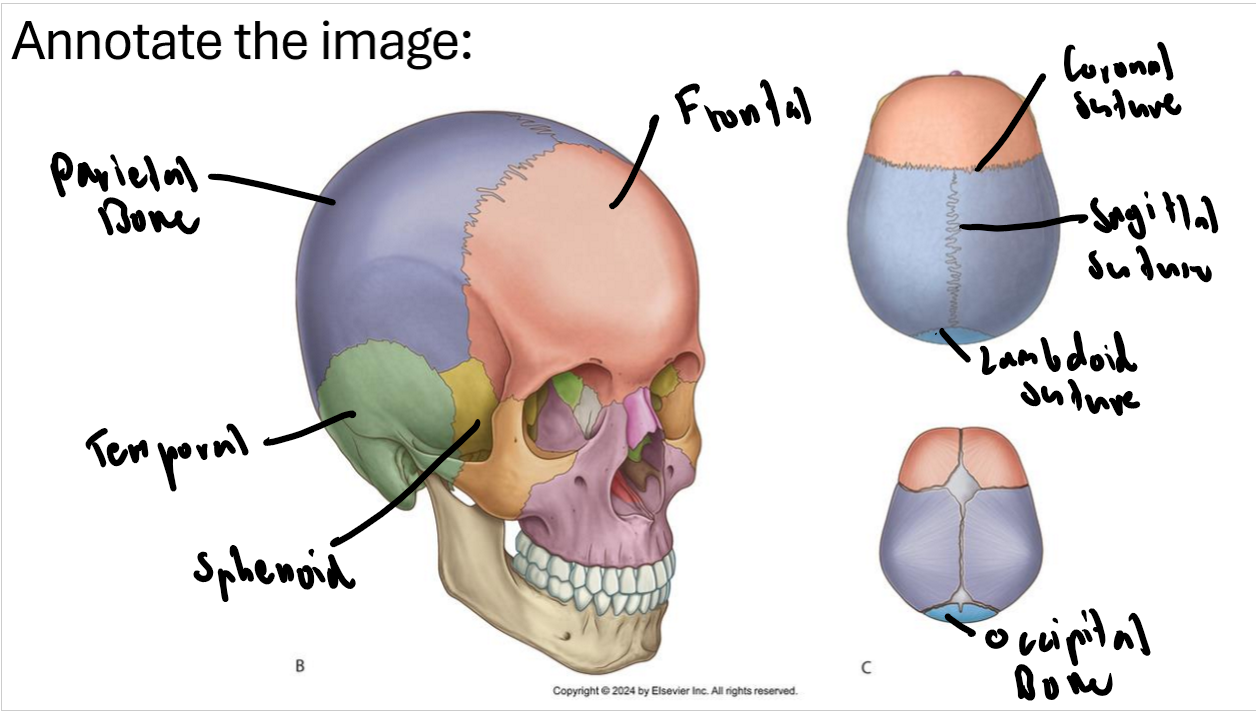
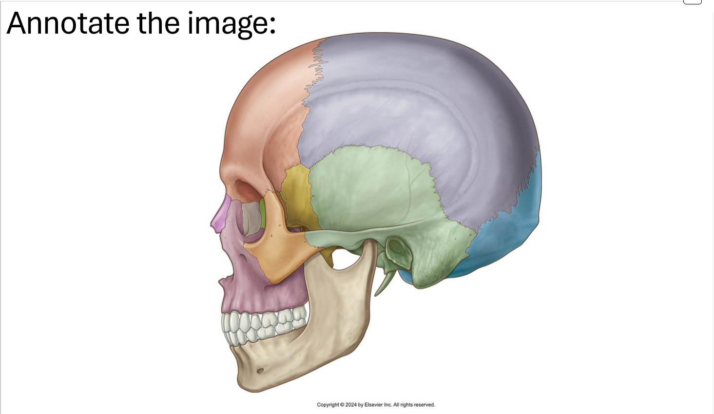
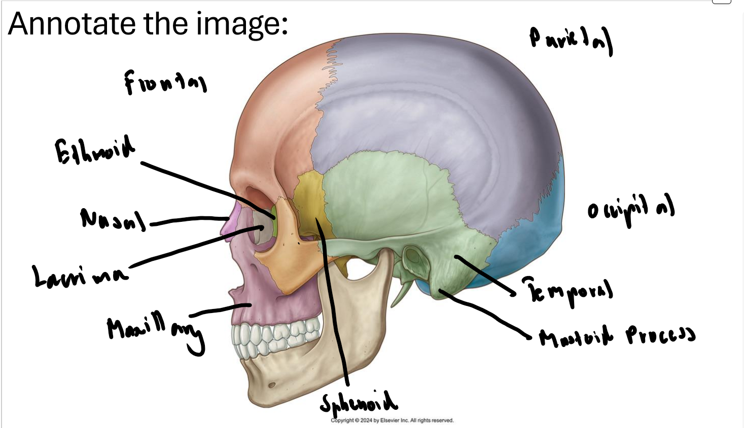
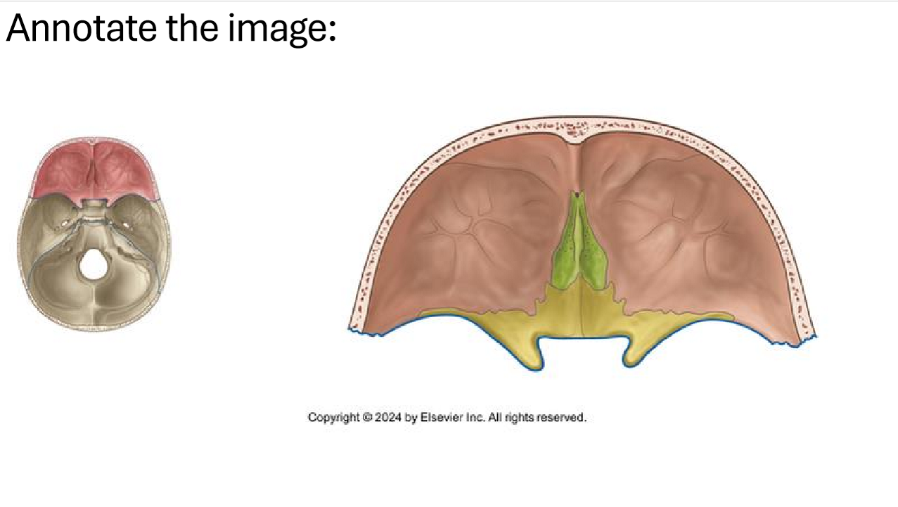
What bones make the anterior cranial fossa?
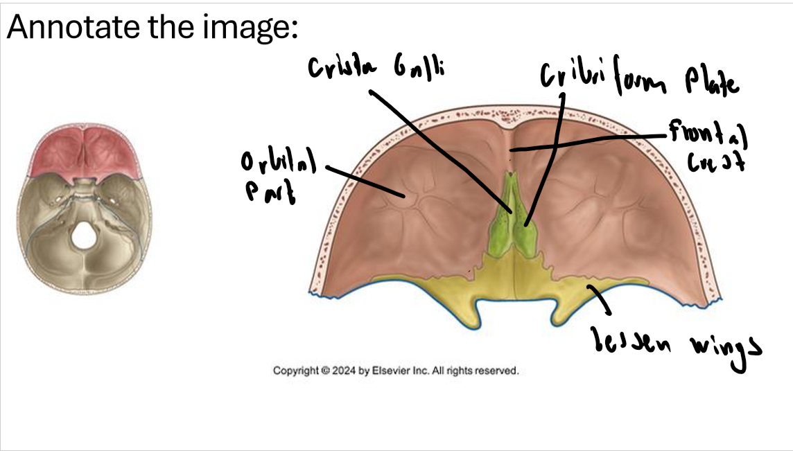
Frontal, ethmoid and sphenoid
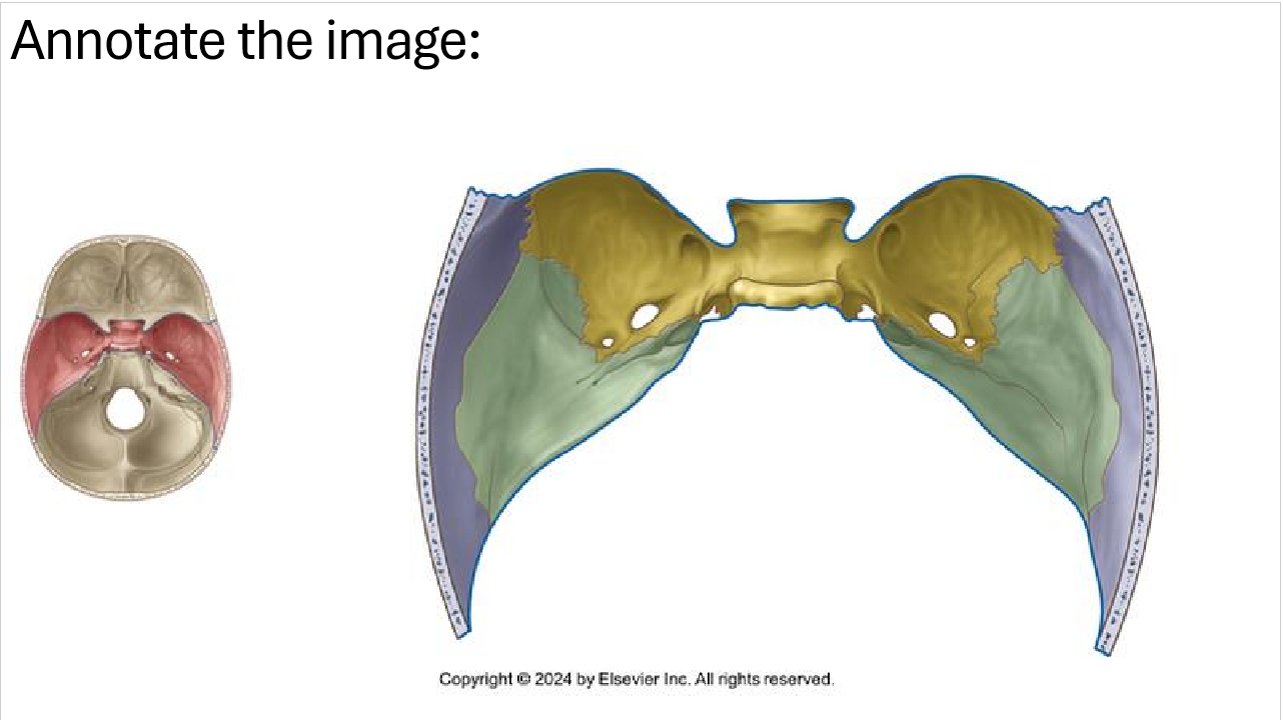
What bones make the middle cranial fossa?
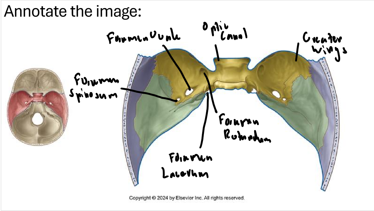
Parietal, sphenoid and temporal
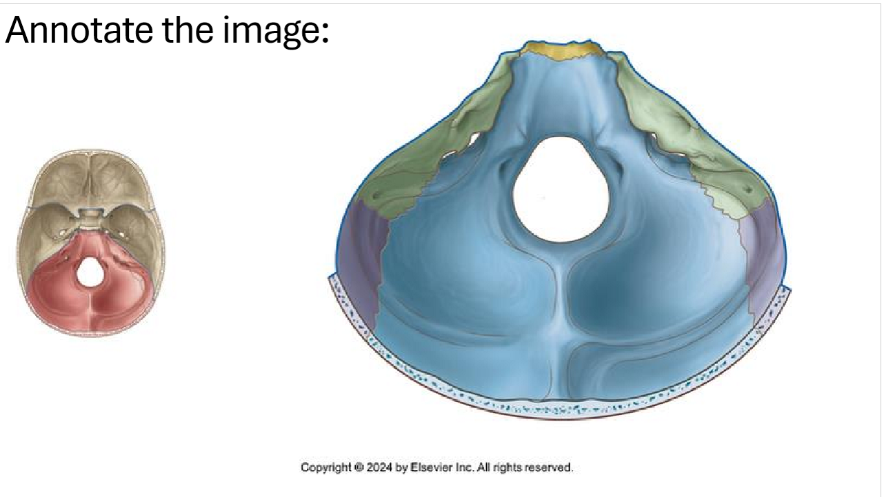
What bones make the posterior cranial fossa?
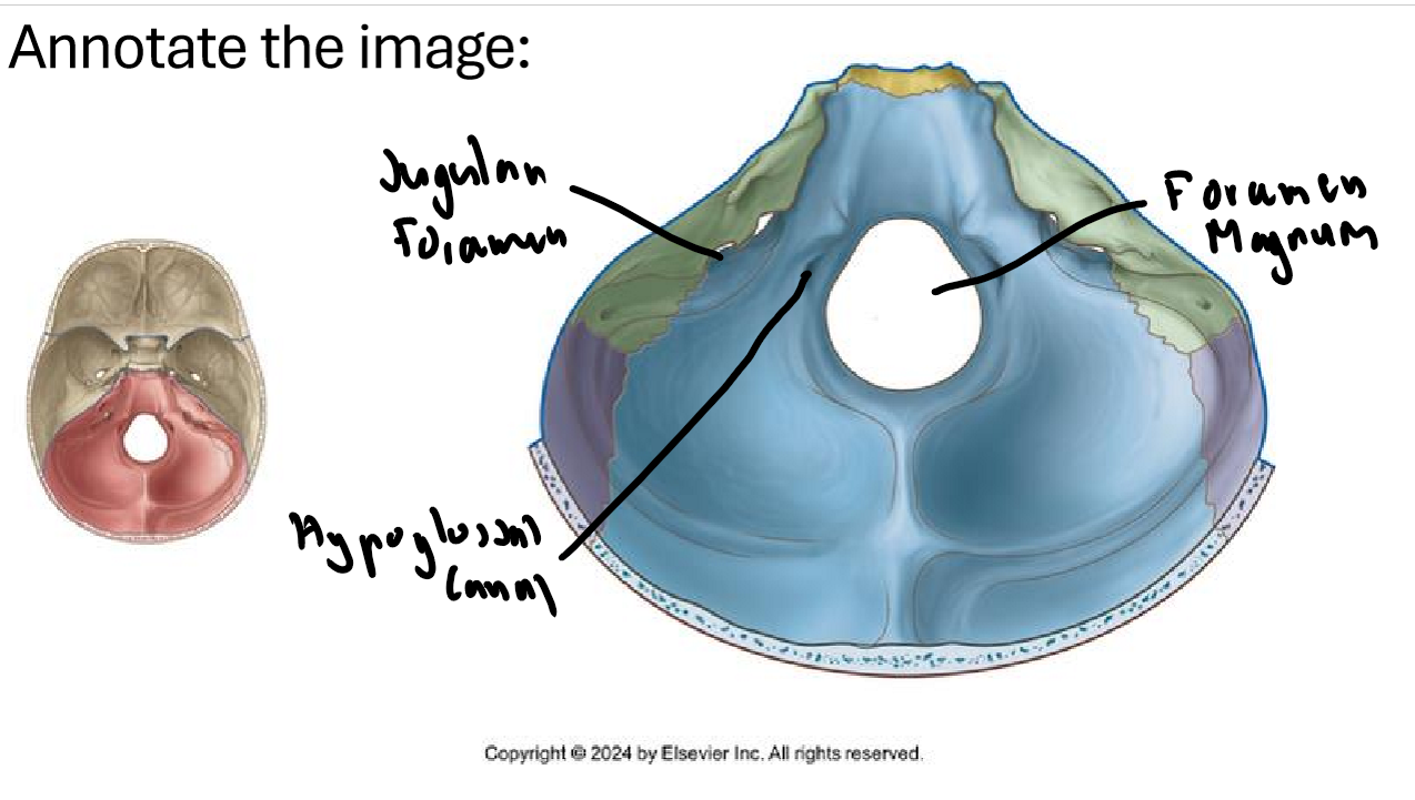
Occipital, temporal and parietal
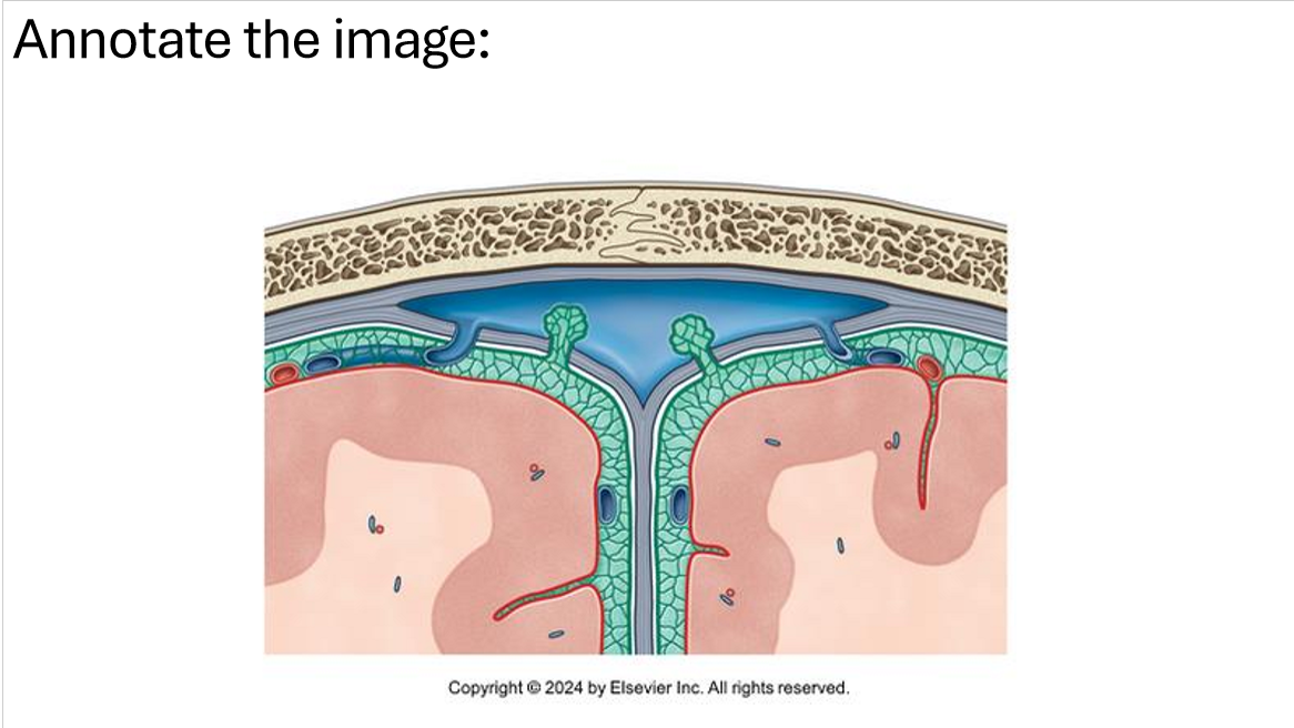
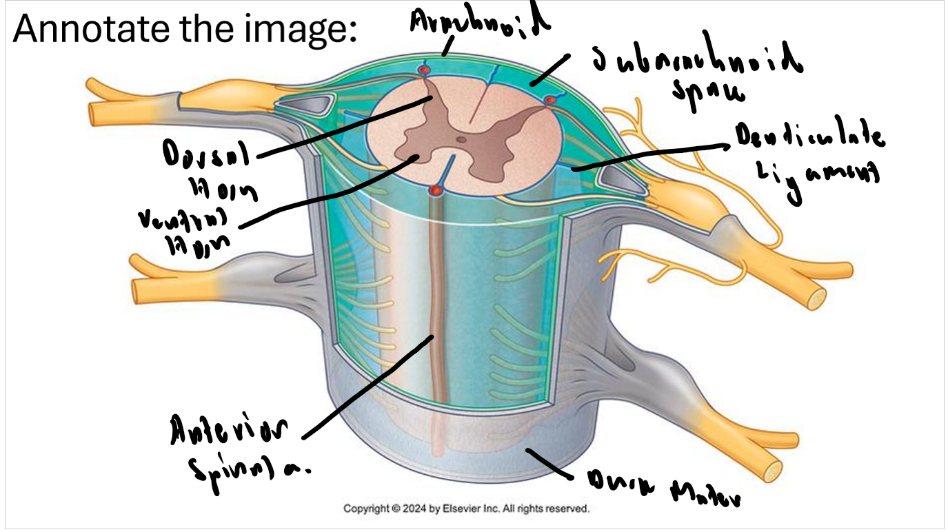
Name the meninges
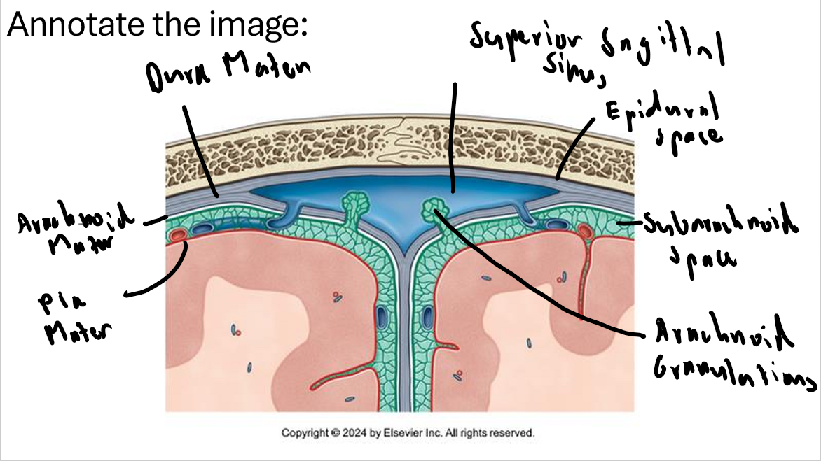
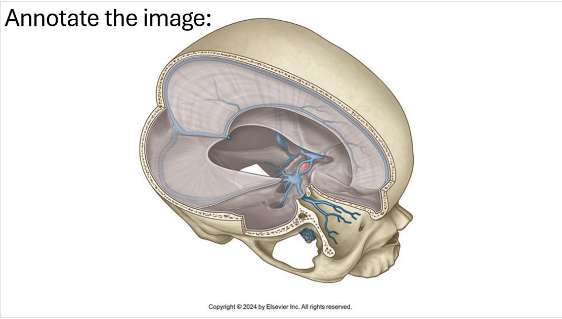
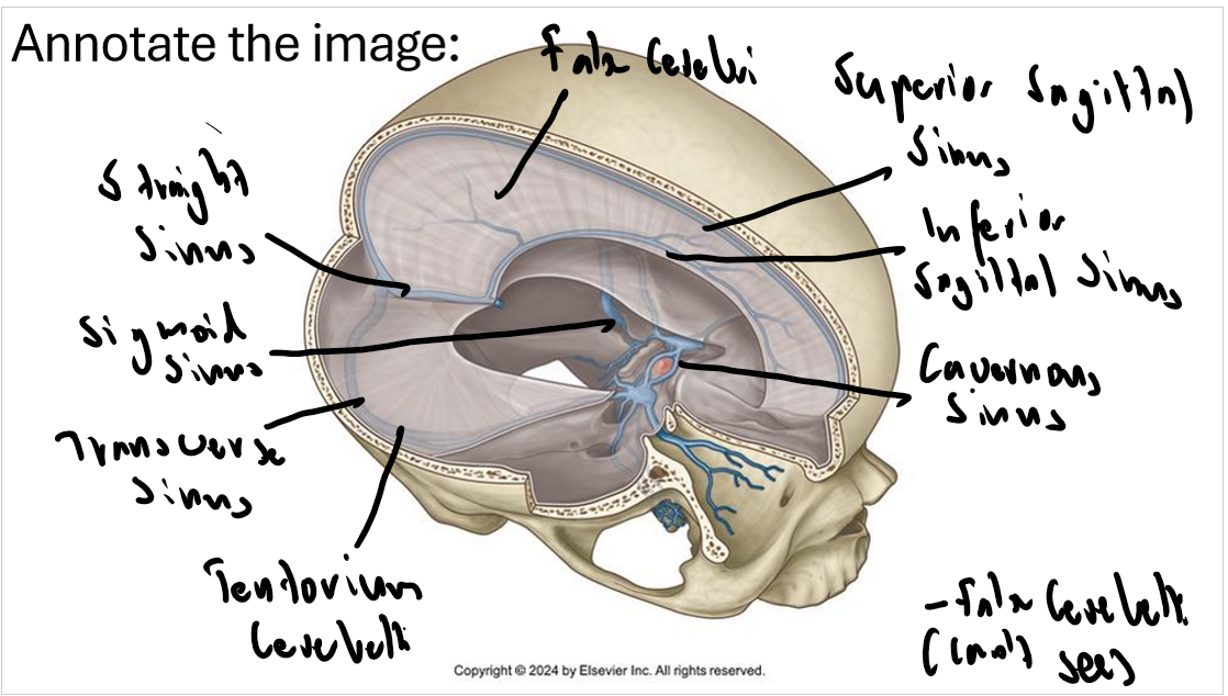
Name all the sinuses and dural folds.
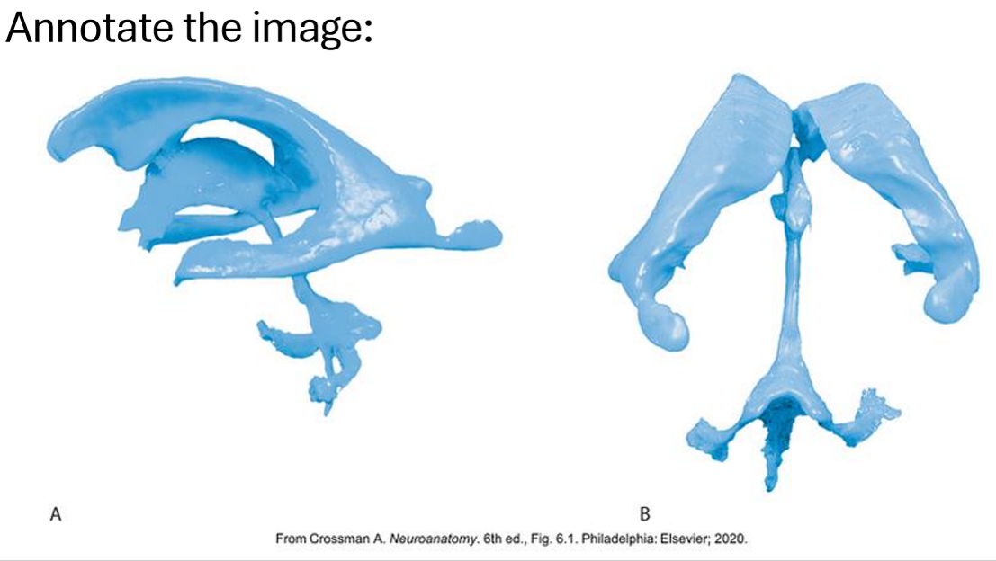
Name the ventricles and explain the drainage of CSF?
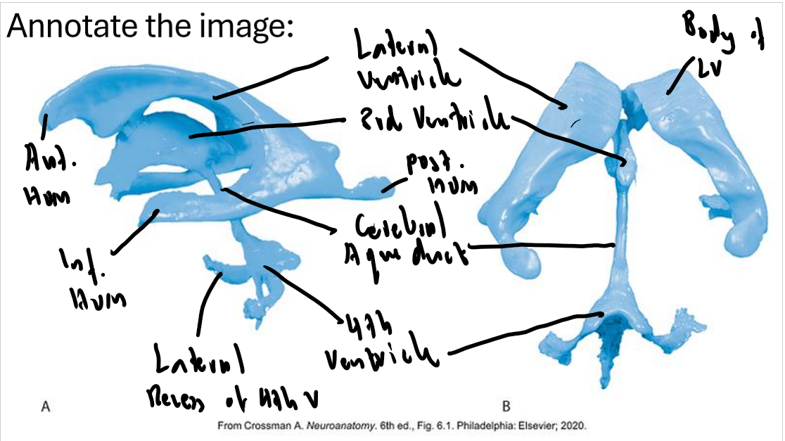
CSF is produced in the choroid plexus and first enters into the lateral ventricles
It then drains into the 3rd ventricle via the interventricular foramen
It then drains into the 4th ventricle via the cerebral aqueduct
CSF then enters the subarachnoid space via the median and lateral apertures and central canal.
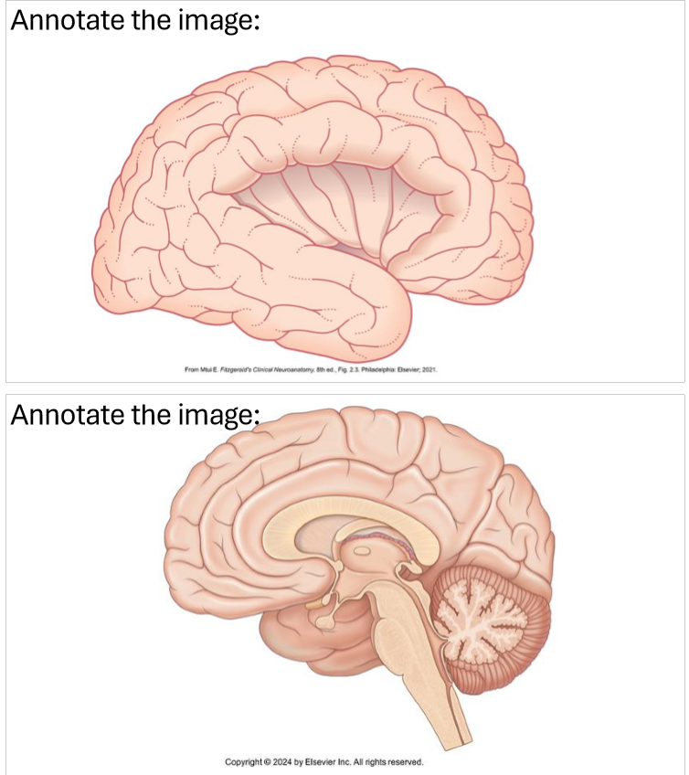
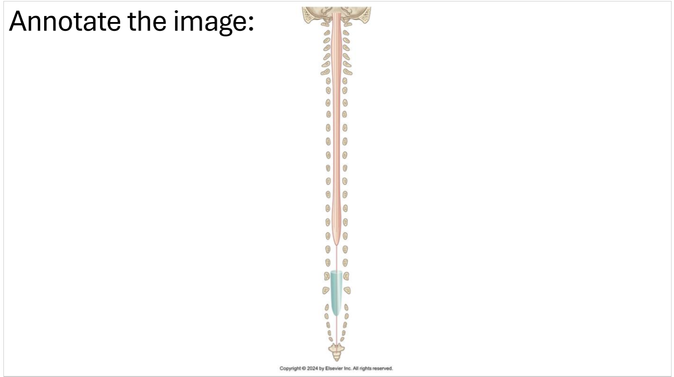
What is the function of the corpus callosum and medulla oblongata?
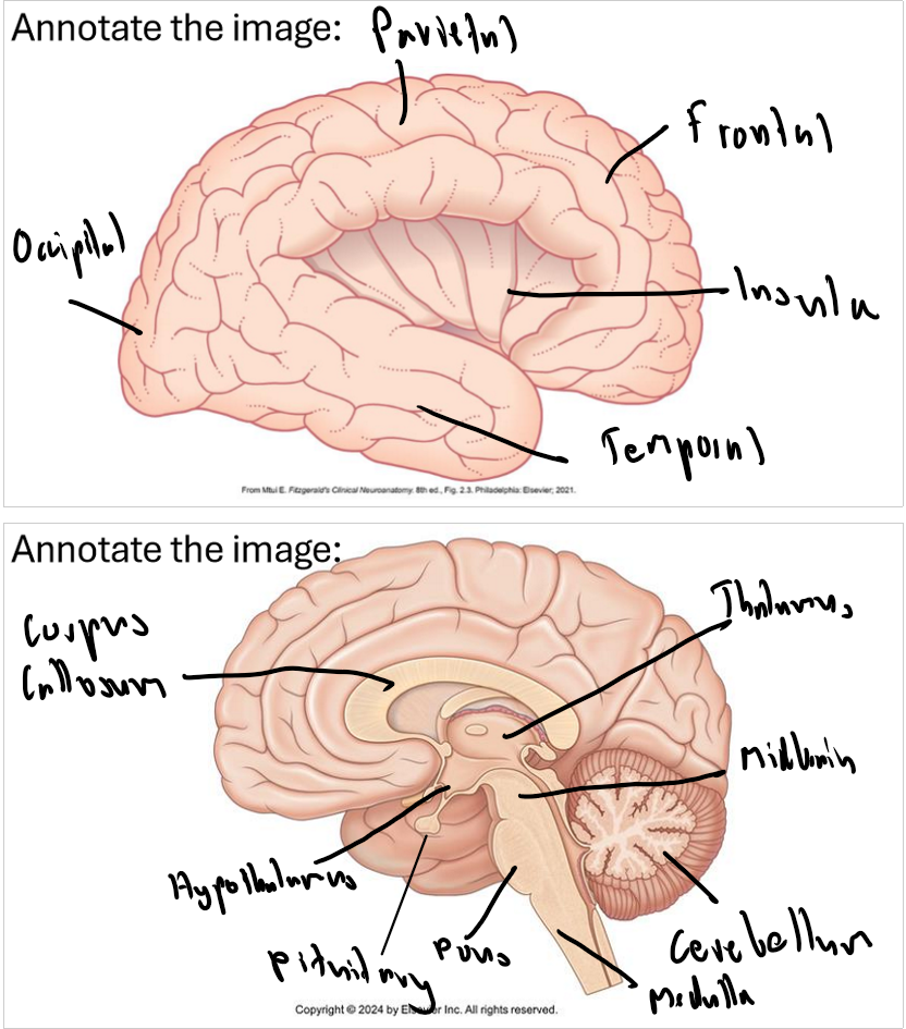
Corpus callosum act as an information highway, enabling rapid communication between the two halves of the brain to coordinate sensory, motor, and cognitive functions
Medulla oblongata is the center for controlling involuntary functions such as rhythmic breathing, heart rate and reflexes such as coughing, sneezing and vomiting.
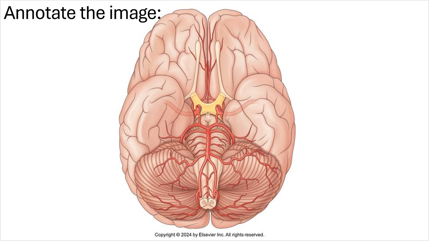
What blood vessel area supplies:
The posterior side of the brain
The lateral side of the brain
The anterior side of the brain

Posterior: PCA (vertebral a.)
Lateral: MCA (internal carotid a.)
Anterior: ACA (internal carotid a.)
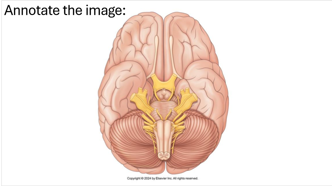
Name and identify the cranial nerves. What are their functions and how to classify this?
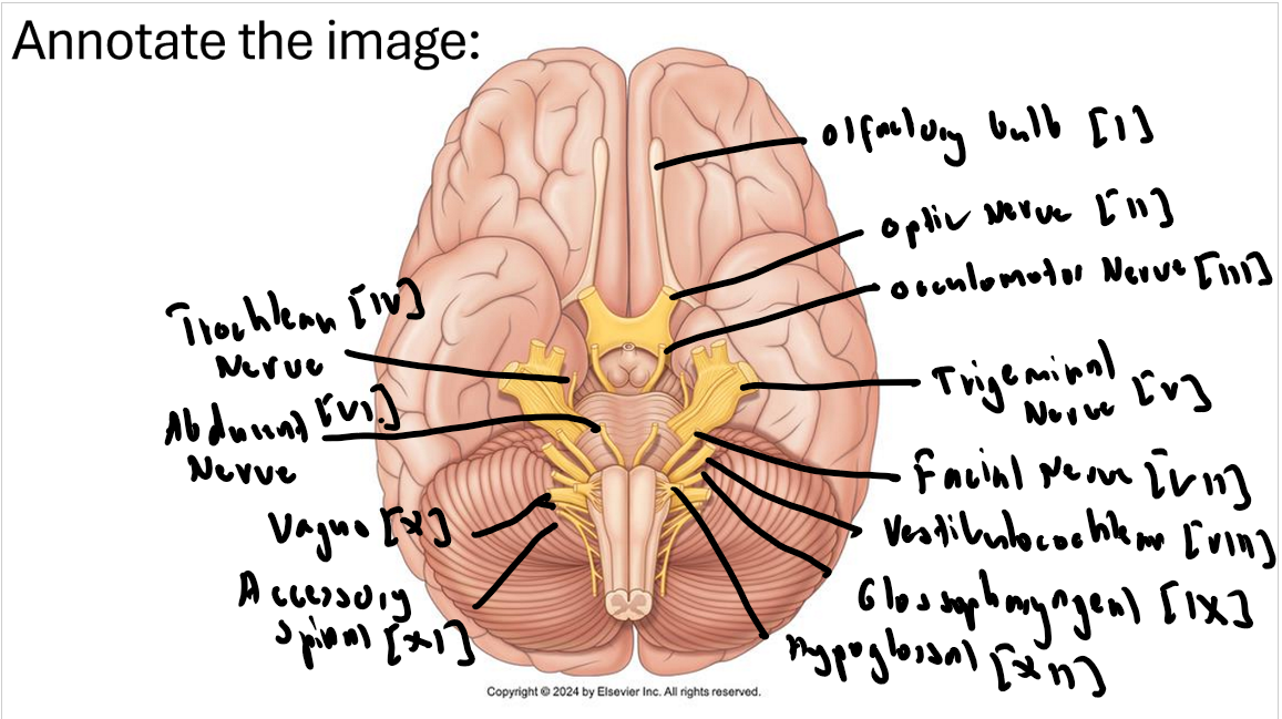
Olfactory Nerve: Smell in the nose
Optic Nerve: Transmitting visual info from retina to the brain
Occulomotor Nerve: Most eye movements and pupil constriction
Trochlear Nerve: Controls superior oblique muscle, move eye downward and inward (toward nose).
Trigeminal Nerve: Three divisions:
-Ophthalmic: somatic sensation in the forehead, bridge of nose, nostrils and eye
-Maxillary: somatic sensation in the midface, maxillary teeth and sinus, nasal cavity and hard palate
-Mandibular: somatic sensation in lower face, mandibular teeth, cheek, anterior 2/3 of tongue + muscles of mastication/jaw movementsFacial Nerve: Muscle of facial expressions, salivation, taste, somatic sensations of the ear.
Abducent Nerve: Controls lateral rectus muscle, abduction of the eye (away and outward from nose)
Vestibulocochlear Nerve: Balance and hearing
Glossalpharyngeal Nerve: Innervates one muscle in pharynx (swallowing), salivation and tase (posterior 1/3 tongue), somatic sensation in posterior 1/3 tounge and pharynx
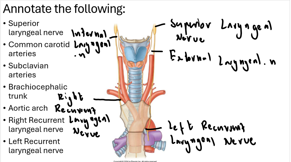
Vagus Nerve: Swallowing and speech, taste in pharynx, somatic sensation in the external ear, pharynx and larynx
Accessory Nerve: Head and pectoral girdle movements (traps and sternocleidomastoid)
Hypoglossal Nerve: Motor innervation of the tongue
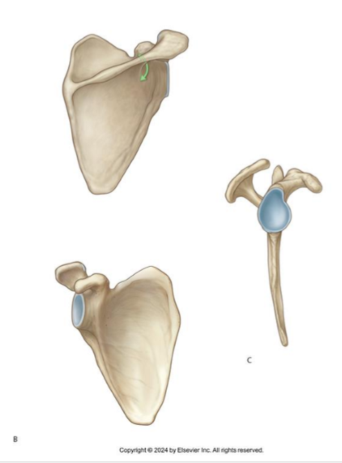
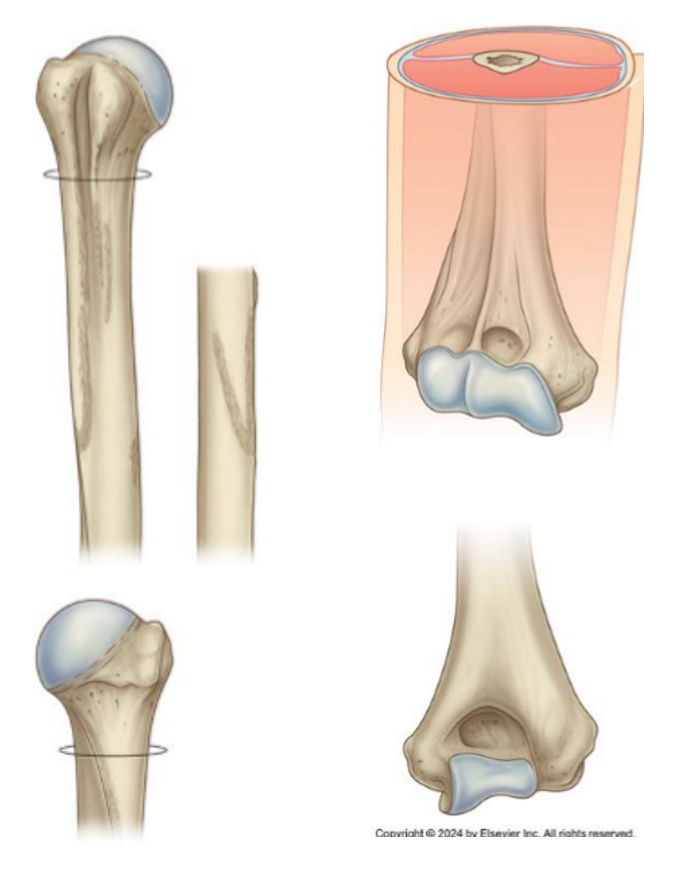
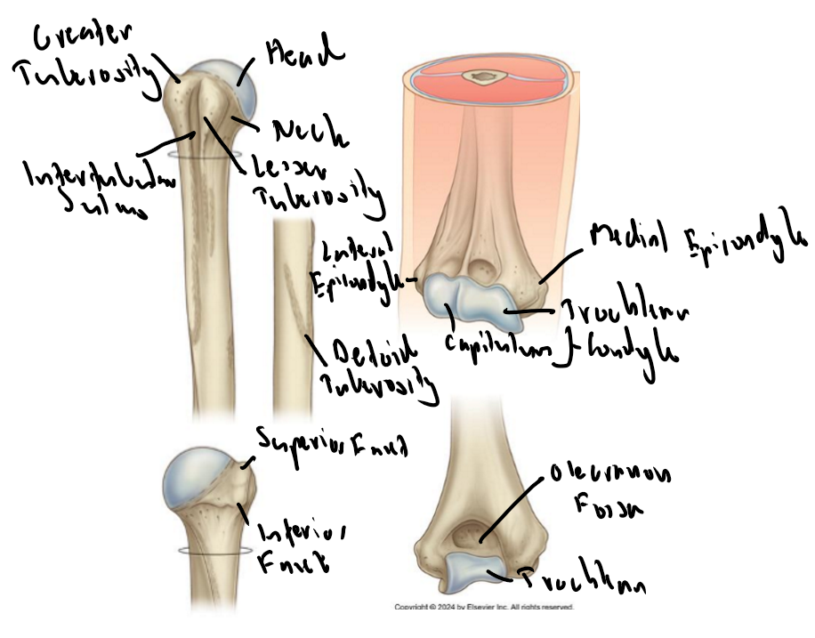
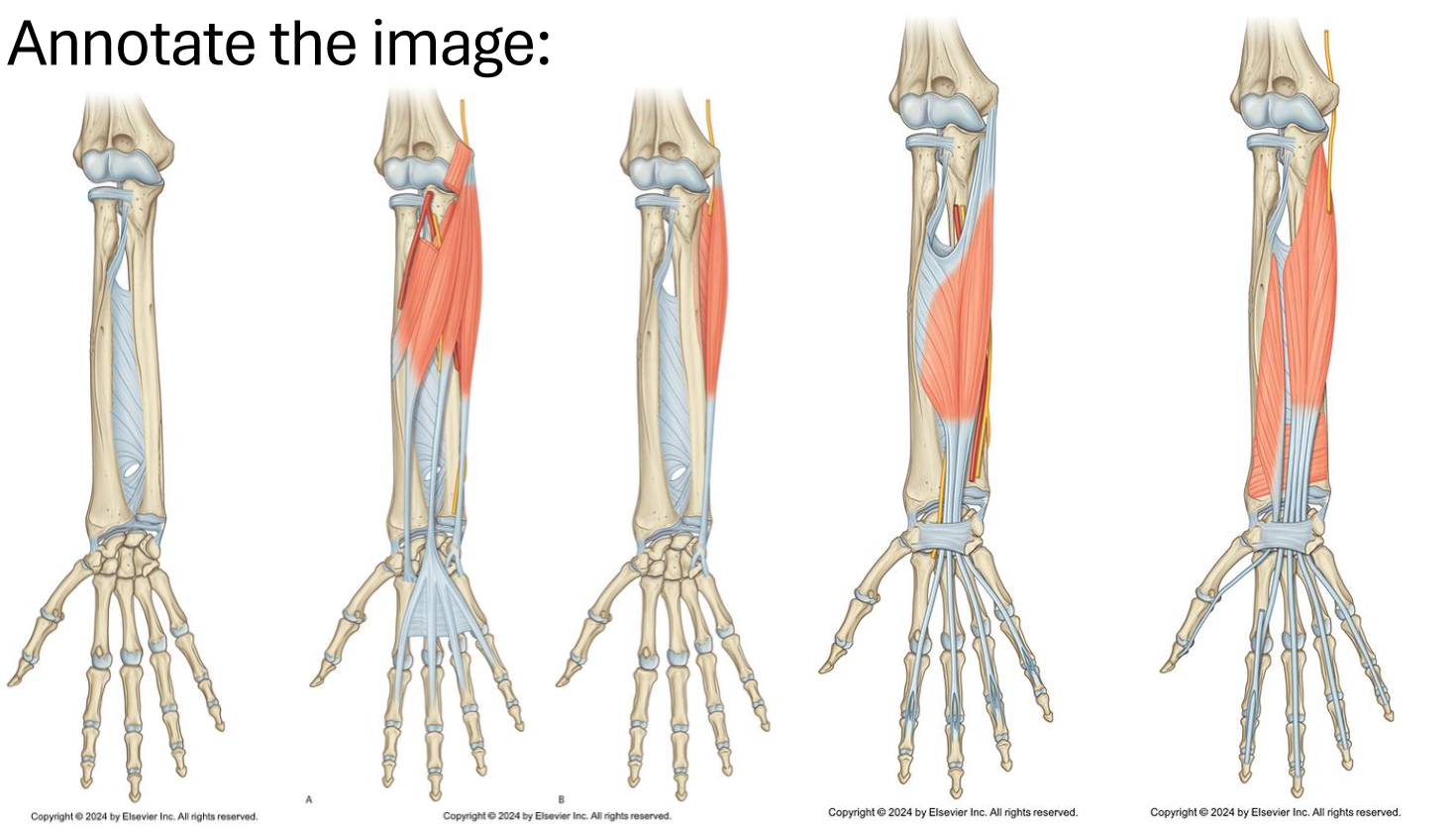
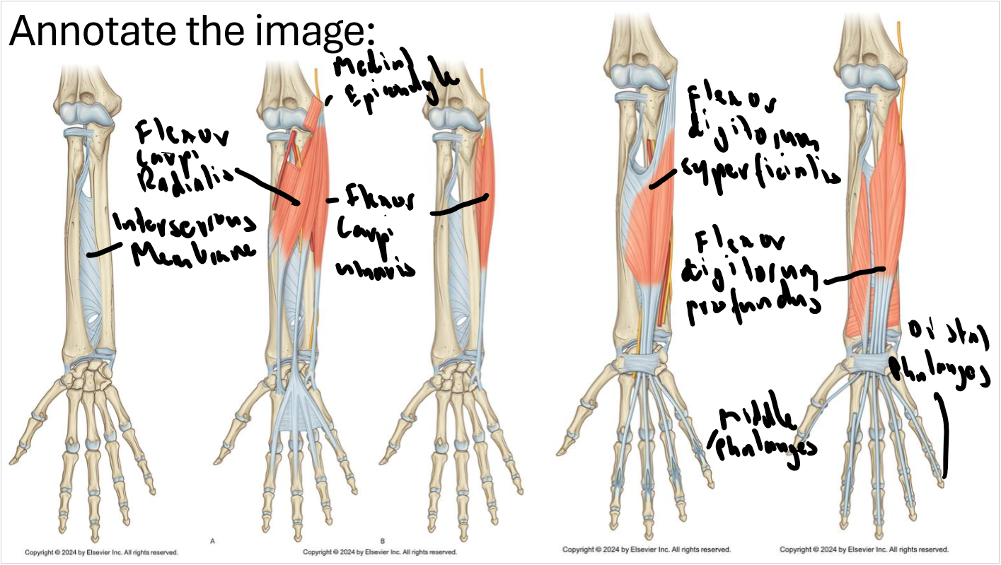
What is significant about the medial epicondyle?
It is the common flexor origin and is larger than lateral epicondyle as it has greater functional importance in movements such as grasping and wrist flexion
Flexor carpi radialis, ulnaris, digitorum superficialis.
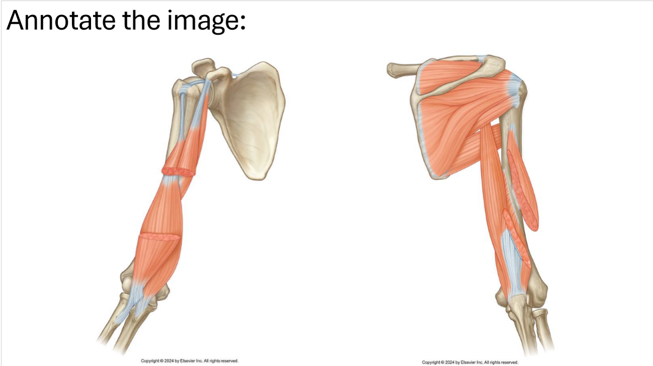
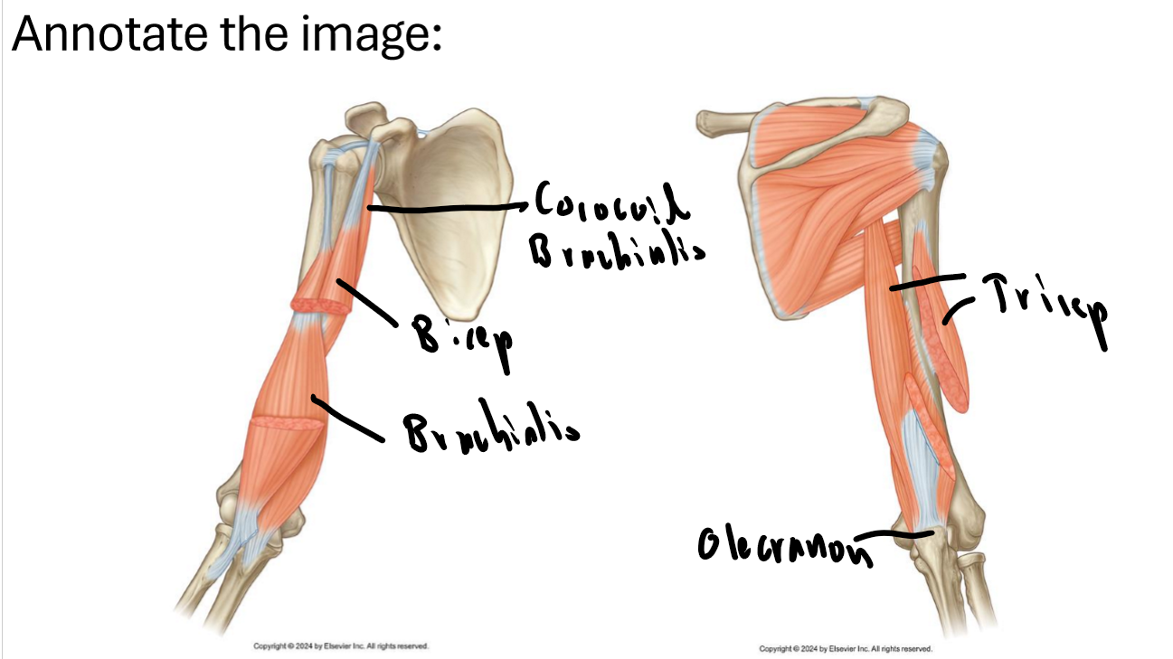
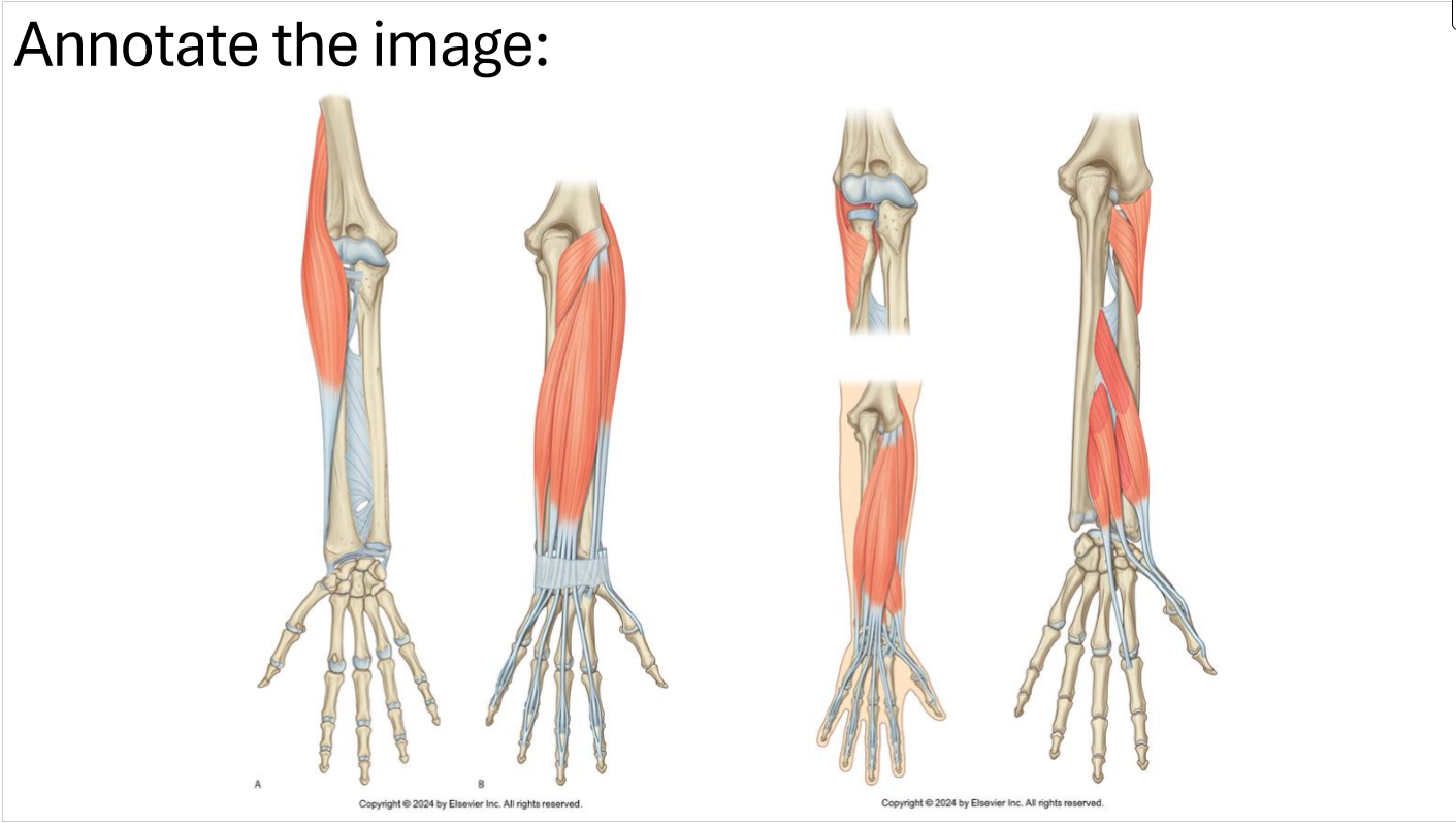
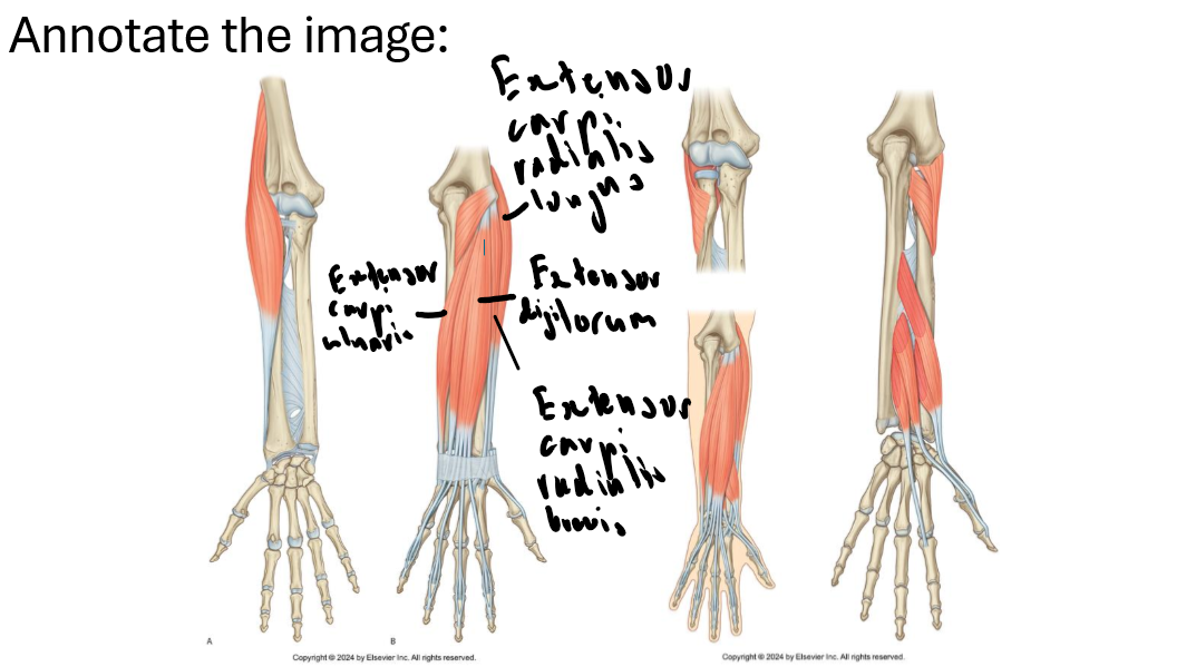
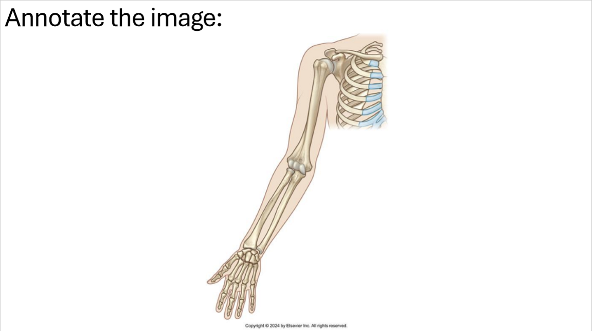
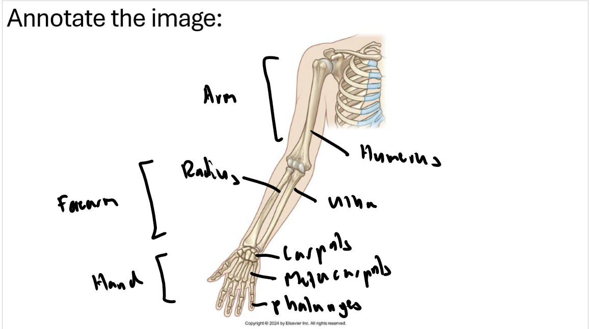
What nerves supply the fore limb compartments and what division do they come from?
Anterior Arm: Musculotaneous nerve (anterior division)
Posterior Arm: Radial nerve (posterior division)
Anterior Forearm: Mostly median, some ulnar (anterior division)
Posterior Forearm: Radial nerve (posterior division)
Intrinsic Hand: Mostly ulnar, some median (anterior division)
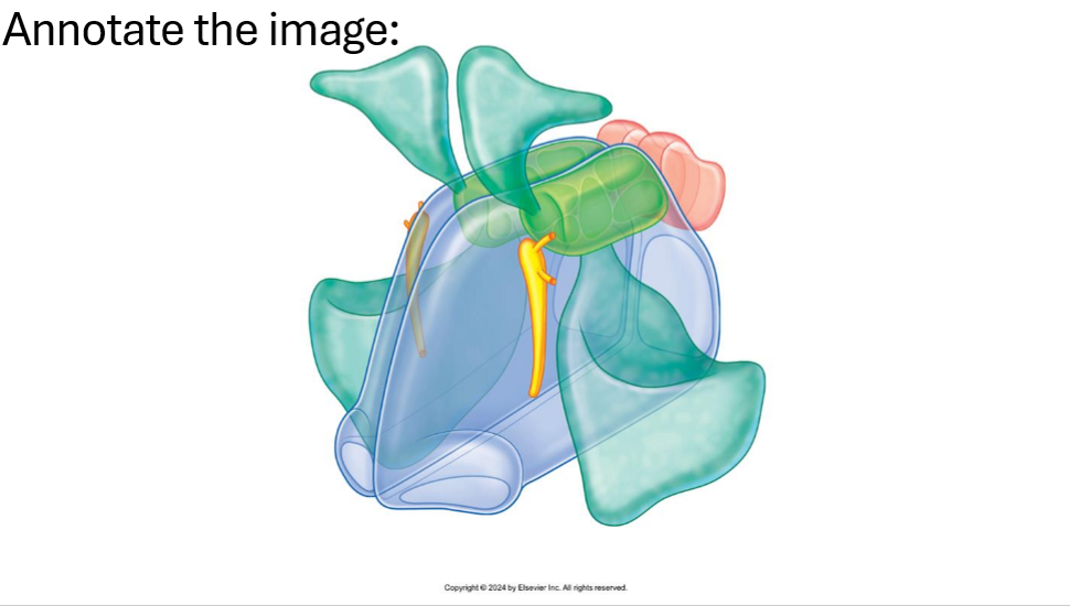
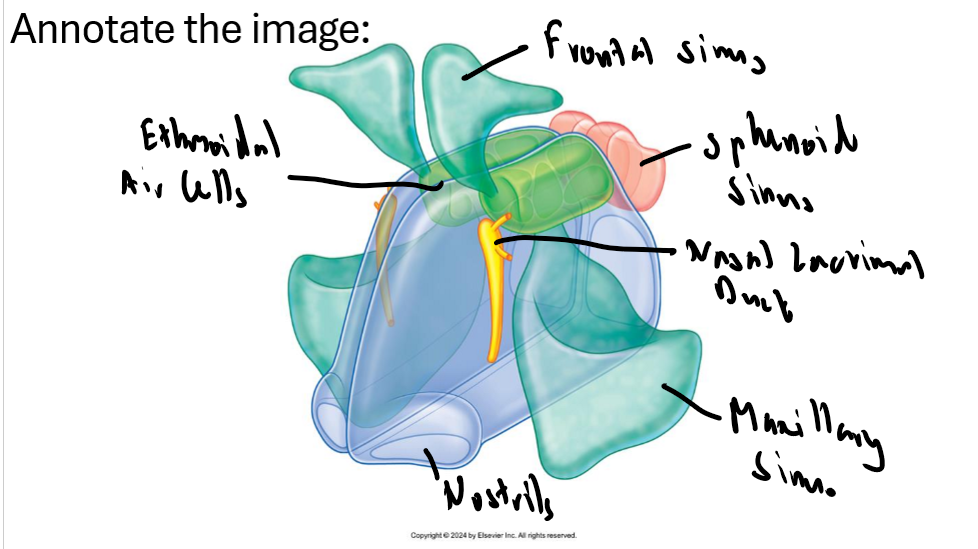
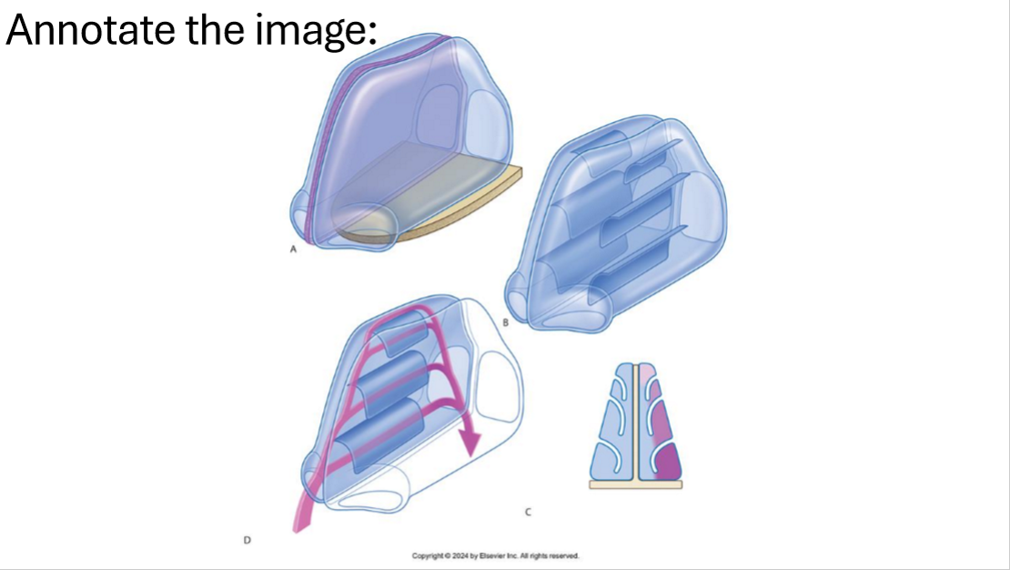
Name the regions of the nasal cavity and there function.
Olfactory region: superior concha area, covered with olfactory epithelium
Respiratory region: middle + inferior concha area, functions to humidify, warm, filter, protect and eliminate debris
Nasal Vestibule: nostrils, contains sweat glands and hair follicles, first line of defense
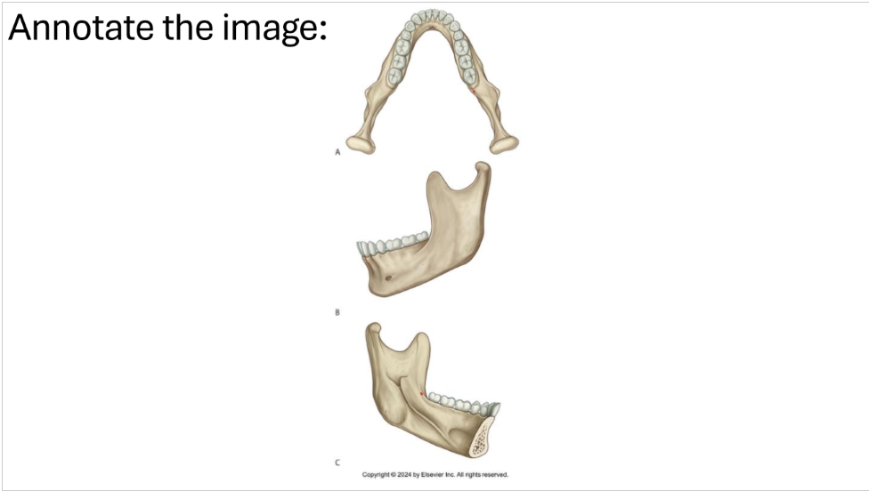
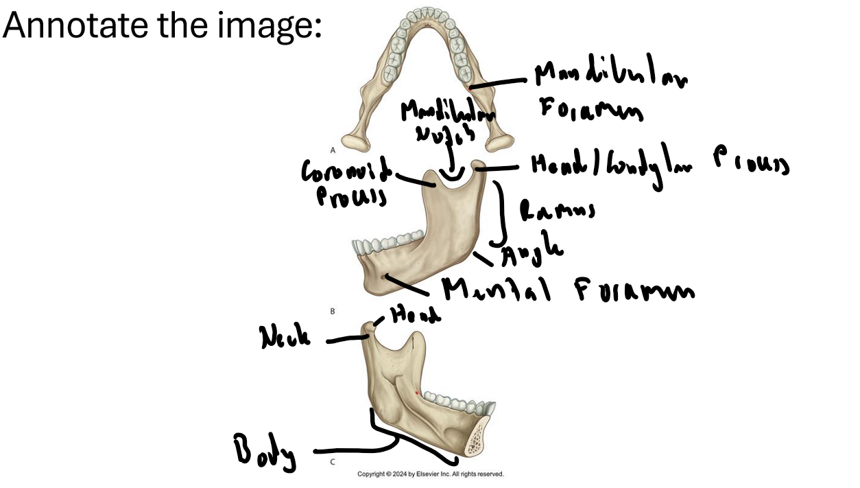
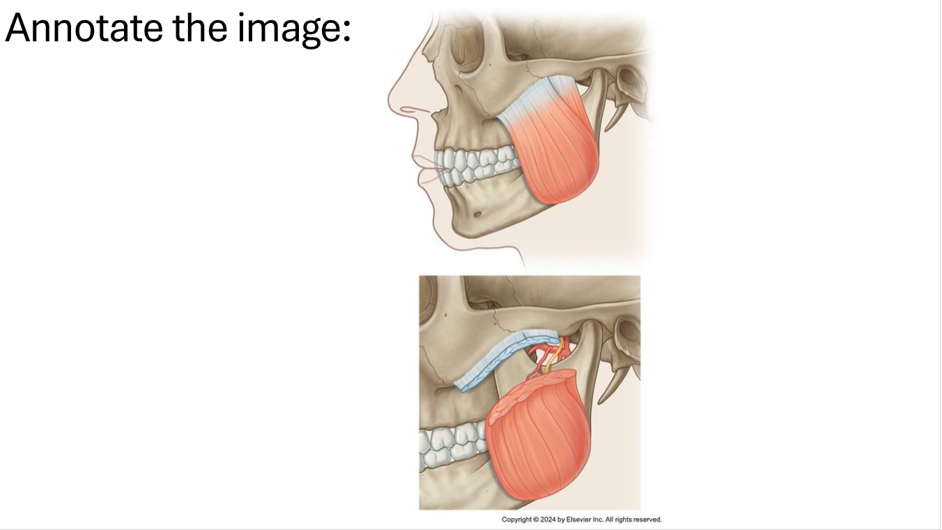
What is the attachments for this muscle and what actions does it do?
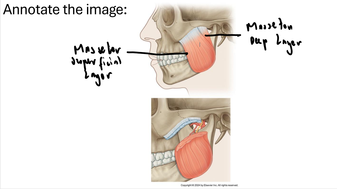
Proximal: Zygomatic arch, Distal: External surface of the mandible (ramus + angle)
Action: Elevation and protrusion
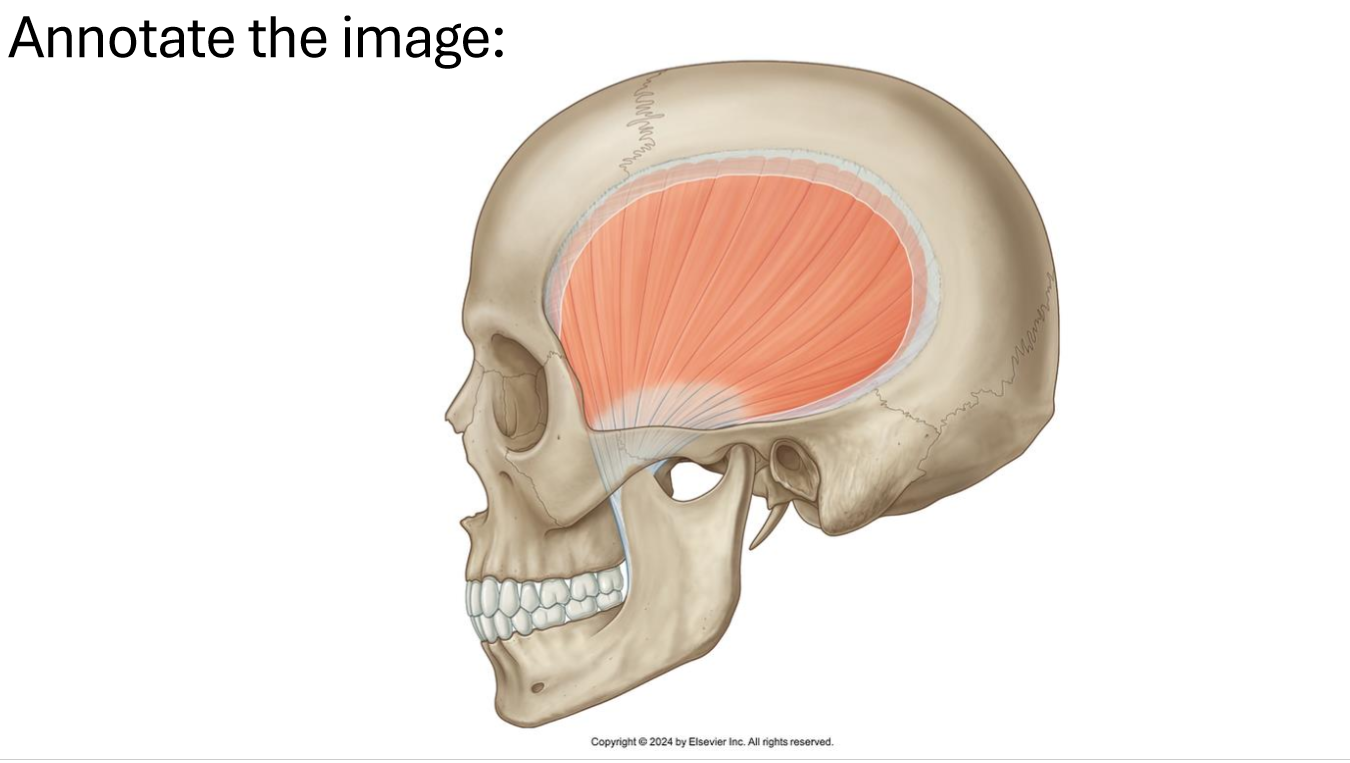
What is the attachments for this muscle and what actions does it do?
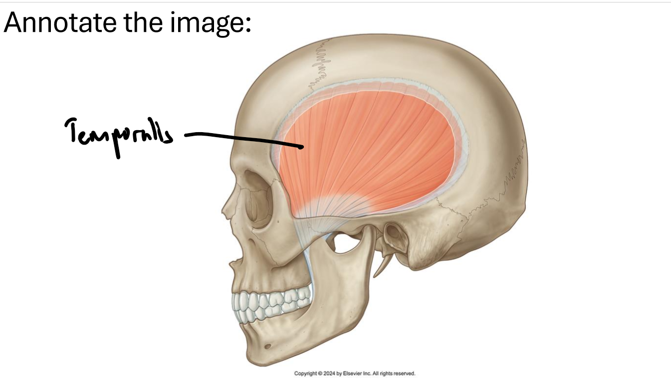
Proximal: Temporal Fossa, Distal: Coronoid process
Action: Elevation, retrusion
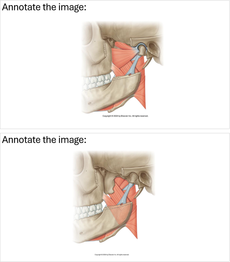
What is the attachments for this muscle and what actions does it do?
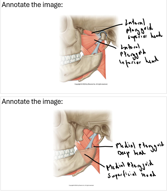
Lateral Pterygoid- Proximal: Pterygoid plate, Distal: Head of mandible
Action: Depression and protrusion
Medial Pterygoid- Proximal: Pterygoid plate, Distal: Internal surface of mandible (ramus + angle)
Action: Elevation and protrusion
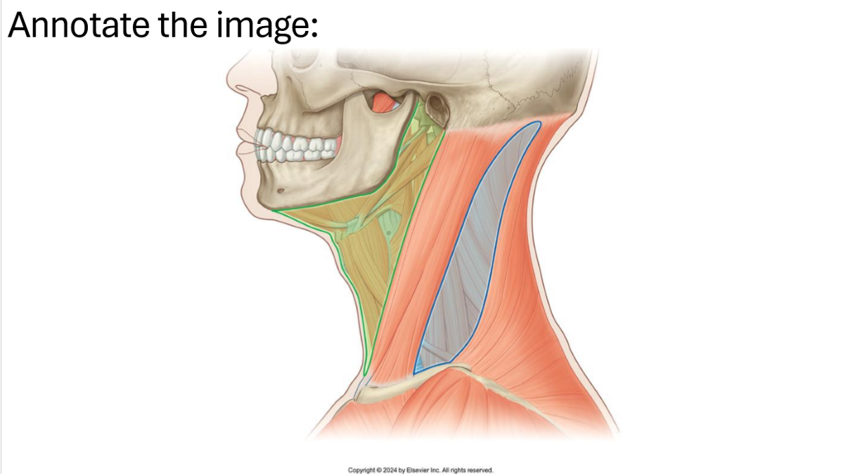
What contents run through each triangle?
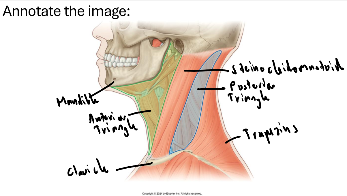
Anterior triangle: Common carotid arteries, internal jugular veins, vagus nerve, lymph nodes, larynx and pharynx
Posterior triangle: Subclavian artery/vein, brachial plexus, phrenic nerve, spinal accessory nerve
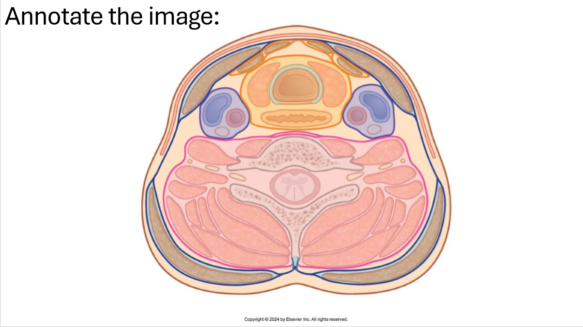
What are the contents of each layer of fascia?
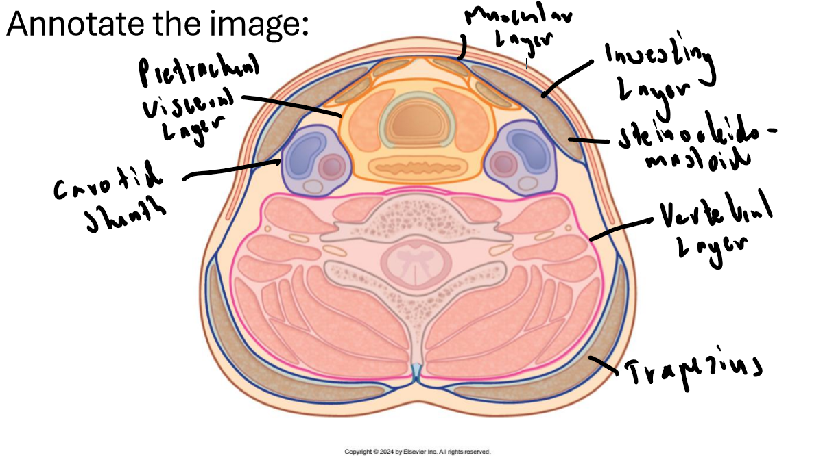
Investing layer: Trapezius and steinocleidomastoid
Visceral layer: Oesophagus, trachea Muscular layer: Infrahyoid muscles
Carotid sheath: Internal jugular vein, Internal + common carotid artery, Vagus nerve
Vertebral layer: vertebrae and deep muscle

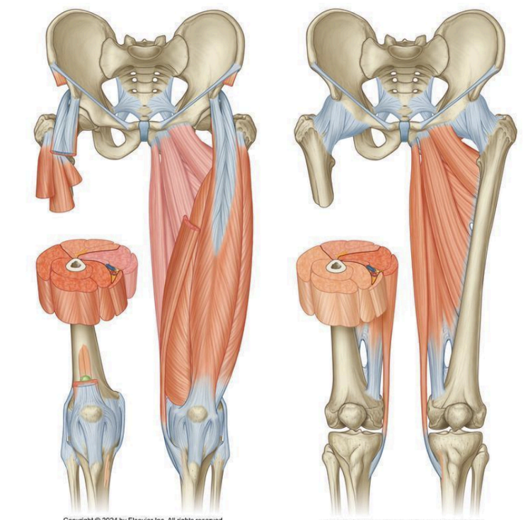
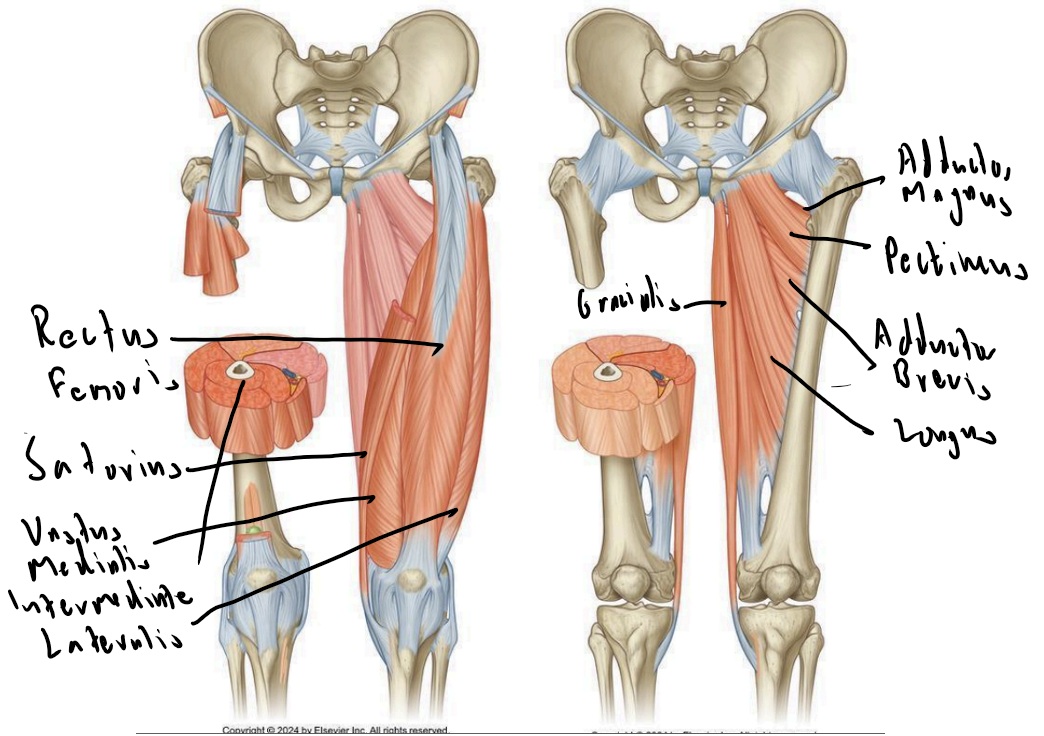
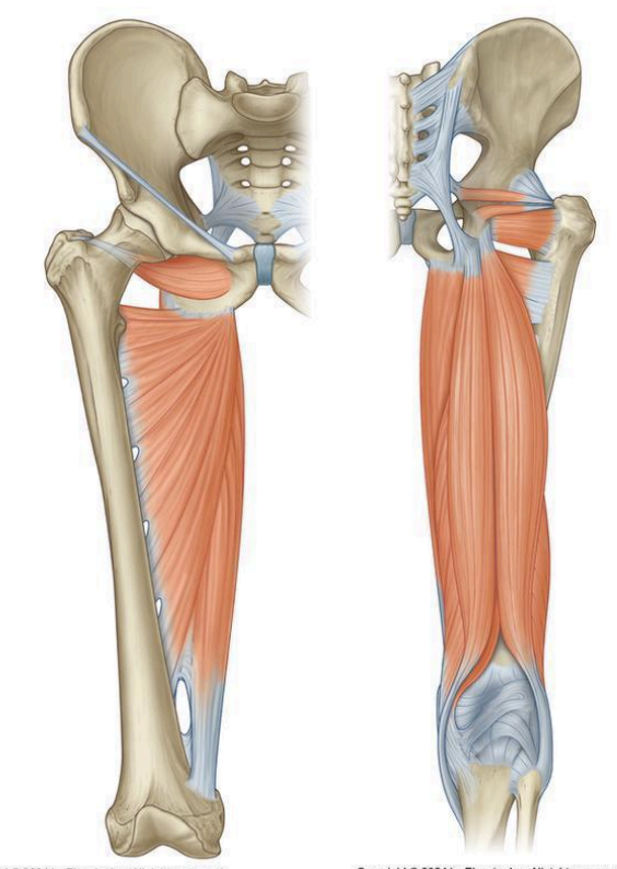
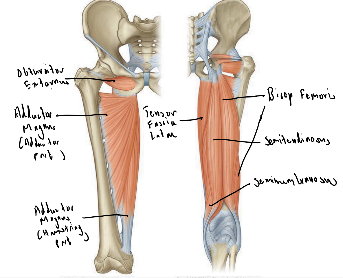
What nerves supply the leg compartments and what division do they come from?
Anterior Thigh: Femoral nerve (dorsal division)
Medial Thigh: Mostly adductor nerve (ventral division)
Posterior Thigh: Mostly tibial division of sciatic nerve (ventral), some common fibular div of sciatic (dorsal)
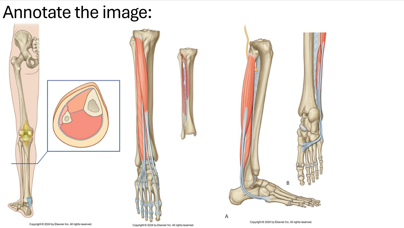
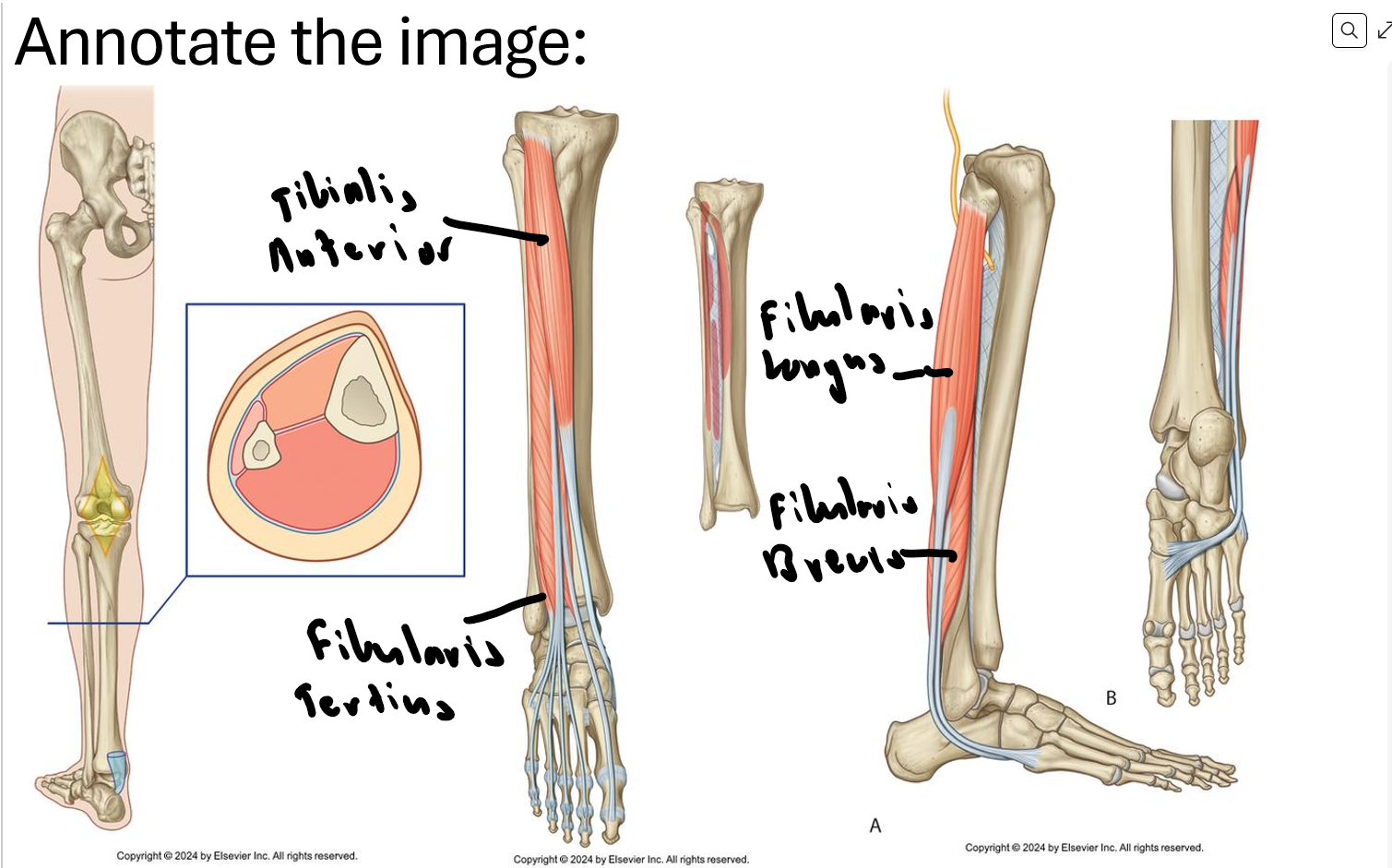
Anterior Leg: Deep fibular nerve (dorsal division)
Lateral Leg: Superficial fibular nerve (dorsal division)
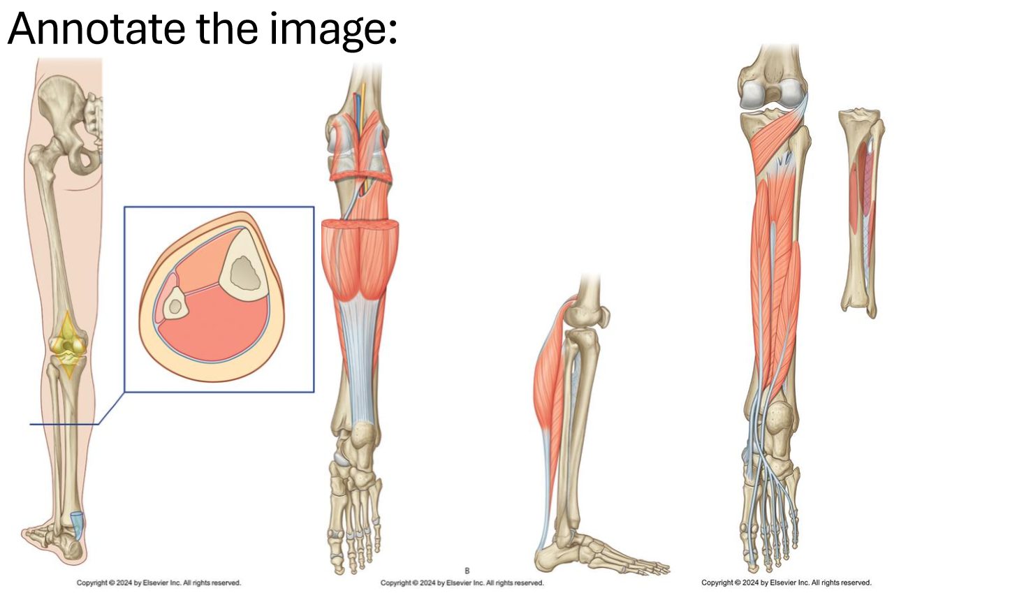
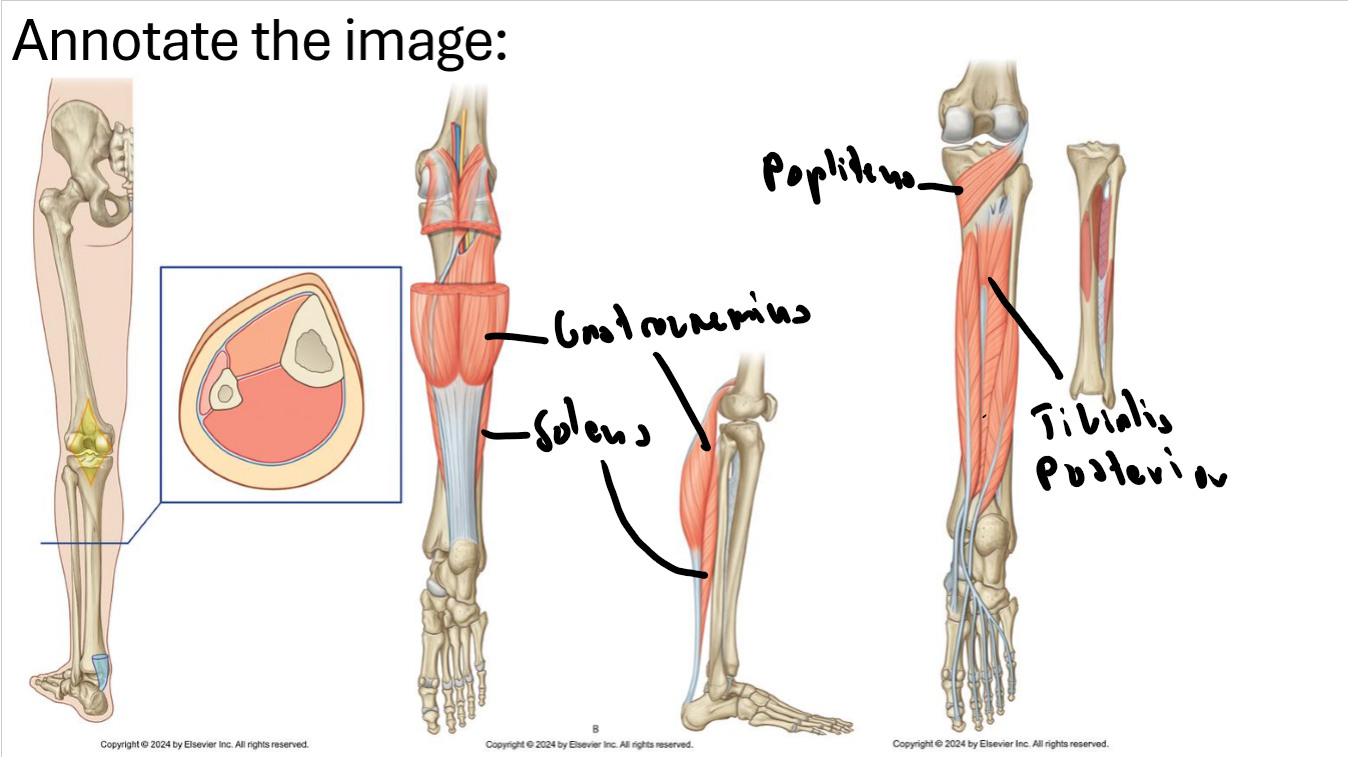
Posterior Leg: (Tibial nerve) (ventral division)
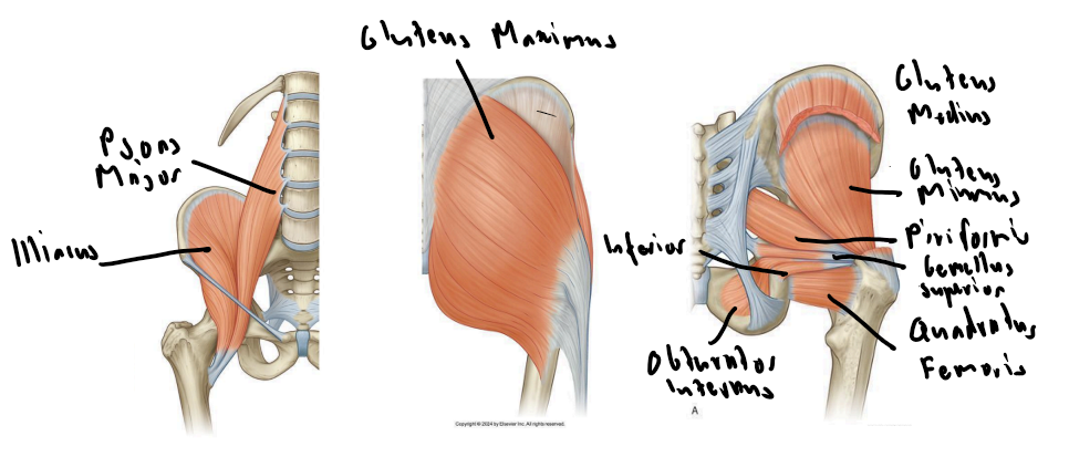
What nerves supply the gluteal region and what division do they come from?
Superior gluteal nerve: Gluteus medius and minimus, tensor fascia latae
Inferior gluteal nerve: Gluteus maximus
Both from the dorsal division of sacral plexus