Echo - Final Exam (Jo's set)
1/110
There's no tags or description
Looks like no tags are added yet.
Name | Mastery | Learn | Test | Matching | Spaced | Call with Kai |
|---|
No analytics yet
Send a link to your students to track their progress
111 Terms
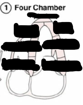
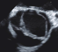
Identify this image.
A4C
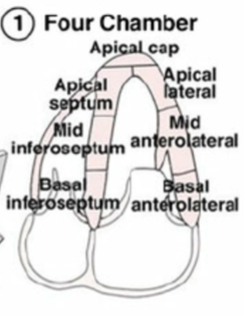
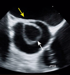
Identify this image.
A2C

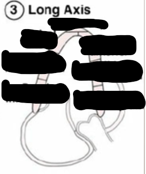
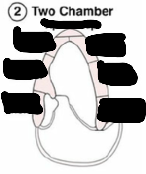
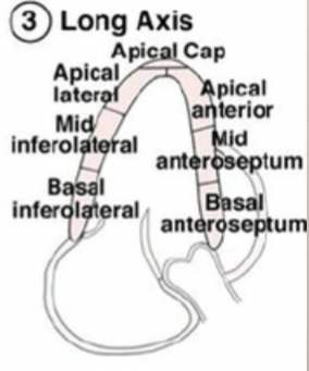
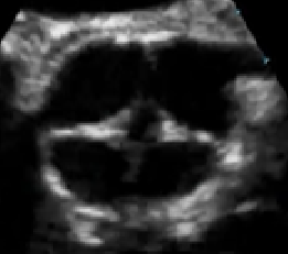
Identify this image.
A3C or LONG AXIS
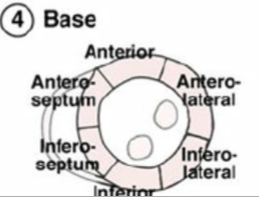
Identify this image.
Base or top
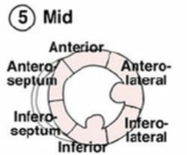
Identify this image.
Mid
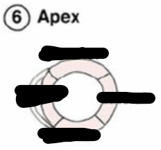
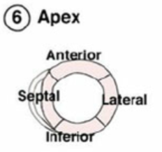
Identify this image.
Apex or bottom
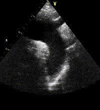
Identify this image.
PLAX
RV
LA
MV
LVOT
AV
Aortic root
Descending AO
Identify this image.
PLAX RVIT
A. RA
B. TV
C. RV
Identify this image.
PLAX RVOT
RV
PV
Main pulmonary artery
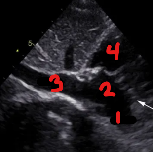
Identify this image.
PSAX AV Basal
Descending aorta
LA
IAS
RA
TV
RVOT
PV
Main pulmonary artery
Right coronary cusp
Noncoronary cusp
Left coronary cusp
Identify this image.
PSAX MV level

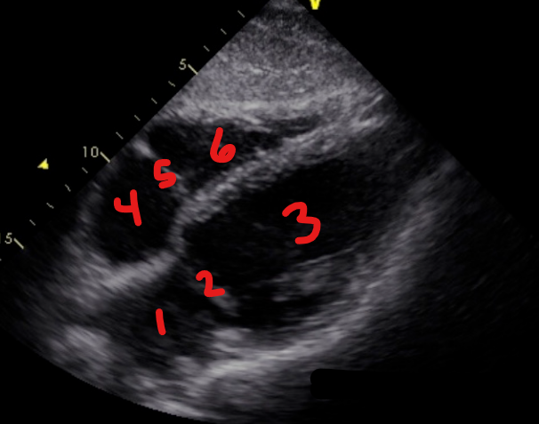
Identify this image.
PSAX LV
Papillary muscle
LV
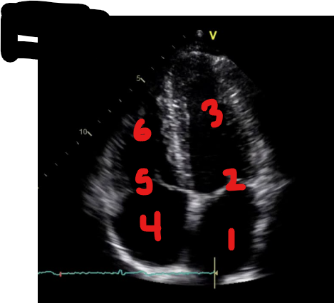
Identify this image.
A4C
LA
MV
LV
RA
TV
RV
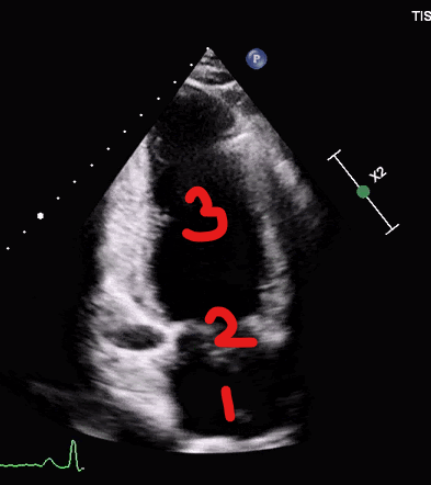
Identify this image.
A2C
LA
MV
LV
Identify this image.
A5C
LA
LV
RA
RV
Ao
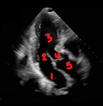
Identify this image.
A3C
LA
MV
LV
AV
Ao
Identify this image.
Suprasternal view of descending aorta
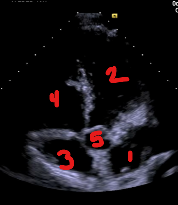
Identify this image.
IVC Subcostal
SVC
RA
IVC
RV
Identify this image.
Subcostal four chamber
LA
MV
LV
RA
TV
RV
Where is the heart located?
Posterior to sternum within middle mediastinum
What is the most anterior chamber of the heart?
RV
What is the order of the layers of the heart?
Endocardium: Innermost layer
Myocardium: Middle layer
Epicardium: Outermost layer
What is the pericardium?
Membrane that lines pericardial cavity and encases heart
What is the crux of the heart?
Posterior portion of heart where all 4 chambers meet
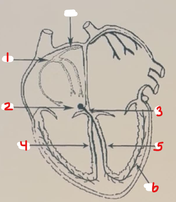
What is the order of blood circulation through the heart?
SVC/IVC
Right atrium
Tricuspid valve
Right ventricle
Pulmonary valve
Pulmonary artery
Lungs
Pulmonary veins
Left atrium
Mitral valve
Left ventricle
Aortic valve
Aorta
Body
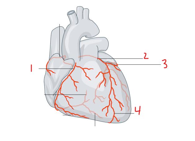
Identify this image.
RCA
LCA
Left circumflex artery
Left anterior descending artery
What is the normal firing rate of the conduction system of the heart?
SA node = 60-100 bpm
AV node = 40-60 bpm
Identify this image.
SA node
AV node
Bundle of HIS
Right bundle branches
Left bundle branches
Purkinje fibers
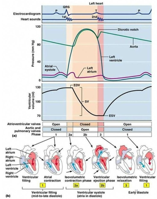
Identify this image.
Isovolumetric contraction
Ventricular systole
Isovolumetric relaxation
Diastole
Atrial systole
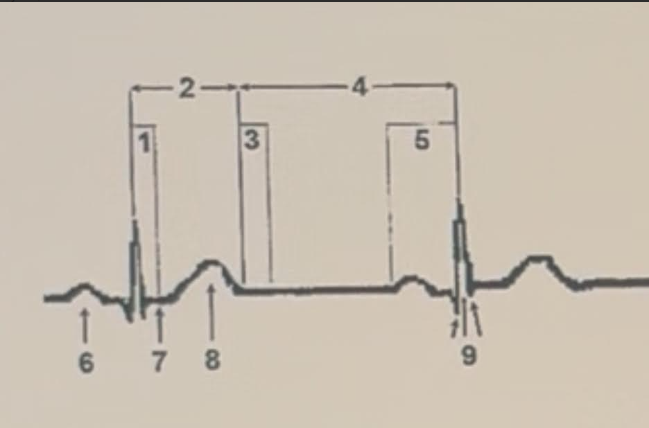
P wave
ST segment
T wave
QRS complex
What are the phases of the action potential curve (APC)?
Phase 0: Depolarization or sodium influx
Phase 1: Potassium influx
Phase 2: Calcium influx
Phase 3: Repolarization (recovery) or potassium outflow
Phase 4: Refractory or potassium influx and sodium outflow
How are the EKG and action potential waveforms related?
P wave: Atrial depolarization
QRS complex: Ventricular depolarization
T wave: Ventricular repolarization
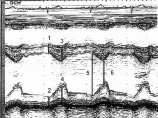
Identify this image.
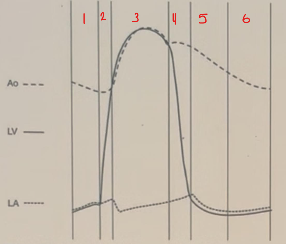
Phases of cardiac cycle
Ventricular filling and atrial contraction
Isovolumetric contraction
Ejection
Isovolumetric relaxation
Rapid ventricular filling
Diastasis
Identify this image.
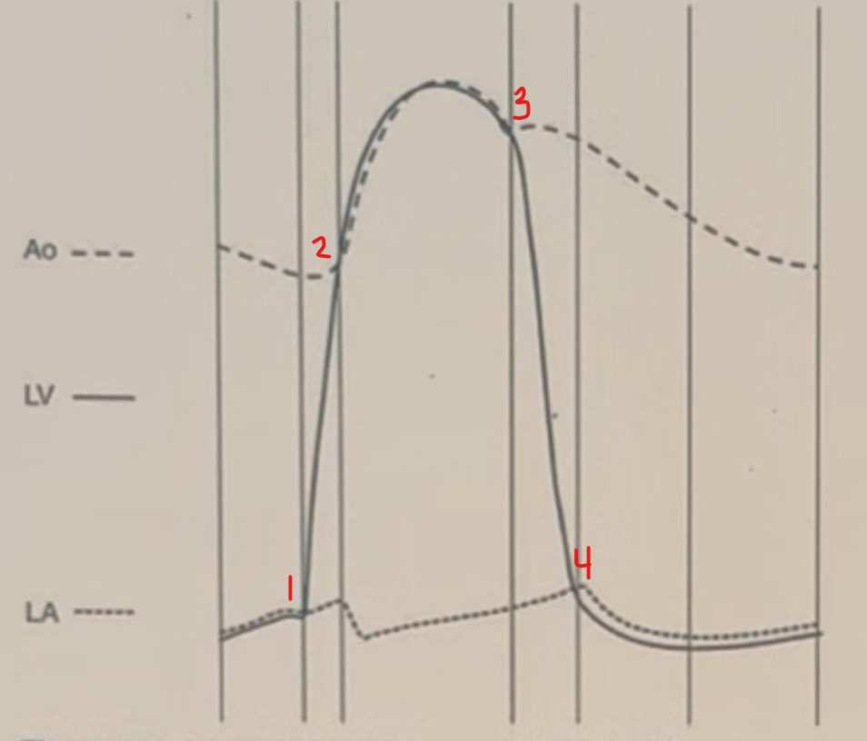
Valvular events
MV closure
AV opening
AV closure
MV opening
What is the role of each segment of the heart during diastole?
Atria: Relaxation
AV valves: Open to allow blood to fill ventricles
Ventricles: Relaxation
Semilunar valves: Closed to prevent backflow of blood
What is the role of each segment of the heart during systole?
Atria: Contraction
AV valves: Closed to prevent backflow
Ventricles: Contraction
Semilunar valves: Open to allow flow to great vessels
What transducer should be used for an adult echocardiogram?
2.5 - 5 MHz
What are the patient positions used for echocardiograms?
Left lateral semidecubitus
Supine
Right lateral decubitus (Pedoff)
Where is the suprasternal window located?
Suprasternal notch
Where is the subcostal window located?
Midline and beneath costal margin
Where is the apical window located?
Over cardiac apex
Where is the parasternal window located?
Area bounded superiorly by left clavicle, medially by sternum, and inferiorly by apical region
Where is 12 o’clock located in echo?
Patient’s head
What are the long-axis views (PLAX)?
LV in sagittal plane
RV inflow (TV) achieved by inferior angulation
RV outflow (PV) achieved by superior angulation
Where is notch pointed in PLAX?
Patient’s right shoulder or 10 o’clock
Where is the notch pointed in PSAX?
Patient’s left shoulder or 2 o’clock
What are the short-axis views (PSAX)?
PSAX LV apex achieved by inferior angulation
PSAX MV achieved by superior angulation
PSAX AV basal achieved by superior angulation
Where is the notch pointed for apical views?
Toward bed or 3 o’clock
What are the apical views?
A4C
A5C achieved by superior angulation
A2C achieved by rotating probe counter-clockwise
Apical long or A3C achieved by rotating probe counter-clockwise
Where is the notch pointed for subcostal views?
Towards bed or 3 o’clock
What are the subcostal views?
Subcostal 4C
Subcostal IVC achieved by rotating probe counter-clockwise
Identify this image.
A4C
Identify this image.
A2C
Identify this image.
A3C or LONG AXIS
Identify this image.
Base or top
Identify this image.
Mid
Identify this image.
Apex or bottom
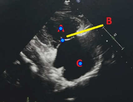
Identify this image.
PLAX
RV
LA
MV
LVOT
AV
Aortic root
Descending AO
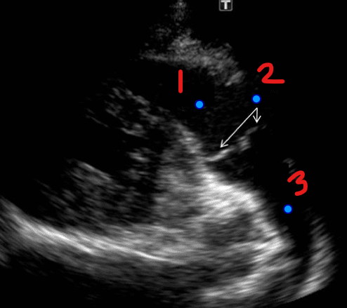
Identify this image.
PLAX RVIT
A. RA
B. TV
C. RV
Identify this image.
PLAX RVOT
RV
PV
Main pulmonary artery
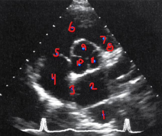
Identify this image.
PSAX AV Basal
Descending aorta
LA
IAS
RA
TV
RVOT
PV
Main pulmonary artery
Right coronary cusp
Noncoronary cusp
Left coronary cusp
Identify this image.
PSAX MV level
Identify this image.
PSAX LV
Papillary muscle
LV
Identify this image.
A4C
LA
MV
LV
RA
TV
RV
Identify this image.
A2C
LA
MV
LV
Identify this image.
A5C
LA
LV
RA
RV
Ao
Identify this image.
A3C
LA
MV
LV
AV
Ao
Identify this image.
Suprasternal view of descending aorta
Identify this image.
IVC Subcostal
SVC
RA
IVC
RV
Identify this image.
Subcostal 4C
LA
MV
LV
RA
TV
RV
Which blood flow abnormalities can cause a murmur?
Left heart disease such as AS, AR, MS, and MR
Intracardiac shunt such as an ASD, VSD, or PDA
Right heart disease such as PS, PR, TS, TR
Normal echo seen as a flow murmur with regurgitation
When performing an echo, what are questions that should be asked about the LV?
Is the EF normal?
Is the LV normal in size or dilated?
Are there any wall motion abnormalities?
When performing an echo, what are questions that should be asked about the valves?
Are the valves thick or stenotic?
Do the valves have normal mobility?
When performing an echo, what are questions that should be asked about the right side of the heart?
Is the size and function normal?
Are the pressures normal?
When performing an echo, what are questions that should be asked about other anatomy?
Is there pericardial effusion present?
Is the IVC dilated?
Are there any masses?
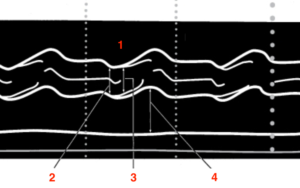
What is m-mode?
Function that allows for identification of thin moving structures such as endocardium
What are the uses for m-mode?
Timing of rapid cardiac motion
Precise measurements of cardiac dimensions
Further evaluation of structures seen on 2D imaging
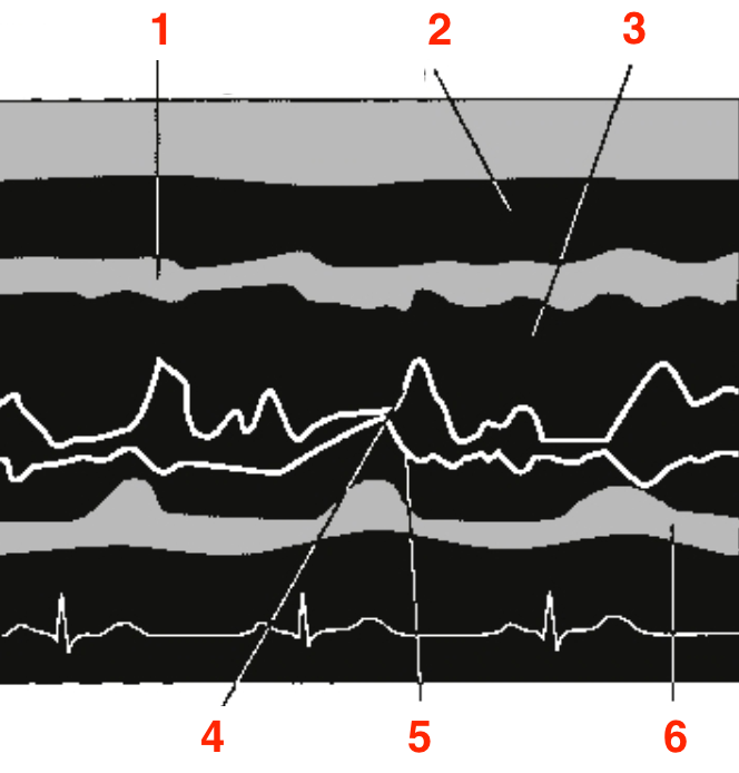
Identify this image.
PLAX image showing LA and AV
RV
Aortic root
AV leaflets opening
LA
Identify this image.
PLAX or PSAX image showing MV
IVS
RV
LVOT
Anterior MV leaflet
Posterior MV leaflet
Posterior wall
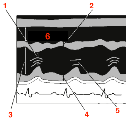
Identify this image.
PLAX or PSAX image showing LV
LV end systolic dimension
IVS
LV end diastolic dimension
LV posterior wall
Chordae
RV
Identify this image.
IVSDd
PWDd
IVSDs
PWDs
LVIDd
LVIDs
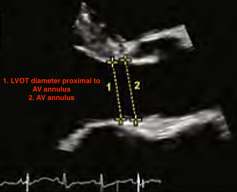
How do you measure the LVOT?
Measure in PLAX when AV is open so LVOT is at largest diameter and place calipers proximal to AV annulus from inner to inner
How do you measure LV volume and LVEF?
Measure in A4C and A2C using modified Simpson’s at end-systole and end-diastole while tracing blood pool interface
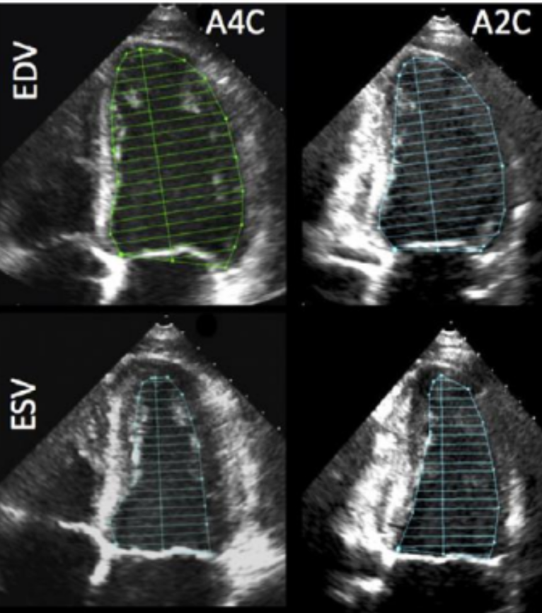
What structures should be excluded from LV volume and LVEF measurement?
Papillary muscles
What is the formula for LVEF?
LVEF = LVEDV - LVESV / LVEDV
What is the difference between LV enlargement and LV hypertrophy?
LV enlargement: Widening of LV chamber OR combination of increased wall thickness and chamber size
LV hypertrophy: Increase in thickness of LV muscular walls
What is diastolic function?
How well ventricles relax
What is the criteria needed to assess LV diastolic function in patients with normal LVEF?
Average inflow velocities (E’)
Septal and lateral mitral annulus early diastolic velocity (e’)
Peak TR velocity
LA volume index
What PW images should be taken of AV?
A5C or A3C with PW sample volume placed 5 mm proximal to AV in center of LVOT
Trace to measure peak velocity and VTI
What CW images should be taken of AV?
A5C or A3C with CW sample volume placed through AV
Trace to measure peak velocity, peak gradient, mean gradient, and VTI
What is the continuity equation for aortic valve area (AVA)?
NEED TO FIND LVOT CSA PRIOR BY USING CSA LVOT EQUATION***
AVA = (CSA LVOT x VTI LVOT) / VTI AV
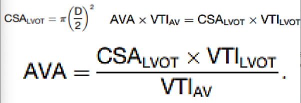
What measurements impact aortic valve area (AVA) calculation?
Inaccurate LVOT diameter (squared value)
Inaccurate transvalvular sampling or not obtaining highest velocity
What is aortic stenosis (AS)?
Narrowing of AV due to congenital, degenerative or calcific, and rheumatic conditions
What criteria could be used to determine severity of AS other than aortic valve area (AVA)?
Peak velocity
Mean gradient
Velocity ratio
Indexed AVA
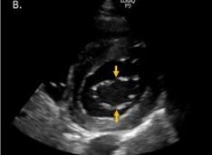
Identify this image.
Bicuspid AV seen as football shape or two leaflets
What are the associated anomalies of a bicuspid AV?
Aortic dilation
Aortic aneurysm
Aortic dissection
Identify this image.
Unicuspid AV seen as one solitary opening or one cusp
What are the associated anomalies of an unicuspid AV?
Stenosis
Identify this image.
Quadricuspid AV seen as X shape in PSAX or four leaflets
What are the associated anomalies of a quadricuspid AV?
Regurgitation
What should be measured with PW when evaluating the MV for diastolic function?
Peak E velocity
Peak A velocity
Early diastolic DT