Comp Vert FINAL EXAM HURRAY!!!
1/82
Earn XP
Description and Tags
good luck everyone
Name | Mastery | Learn | Test | Matching | Spaced | Call with Kai |
|---|
No analytics yet
Send a link to your students to track their progress
83 Terms
What are the two ways that blastopore cells may develop?
Some ingress to form the primary mesenchyme, and some invaginate to form the archenteron
What forms the primary mesenchyme?
In embryo, some cells from the blastopore may ingress to form the primary mesenchyme
What forms the archenteron?
In embryo, some cells from the blastopore may ingress to form the archenteron
What forms the lower part of the anal cavity? (primordial anus)
The proctodeum
What is the proctodeum?
The primordial anus
What doe the archenteron eventually become?
The cavity that eventually forms the digestive tract
What eventually becomes the cavity that becomes the digestive tract?
The archenteron
What becomes the primordial mouth?
The stomodeum
What is the stomodeum?
The primordial mouth
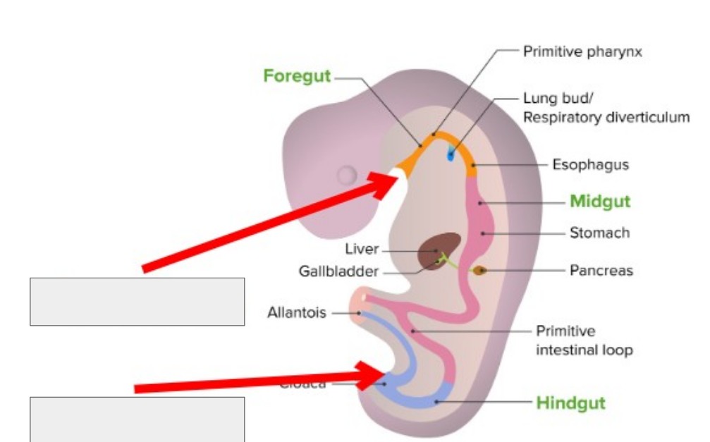
What two embryological features are being pointed to in this image?
The stomodeum and blastopore
Which germ layer forms the internal lining of the GI tract and accessory organs of the digestive system?
The endoderm
What role does the endoderm play in the development of the digestive system?
It forms the internal lining of the GI tract and accessory organs
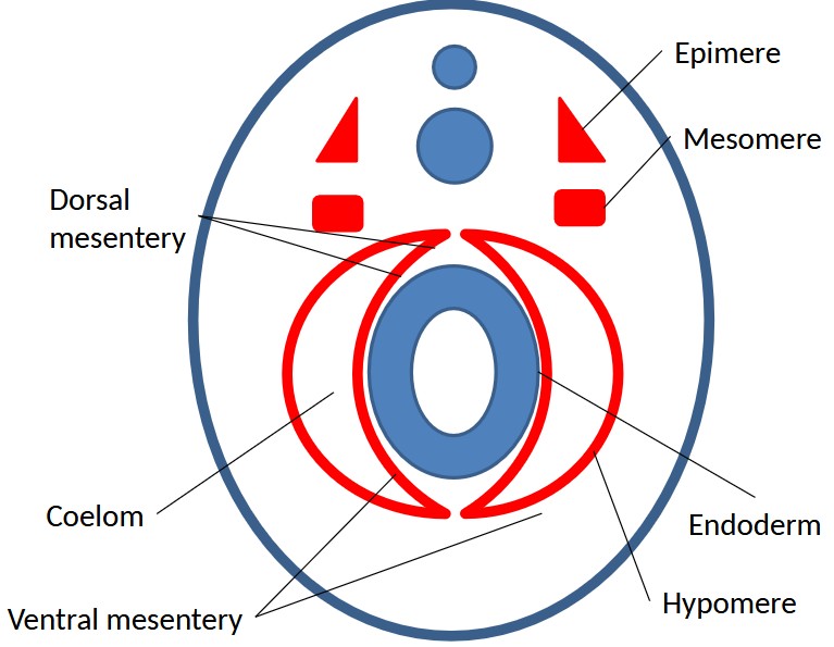
What is the distinction between the splanchnic and parietal hypomere?
The splanchnic hypomere is associated with the internal organs and the parietal hypomere is associated with the body wall.
Which germ layer develops the walls of the GI tract, visceral, and parietal membranes?
The mesoderm
What role does the mesoderm play in the development of the digestive system?
For the splanchnic hypomere, it forms the walls of the GI tract and the visceral membrane.
For the parietal hypomere, it forms the parietal membrane.
Label this diagram and, if applicable, describe each part.
Dorsal mesentery- attaches to the curve of the stomach, keeps the digestive organs separate from digestive wall
Epimere - dorsal mesoderm, epaxial muscles
Mesomere - intermediate mesoderm, kidney tubules and excretory glands
Hypomere - lateral mesoderm, heart, lymphatic system
What are the accessory organs in the digestive system?
liver: forms from a diverticulum from the foregut
gall bladder: develops froms a diverticulum which forms a bile duct
pancreas: Forms from a diverticulum from the duodenum and migrates
What are the two gland types inside the pancreas?
Exocrine glands- acinar cells secrete digestive enzymes, ions, and water into the pancreatic duct which goes to the duodenum
Endocrine glands- cells called Islets of Langerhan secrete hormones into blood vessels (primarily insulin and glucagon)
What do the exocrine glands in the pancreas do?
Acinar cells secrete digestive enzymes, ions, and water into the pancreatic duct which goes to the duodenum
What do the endocrine glands in the pancreas do?
Islet of Langerhan cells secrete hormones into blood vessels (primarily insulin and glucagon)
What is the difference between insulin and glucagon?
insulin lowers your blood sugar and glucagon raises it
Which gland in the pancreas doesn’t have a duct and secretes hormones like insulin and glucagon?
Endocrine glands
Which gland in the pancreas secretes ions, digestive enzymes, and water into the pancreatic duct?
Exocrine gland
What is the purpose of the mesentery in the development of the digestive system?
It anchors the intestines to the abdominal wall to prevent them from twisting or collapsing.
It carries blood vessels, nerves, and lymphatics to the intestines.
What are the major cavities that we need to know?
The pleural cavity, pericardial cavity, and peritoneal cavity
What is the pleural cavity?
embryological origin
location
purpose
layers
Embryological origin
Lateral plate mesoderm
Location
Surrounds each lung, located between the lungs and the chest wall
Purpose
It keeps the lungs against the chest wall, allowing for more efficient expansion during inhalation
Layers
Visceral pleura: covers the lungs
Parietal pleura: lines inner chest wall and diaphragm
Which cavity exists between the lungs and the chest wall?
Pleural cavity
What are the two layers of the pleural cavity and what do they do?
The visceral pleura lines the lungs and the parietal pleura lines the chest wall and diaphragm. They both create negative pressure to allow for more efficient expansion during inhalation.
What is the Pericardial cavity?
embryological origin
location
purpose
layers
Embryological origin
Lateral plate mesoderm
Location
The space between the two layers of the serous pericardium surrounding the heart.
Purpose
Reduces friction from heartbeats and protects the heart from over exertion
Layers
Visceral pericardia is directly adhered to the heart muscle
Parietal pericardia lines the inner surface of the fibrous pericardium
What is the Peritoneal cavity?
embryological origin
location
purpose
layers
Embryological origin
Lateral plate
Location
Lines the abdominopelvic region
Purpose
Allows for friction-free movement of abdominal organs
contains antibodies and leukocytes to prevent infection
Layers
Visceral peritoneum covers the organs
Parietal peritoneum lines the abdominal wall
Which cavity surrounds the heart?
Pericardial cavity
What are the two layers of the pericardial cavity?
Visceral pericardia is adhered to the actual heart muscle and parietal pericardia lines the fibrous outer layer
Which cavity lines the abdominopelvic region and what are the layers
The peritoneal cavity
Visceral peritoneum lines the organs, parietal peritoneum lines the abdominal wall
What are the four “Tunics” of the digestive tract?
Mucosa, submucosa, mucularis externa, and serosa
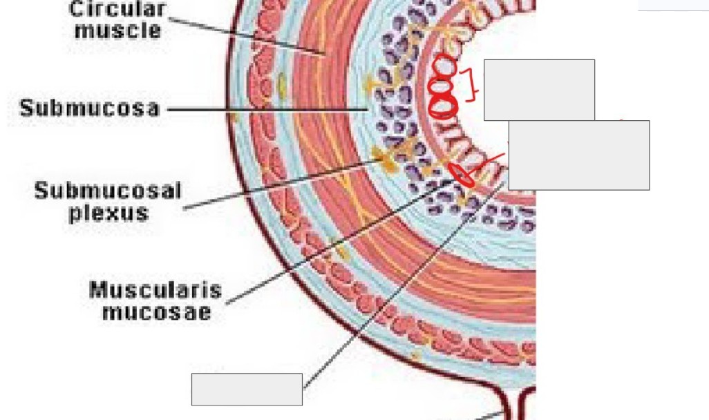
Label
What is the mucosa tunic of the digestive tract? What is its purpose?
It is the innermost layer, extending from mouth to anus.
It is made of epithelial tissue.
It is responsible for absorption of nutrients and secretion of enzymes, mucus, and hormones.
What is the submucosa tunic of the digestive tract? What is its purpose?
It is between the mucosa and mucularis externa, and is made of connective tissue, blood vessels, and nerves.
The layer of nerves is responsible for regulation and secretion of glands. The nerves send signals to the glands based on what is/isn’t being absorbed.
The connective tissue provides support for the tract and allows for flexibility.
The blood vessels support the mucosa and transport absorbed nutrients.
What is the mucularis externa tunic of the digestive tract? What is its purpose?
It is comprised of smooth muscle, circular muscle, and longitudinal muscle.
It is responsible for the contractions of the GI tract, moving the food throughout it.
What is the serosa tunic of the digestive tract? What is its purpose?
It is made out of loose and dense connective tissue.
It secretes lubricant around your organs so that there is less friction when the organs move around.
Which tunic of the digestive tract secretes lubricant to prevent friction?
The serosa
Which tunic of the digestive tract is responsible for moving the food throughout?
Mucularis externa
Which tunic of the digestive tract provides structure for the mucosa, transports absorbed nutrients, and tells glands when to secrete?
Submucosa
Which tunic of the digestive tract absorbs nutrients and secretion of mucus, enzymes, and hormones?
Mucosa
What kind of epithelial tissue does the mouth/pharynx have to have and why?
Stratified squamous because of the amount of food that passes through it.
What kind of epithelial tissue does the stomach have to have and why?
Simple columnar because it needs to protect the rest of your organs from stomach acid, bacteria, etc.
There is a lot of mucus secreted because of the acid.
What kind of epithelial tissue do the intestines have to have and why?
Simple columnar with microvilli, because it aids in the moving of food.
What are the bones that make up the oral cavity?
Dermal bones make up the lower portion of the oral cavity and the chondrocranium makes up the upper portion
Describe what goes into tooth development
Dermal papillae
This is your gum line
Fleshy, highly vascularized connective tissue
Ameloblast
comes from the ectoderm and creates tooth enamel
enamel does not regrow
Odontoblast
creates dentin
comes from mesoderm
dentin is the bottom part of your tooth
What are the two kinds of tooth shape?
Homodont and heterodont
What are the terms for whether or not teeth get replaced when lost?
Polyphyodonty- teeth get replaced when lost (like sharks)
Diphyodonty- teeth do not get replaced when lost
What are the different kinds of tooth attachment? Example species
Acrodont- not deep in the gum line, less bleeding when teeth break or are lost. Sharks, fish
Pleurodont- partially in the gum line, amphibians and lizards
Thecodont - goes deeply into the jaw, lots of vascularized tissue involved. archosaur lineage, mammals
Vomerine - actually fuse with the jaw fish, amphibians generally covered by a layer of mucus
Which type of tooth attachment has the tooth fused to the jaw?
Vomerine
Which kind of tooth attachment is deeply rooted in the gum tissue?
Thecodont
Which kind of tooth attachment is partially entrenched in the gum tissue
Pleurodont
Which kind of tooth attachment is on the surface of the gum tissue
Acrodont
What are teeth that are replaceable called?
Polyphyodonty
What are teeth that do not get replaced when lost called?
Diphyodonty
What are the three main glands in the oral cavity?
Mucus, salivary, and venom
What are mucus glands in the oral cavity?
They are in the epithelial layer of the buccal cavity. They lubricate food and are also used for filter feeding
They help maintain a healthy oral cavity by neutralizing toxins
What are salivary glands?
They are only present in tetrapods because we chew
They moisten food and produce amylase which breaks down complex carbohydrates
What are venom glands?
They can produce hemotoxins or neurotoxins
Neurotoxins destroy nerves and hemotoxins destroy organs, red blood cells, etc
They immobilize prey and are also used defensively
Venom also has digestive enzymes to help start the digestive process early
Describe the tongue of fish
derived from the floor of the mouth/pharynx
“soft’ tongue
may develop keratinized teeth
Describe the tongue of tetrapods
muscular tongue
derived from hypobranchial musculature
rests on the hyoid apparatus
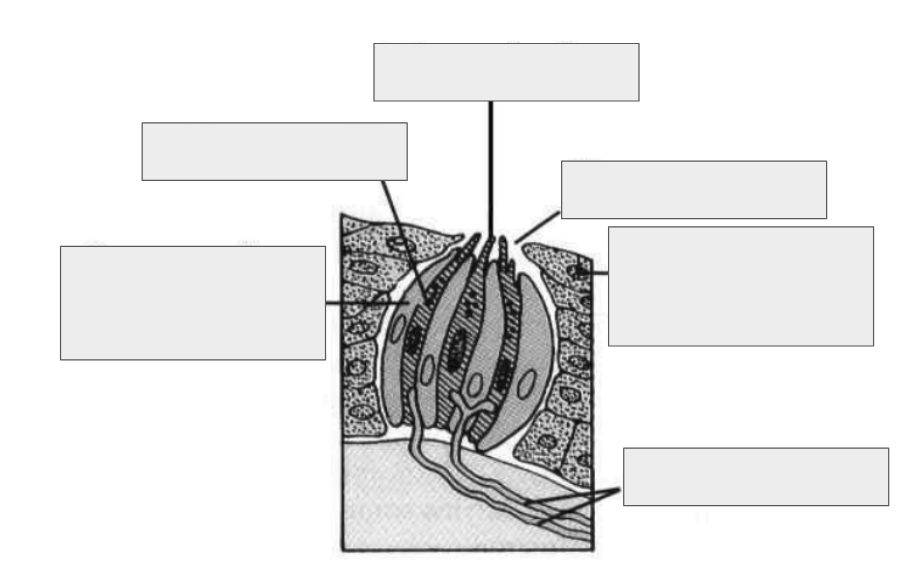

Label
nerve fibers
supporting cell
oral epithelium
taste pore
taste cell
taste hair
What are some special tongues?
Frog
lingual feeding - uses tongue to catch prey
Snake
Jacobson’s organ - tongue is used to pick up chemicals and jacobson’s organ identifies the smell
Woodpecker
Hyoid apparatus allows woodpecker to protrude its tongue
What is the pharynx?
extension of the mouth
embryological source of many organs
source of gill slits
What is the esophagus?
reduced in anamniotes
muscular tube that connects the mouth to the stomach
in crocodilians and birds, there is an extension of the esophagus called the crop that keeps an excessive amount of food from entering the esophagus
What is the stomach?
originally functioned as a storage organ which would gradually release food into the intestine
now has a second phase of digestion that involves chemical and mechanical digestion
develops from the foregut (endoderm)
What is found along the lining of the stomach?
Gastric pits above gastric glands
What is a gastric gland? What cells are within them and what do they do?
simple columnar epithelium
Mucous cells
secrete alkaline mucin which forms a protective layer against the gastric acid of the stomach
neutralizes acidity
contains tight junctions to prevent acid from going into muscle tissue
Mucous neck cells
beneath mucous cells
secrete acidic mucin
believed to act as a protective barrier from the alkaline mucin
D cells
main stomach enteroendocrine cells
secrete somatostatin which prevents gastric acid secretion
main function is to inhibit HCl secretion
G cells
secrete gastrin which stimulates gastric acid secretion
works to stimulate HCl secretion
Parietal cells
secrete HCl and intrinsic factor
HCl (gastric acid) acts to cleave pepsinogen
Intrinsic factor stimulates absorption of B12
Chief Cells
secrete pepsinogen
cleaved by HCl to create pepsin
pepsin denatures proteins
What is the purpose of mucous cells in the gastric pit?
form outermost layer
secretes alkaline mucin, which creates a protective layer against gastric acid
neutralizes acidity
has tight junctions to prevent acid or food particles from reaching muscle tissue
What is alkaline mucin?
A protective mucus secreted by mucous cells that protect the gastric pit and stomach lining from gastric acid
What is HCl?
Gastric acid
What are Mucous neck cells and what do they do?
They are beneath the mucous cells in the gastric pit. They produce acidic mucin. Function unknown but assumed to produce protective layer from alkaline mucin
What are D cells? What do they do?
main stomach enteroendocrine cells
inhibits secretion of HCl (gastric acid) by producing somatostatin
What are G cells? What do they do?
G cells act in opposition of D cells.
They produce gastrin which stimulates the production of HCl.
What are parietal cells? What do they do?
Parietal cells are in the gastric pit and they are the cells that the D and G cells act upon. They secrete gastric acid and intrinsic factor.
Gastric acid cleaves pepsinogen to make pepsin which denatures proteins.
Intrinsic factor stimulates the absorption of B12.
What are chief cells? What do they do?
Secrete pepsinogen, which is cleaved by gastric acid to become pepsin, which denatures proteins.
How do proteins get denatured?
When a protein loses its shape, it loses its function. Different denaturing agents can corrupt the shape of a protein. In the stomach, the denaturing agents are pepsin and gastric acid.
What are the four major parts of the stomach?
Cardiac region
fundus
body
Pyloric region
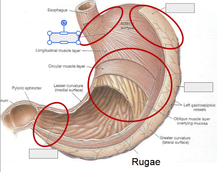
Label the regions of the stomach
What is the cardiac region of the stomach?
portion surrounding the opening from the esophagus into the stomach
What is the fundus of the stomach?
farthest from the pyloric sphincter
enlarged part above the cardiac orifice and is inferior to the diaphragm