Histology of the GI tract
1/35
There's no tags or description
Looks like no tags are added yet.
Name | Mastery | Learn | Test | Matching | Spaced | Call with Kai |
|---|
No analytics yet
Send a link to your students to track their progress
36 Terms
What is histology?
The study of the microanantomy of tissues & organs
Stains and dyes are used too make structures visible
Used in understanding structure and function of tissues
Used in diagnosis and staging of disease
What is epithelium and its function?
A type of tissue primarily involved in 3 functions:
Lining and protection of body surfaces and cavity
Secretion
Absorption
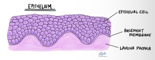
Basic structure of epithelium
Epithelial cell layer
Epithelial cells are anchored to the basement membrane
Basement membrane is supported by the laminate propria
How can epithelium differ?
Epithelial cell layer can vary in:
The number of epithelial cells layers
The shape of the epithelial cells
Some epithelial cell layers have surface specialisations
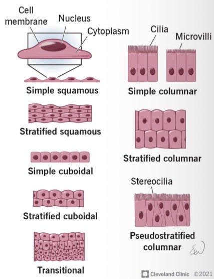
Epithelium types - simple squamous
Simple squamous epithelium
Simple = one cell layer
Squamous = flattened cell shape
This thin epithelial layer can be found in the lung alveoli → permits gas exchange
Epithelium types - simple cuboidal
Simple cuboidal epithelium
Cuboidal = cube shaped cell
Permits secretion and absorption e.g. kidney
Epithelium types - simple columnar
Simple columnar epithelium
Columnar = column shaped
Permits secretion and absorption e.g. small & large intestine
Epithelium types - stratified squamous
Stratified squamous epithelium
Stratified = multiple layers
Squamous = flattened cell shape
Primarily responsible for protection from abrasion
How does the epithelium protect?
Protects from friction e.g. epidermis of skin
Some epithelium are specialised to protect against stretch e.g. epithelium of utters and bladder
The mucous secreted by gastrointestinal and respiratory epithelium protects from toxic and pathogenic material
Immune cells found in lamina propria
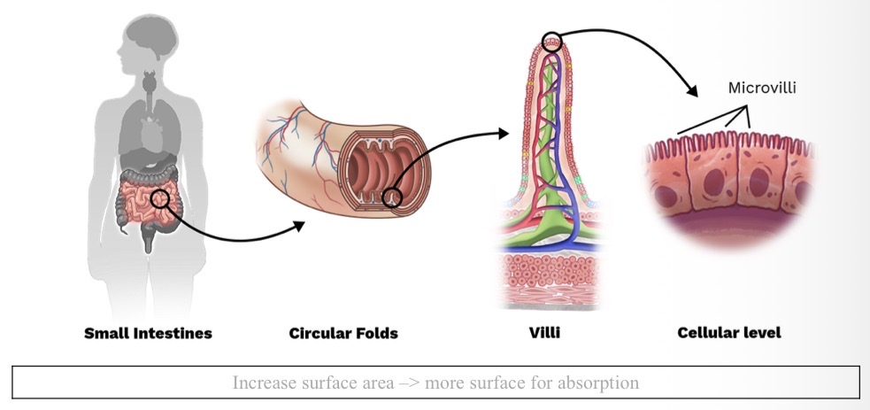
How does epithelium facilitate absorption?
Selective absorption = allowing absorption selectively of only certain molecules
Molecule absorption regulated based on molecule size, charge and molecular structure
Tight junctions - leak-proof gaps between cells → provide cell polarity
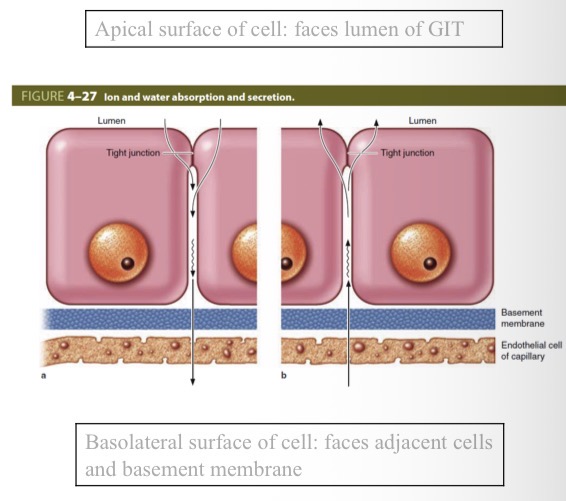
Villi and microvilli - surface specialisations
How do epithelia secrete?
They have glands which secrete enzymes into the lumen of the GI tract
Glands are specialised for:
Synthesis
Storage
Release
Examples:
Pancreatic enzymes
Sebaceous glands (lipids)
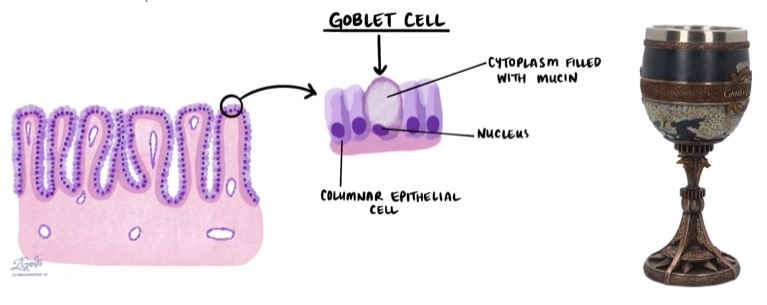
Goblet cells
Mucous secreting cells found in the epithelium
Why does the epithelium have no blood vessels?
Blood vessels located in lamina propria - gas, nutrients & waste diffuse between epithelial cell layer and blood vessels → protects blood vessels from abrasion
Epithelia summary
Tightly packed cell layer(s) anchored by intercellular junctions with a supporting basement membrane
Epithelia provide a protective barrier between the external and internal environments
By doing so they are effective at maintaining cell polarity and selective absorption and secretion
The epithelia can be classified by: number of layers, cell shape, surface specialisations
Glands are derived from epithelia and can be endocrine or exocrine
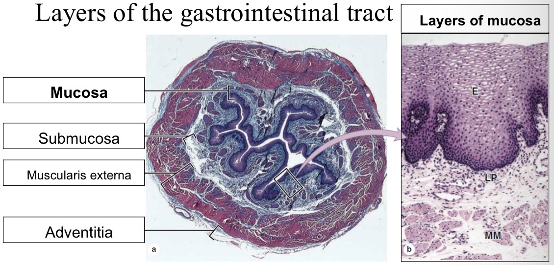
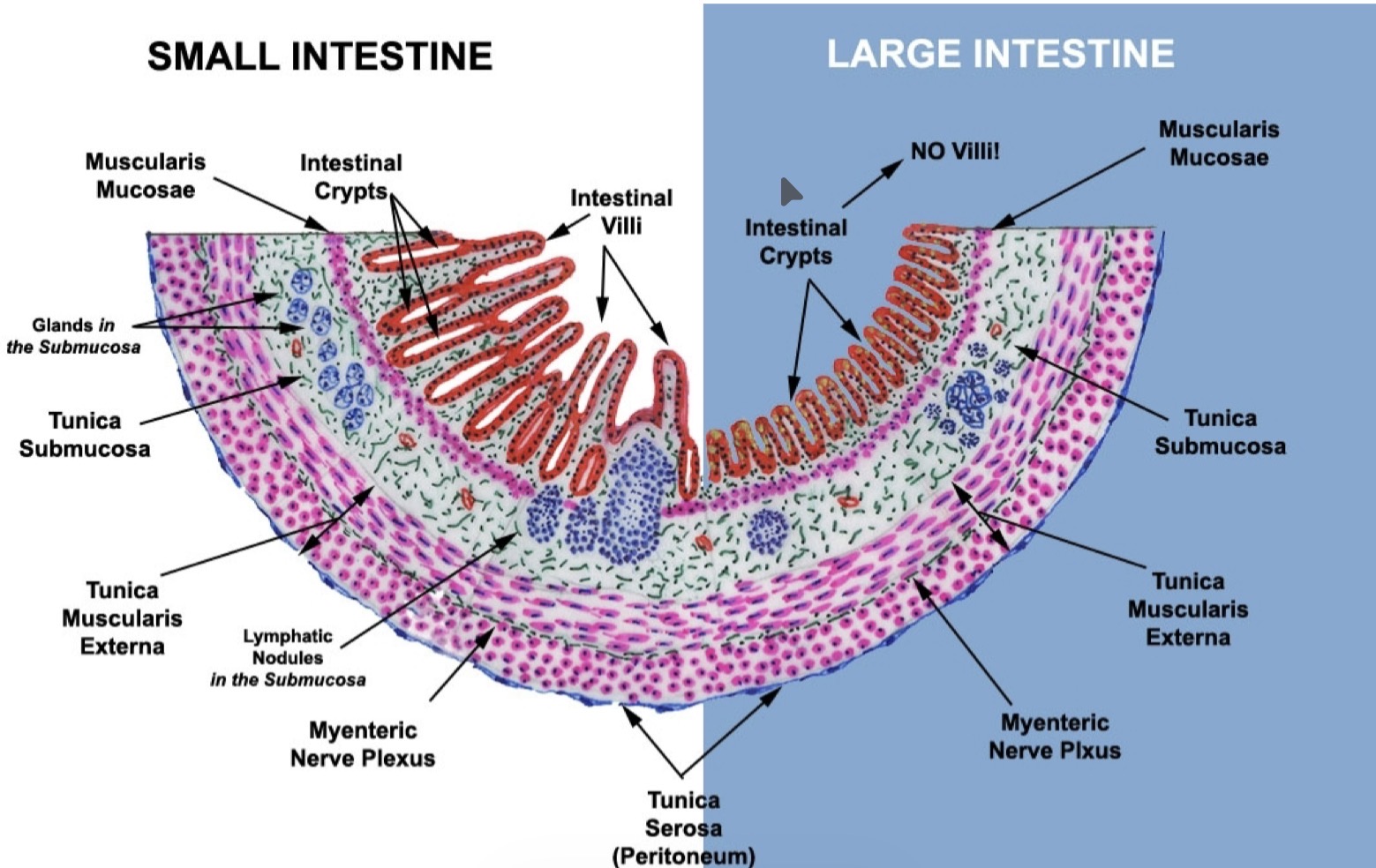
Layers of the GI tract
From innermost to outermost:
Lumen
Mucosa = epithelium + lamina propria + muscularis mucosae
Submucosa = connective tissue and blood vessels (arterioles/venules)
Muscularis externa = circular and longitudinal smooth muscle
Adventitia/serosa = outer connective tissue/visceral peritoneum + major nerves/blood vessels
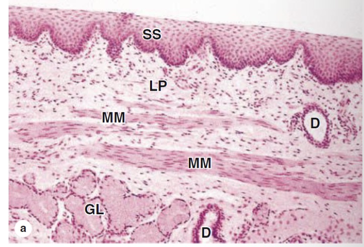
Epithelium of oesophagus
Stratified squamous epithelium
Protects the oesophagus from abrasion
SS = stratified squamous
LP = lamina propria
MM = muscularis mucosae
GL = glands
D = duct
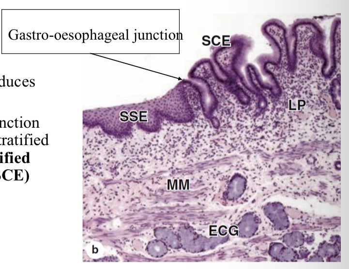
Stomach epithelium
Stomach epithelium → produces acids and enzymes
At gastrointestinal-oesophageal junction there is a transition from stratified squamous (SSE) to stratified columnar epithelium (SCE)
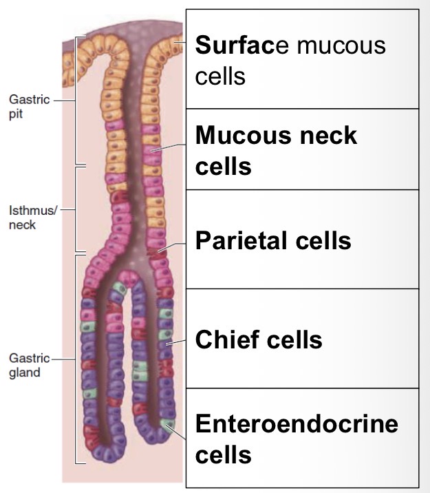
Stomach - gastric pits & gastric glands diagram
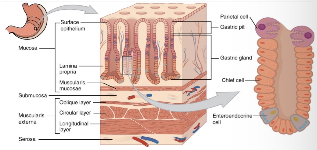
Gastric pits & glands - what are they and what type of cells do they contain?
Gastric pits: opening to the gastric glands, contain mucous cells
Gastric glands: secrete stomach acids, enzymes and local signals
Simple columnar epithelium
Chief cells (pepsinogen and gastric lipase)
Parietal cells (HCl and GIF) HCl activates pepsinogen to pepsin
Mucous cells (rich in HCO3-)
Enters endocrine cells (secret gastrin to stimulate acid secretion)
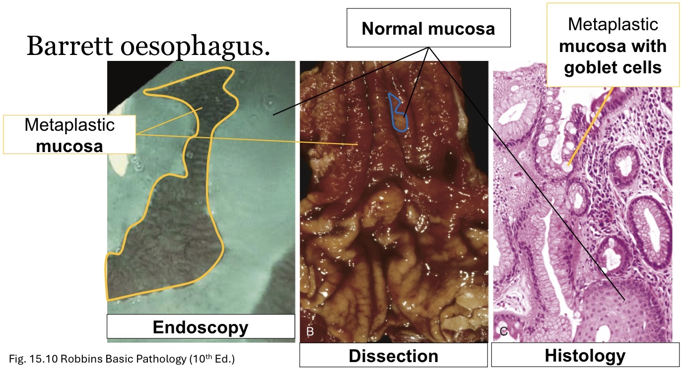
Barrett oesophagus
Heartburn
Gastro-oesophageal reflux disease (GORD)
GORD can lead to metaplastic changes i.e. oesophageal type epithelium becomes stomach type epithelium
Barrett oesophagus is diagnosed histologically
These changes can become cancerous
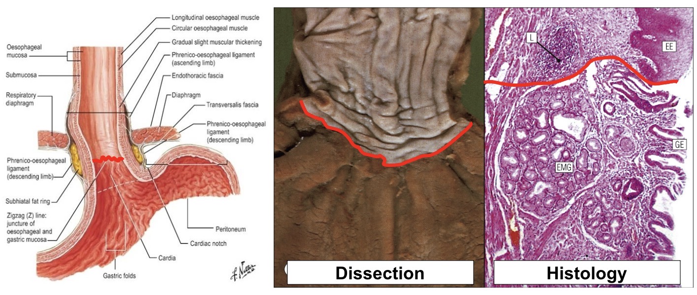
Normal gastro-oesophageal junction image
Barrett oesophagus diagram
Summary of oesophagus and stomach
Oesophagus = stratified squamous epithelium
Stomach = simple columnar epithelium
Gastric pits are invaginations where gastric glands secrete HCl- for example:
Parietal cells = HCl-
Chief cells = enzymes
Enteroendocrine (APUD) cells = local hormone control (gastrin)
Barrett oesophagus
Small intestine structure
Villi (V)
Lamina propria (P)
Submucosa (SM)
Muscularis externa (M)
Serosa (S)
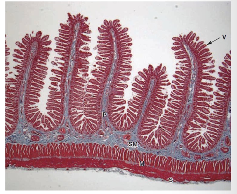
Small intestine
Below the simple columnar epithelial cell layer, the lamina propria occupies the core of each villus
Lamina propria contains blood capillaries and lacteal
Lacteal: absorbs dietary fats and transmits them through the lymphatic system
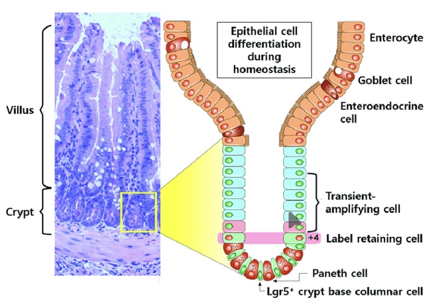
Duodenum
Characteristic features:
Brunner’s glands: collection of mucous secreting glands - help neutralise chyme from stomach
Crypts:
Contain enzyme secreting cells (digestion)
Contain stem cells - replenish the cells of the epithelium
What do crypt stem cells do?
Stem cells of the crypts replenish cells of the villi
Jejunum
Contains no Brunner’s glands
Contain villi and crypts
Highly folded (plicae circulares)
Ileum
Contains the shortest villi of the small intestine
Contains Peyer’s patches - aggregations of lymphoid tissue
Large intestine
Lacks villi but has glands and crypts
Crypts contain mucous secreting cells and stem cells
Simple columnar epithelial cells
Liver histology overview
Cells are organised into hexagonal shaped lobules
Central vein located in the centre of lobule
Portal triad located at edges:
Portal vein branches
Hepatic artery branches
Bile duct branches
Liver histology - blood flow, sinusoids and bile
Blood from the portal vein branches and hepatic artery branches flow through sinusoids toward the central vein
Sinusoids have a fenestrated endothelium which permits exchange between the blood and the adjacent hepatocytes
Bile flows to bile duct through bile canaliculi
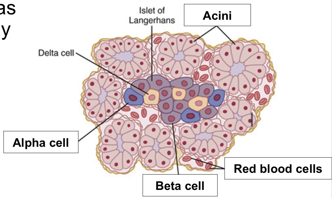
Pancreas histology
Has clusters of exocrine and endocrine cells
Exocrine:
Pancreatic acini
Secrete contents into duct system → duodenum
Endocrine:
Islets of Langerhans
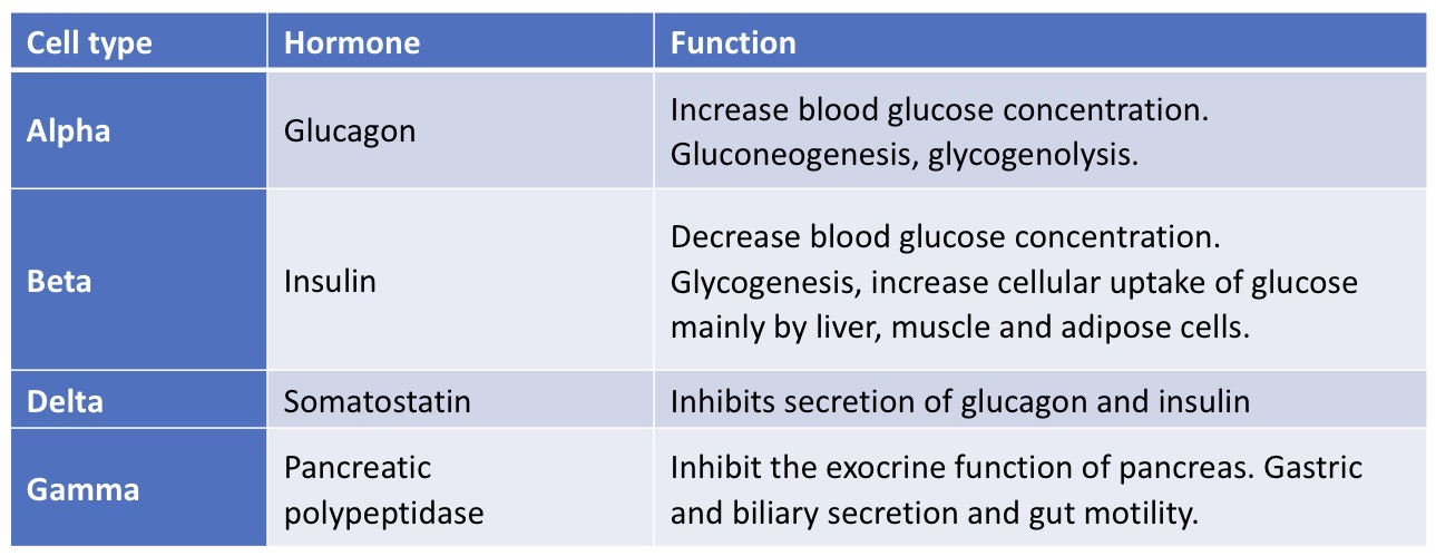
Pancreas histology table - cell type, hormones secreted & function
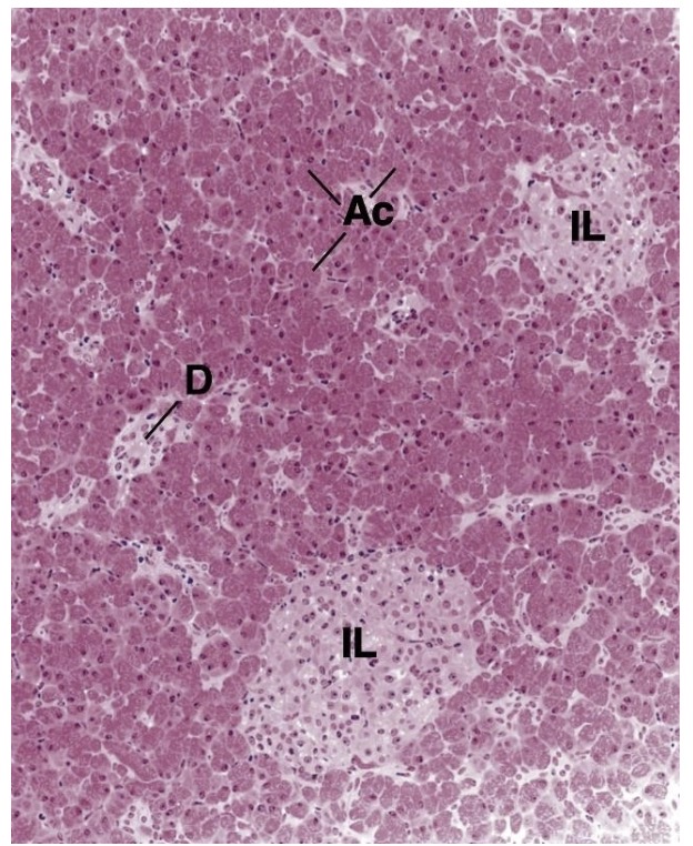
Pancreas histology - tissue diagram
Ac = acini
IL = Islets of Langerhans
D = exocrine duct
Exocrine portion of pancreas is larger