GI Skin Conditions
1/19
There's no tags or description
Looks like no tags are added yet.
Name | Mastery | Learn | Test | Matching | Spaced | Call with Kai |
|---|
No analytics yet
Send a link to your students to track their progress
20 Terms
What are the classic skin findings seen in cirrhosis?
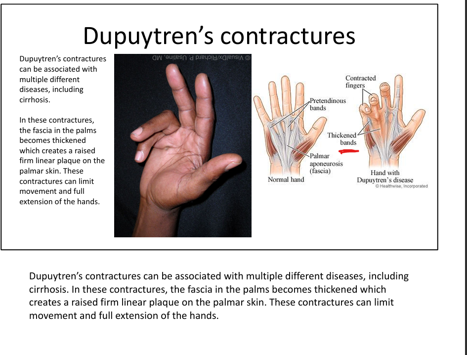
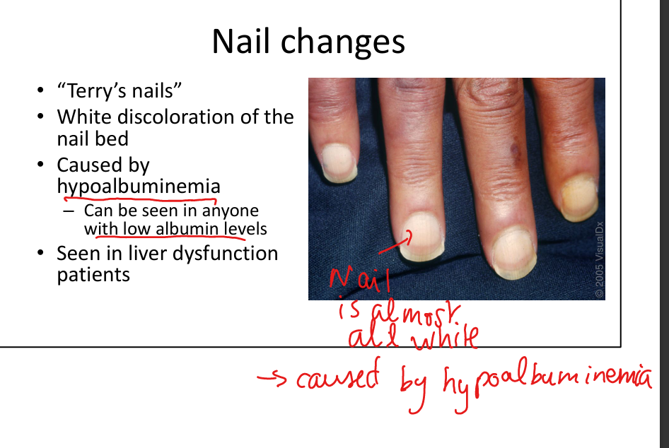
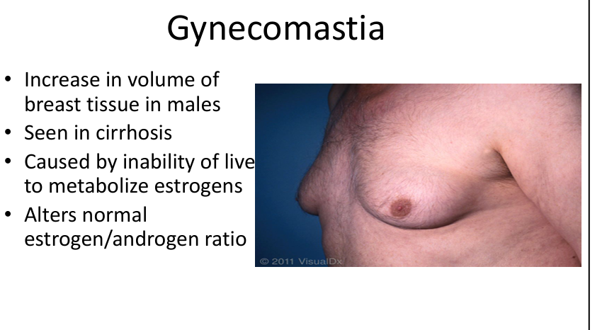
Scleral icterus, spider angiomas, palmar erythema, gynecomastia, sparse body hair, hypogonadism, caput medusae, Terry’s nails, Dupuytren’s contractures.
What causes the skin findings of “feminization” in cirrhosis?
Inability of the liver to metabolize estrogens → increased estrogen levels.
What causes spider angiomas in cirrhosis?
Elevated estrogen and VEGF causing dilated central arteriole with radiating capillaries.

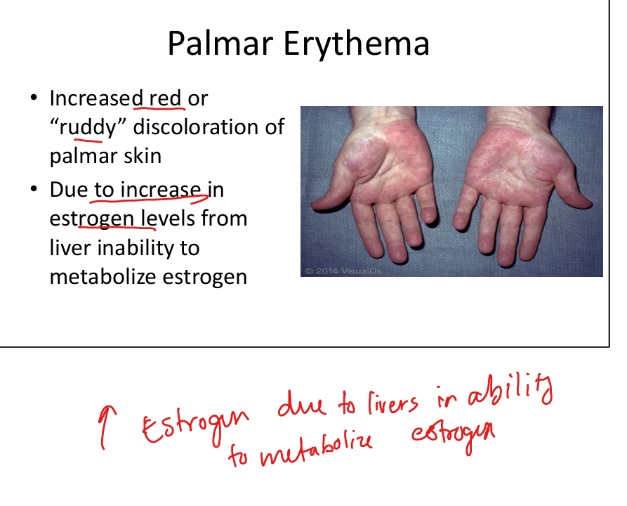
What causes palmar erythema in cirrhosis?
Increased circulating estrogen due to impaired hepatic metabolism.
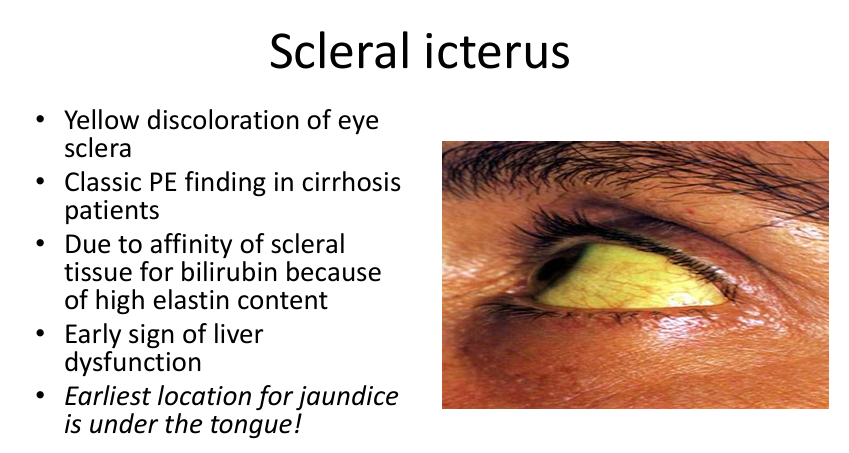
What causes scleral icterus?
Bilirubin deposition in elastin‑rich scleral tissue due to hyperbilirubinemia.
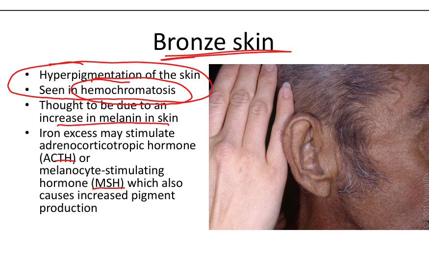
What is the mechanism of bronze skin in hemochromatosis?
Iron overload stimulates ACTH/MSH → increased melanin production.
What is dermatitis herpetiformis associated with?
Celiac disease.
What are the classic skin findings of dermatitis herpetiformis?
Extremely pruritic grouped vesicles/papules on elbows, knees, buttocks.

What is the immunopathology of dermatitis herpetiformis?
Granular IgA deposition in dermal papillae.
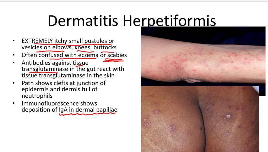
What is the pathophysiologic link between celiac disease and dermatitis herpetiformis?
IgA antibodies against tissue transglutaminase cross‑react with epidermal transglutaminase.

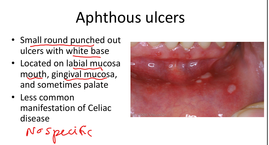
What oral finding can be seen in celiac disease?
Aphthous ulcers.
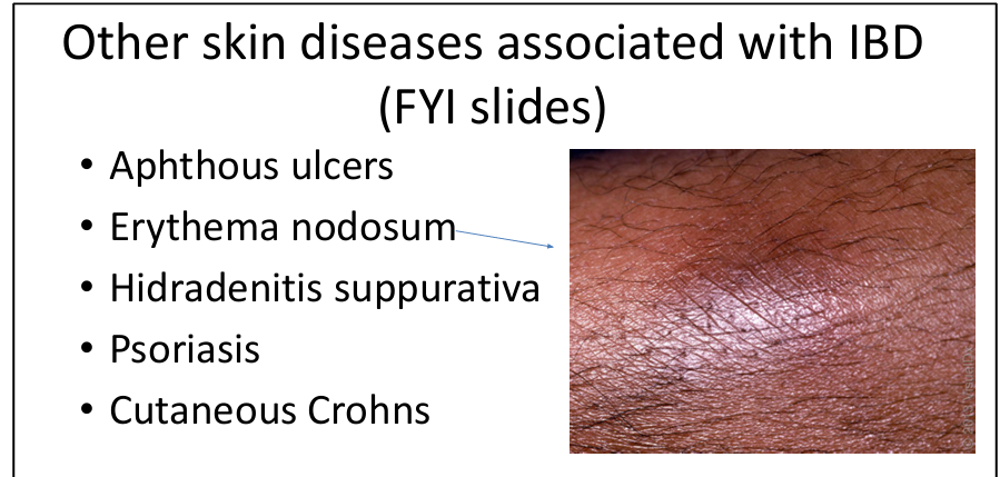
What skin diseases are associated with inflammatory bowel disease?
Pyoderma gangrenosum, erythema nodosum, hidradenitis suppurativa, cutaneous Crohn’s, aphthous ulcers.
What is pyoderma gangrenosum associated with?
Ulcerative colitis (most common) and other IBD.
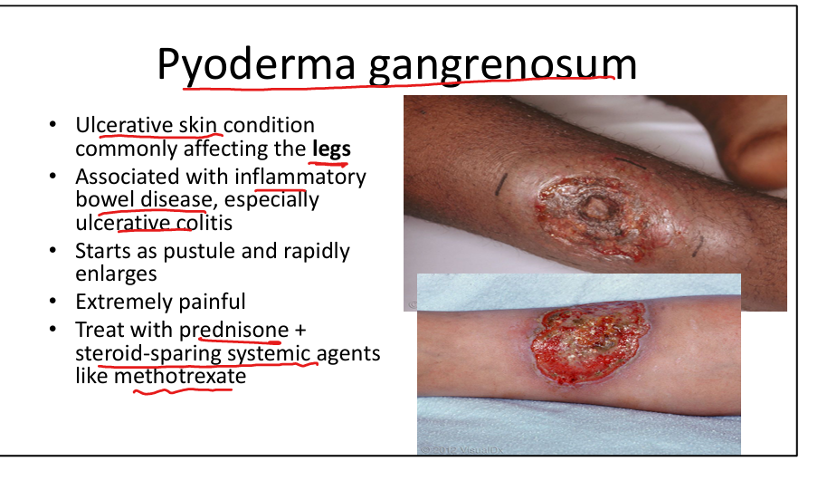
What are the clinical features of pyoderma gangrenosum?
Painful rapidly enlarging ulcer with undermined violaceous border (“cliff‑edge”).

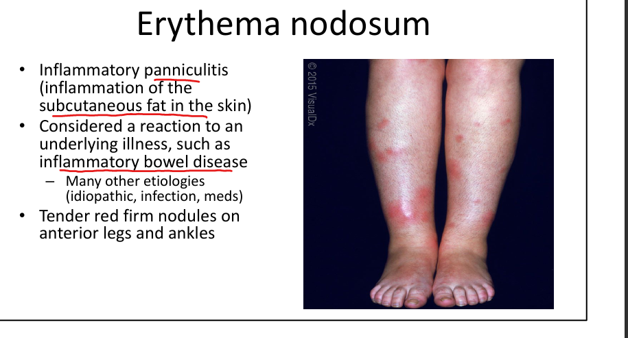
What is erythema nodosum?
Tender red nodules on shins due to septal panniculitis(inflammation of the subcutaneous fat in the skin)
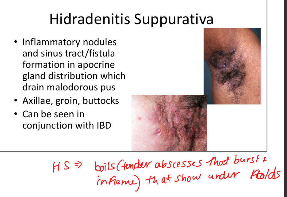
What is hidradenitis suppurativa and how is it related to GI disease?
Chronic inflammatory nodules and sinus tracts in apocrine gland areas (often axillae, groin, buttocks»folds)
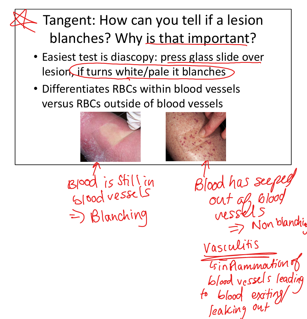
What is leukocytoclastic vasculitis?
Small‑vessel vasculitis with non‑blanching palpable purpura due to neutrophilic destruction of vessel walls.
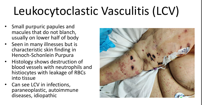
What disease classically presents with IgA‑positive leukocytoclastic vasculitis?
IgA vasculitis (Henoch‑Schönlein purpura).


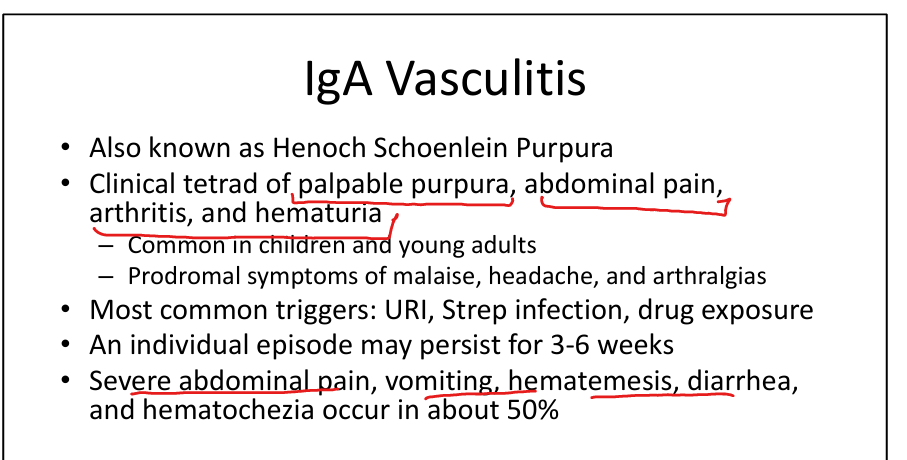
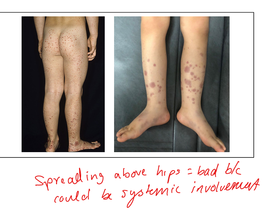
What is the clinical tetrad of IgA vasculitis?
Palpable purpura, abdominal pain, arthritis, hematuria.
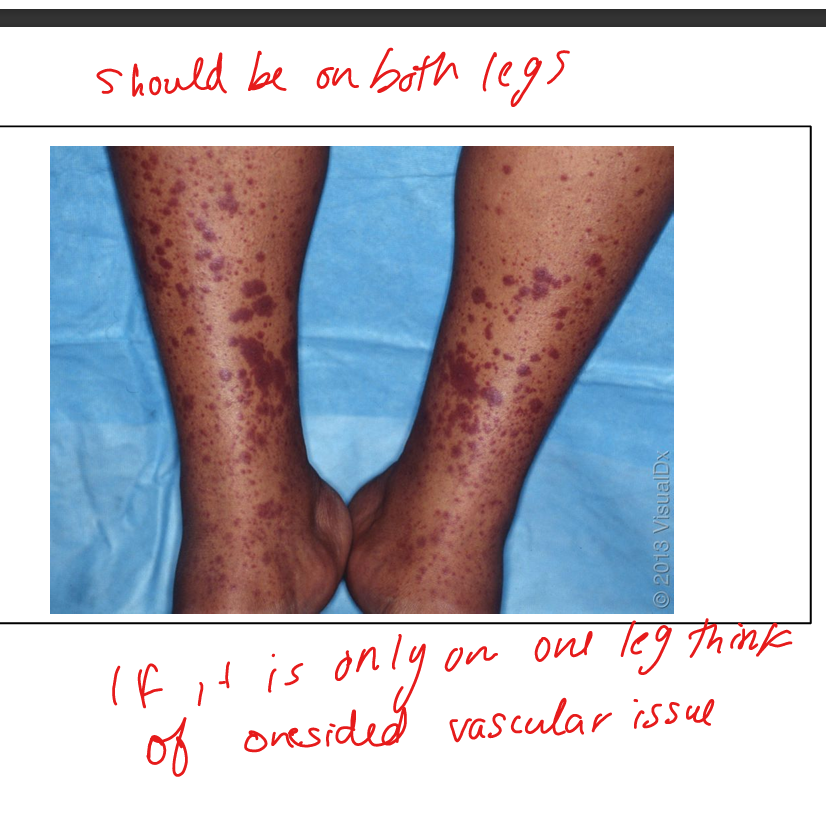
What does a blanching vs nonblanching lesion indicate?
Blanching=turns white/pale when pressed»blood is still in the blood vessels
Nonblanching=stays red when pressed»blood has seeped out of blood vessels»Vasculitis