Control of Eye movement
1/19
There's no tags or description
Looks like no tags are added yet.
Name | Mastery | Learn | Test | Matching | Spaced | Call with Kai |
|---|
No analytics yet
Send a link to your students to track their progress
20 Terms
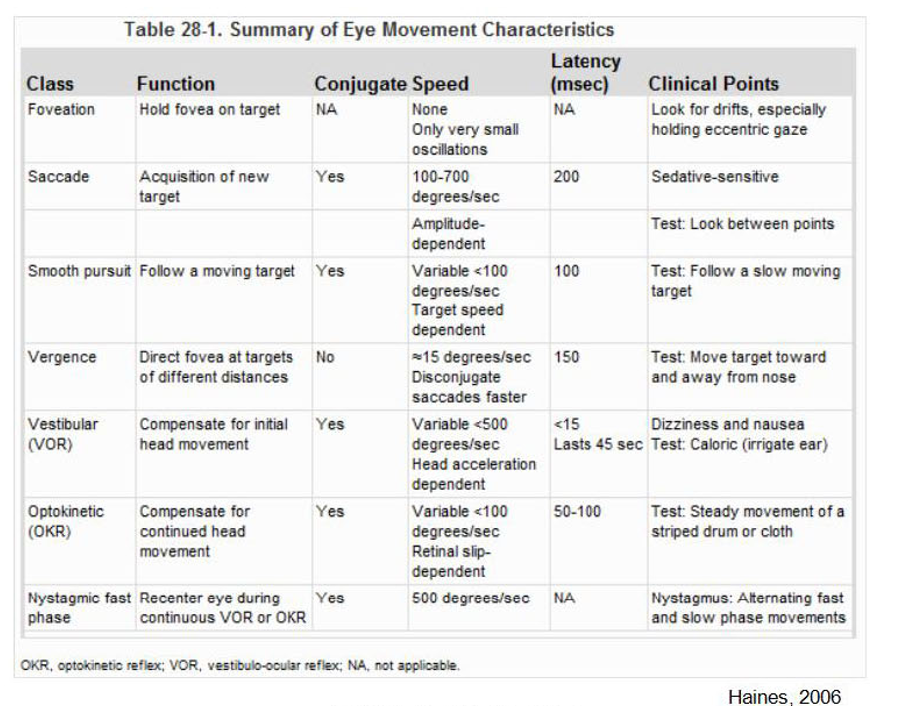
Describe the fovea
What are the three axis of eye movement
Fovea:
area of high visual resolution
area subtending about one degree of visual angle.
The eye moves around three axis of rotation
X axis: vertical movements
Y axis: horizontal movements
Z axis: torsion
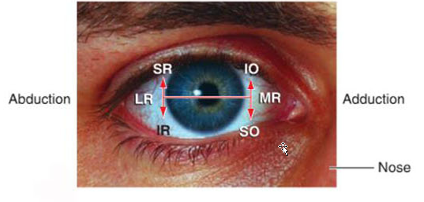
[REVIEW] actions of EOMs
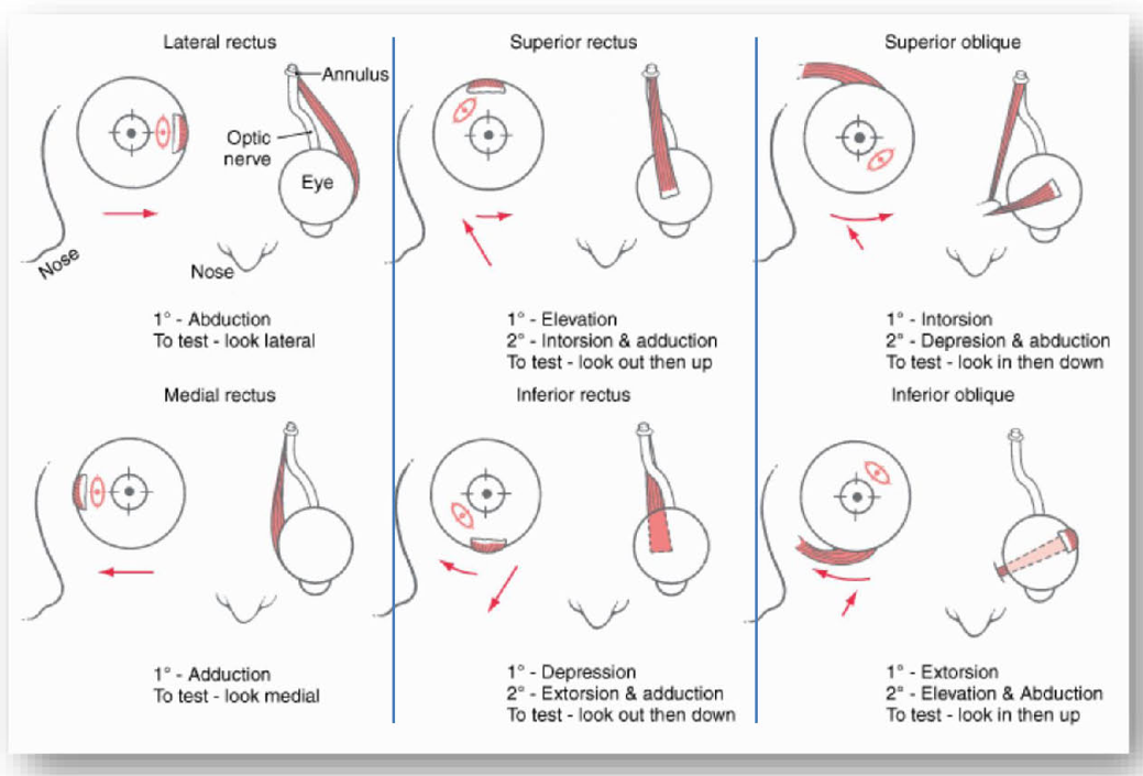
How do we clinically test EOMs
Describe the eye movement mechanics
Resistance?
Requires?
Eye movement mech:
Resistance:
Orbit’s soft tissues + muscles → resistance to movement → sluggish dynamics
Eye movement requires:
Force to overcome viscous drag
Force to maintain eccentric position
THUS: requires pulse and step activity of motor neurons
Describe brainstem’s role in eye movement
Nuclei’s
Gaze Centers
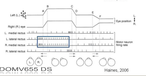
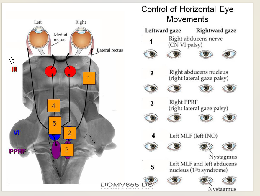
Describe the mech of horizontal eye movements:
Pulse signals
Step signals
Omnipause cells?
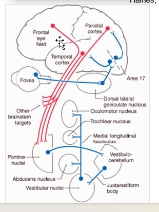
Brainstem:
EOM’s Nuclei:
Oculomotor nucleus
Trochlear nucleus
Abducens nucleus
Nuclei connected to each other via MLF
Gaze Centers:
PPRF: horizontal eye movement
riMLF: vertical eye movements
***P-H; R-V; Potter Had Rhino Virus***
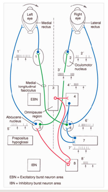
Horizontal eye movement mech:
Pulse Signals:
via excitatory + inhibitory burst neurons in Gaze Centers
Step signals (tonic position)
Nucleus prepositus hypoglossi: horizontal saccades
Interstitial nucleus of Cajal: vertical saccades
***P-H; C-V: Potter Had Crucio Virus***
Omnipause cells
provide tonic inhibition of burst cells during fixation;
pause as saccade trigger
Draw out the circuits for horizontal eye movement
Describe Gaze stabilization
Why needed?
Two systems?
VOR
Afferent
Function
Optokinetic System:
function/Mech
Input
Pathway
Gaze Stabilization:
Why needed?
Head Movement impacts image stabilization.
Two reflex response systems
Vestibulo-ocular reflexes (VOR)
Optokinetic system
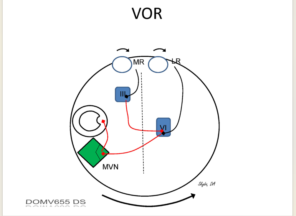
VOR:
A:
vestibular apparatus (ampullary cristae)
coordinated by vestibular nuclei
Function:
Produce Equal/Opposite eye movements to angular head movement.
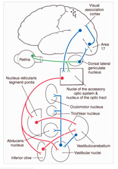
optokinetic system
Function/Mech: compensates for sustained or slow head movements.
Produces slow eye movement matching (direction + velocity) retinal slip w/ rapid repositioning phase (opposite direction).
Optokinetic Nystagmus
Input:
Visual Input is used → infer direction+ speed of head motion
particularly whole field movement of visual scene (retinal slip)
Pathway:
Activates wide-field retinal ganglion cells → nucleus of optic tract + accessory optic nuclei.
Project to vestibular nuclei + vestibulocerebellum (indirectly)
Draw out the circuitry of VOR
Draw out circuitry of optokinetic System
Describe how eye motor neurons uses both pulse/step components
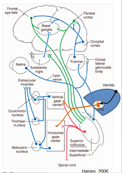
Describe the Superior Colliculus’ Role in eye movement:
Contains
Function
A/E
Describe the function of the cerebellar vermis in eye movement
Superior colliculus
Contains:
retinotopic map of Contra visual space used in directing eye movement
(deeper layers are visuomotor)
Function:
Translate sensory information → motor error signal (desired change in position)
Reflex orienting movements
A/E:
A: cortical eye fields + SNpr
E: brainstem gaze centers + frontal cortex via thalamus (md)
cerebellar vermis
Function:
calibrating saccades
long term adaptation in eye movement control
EX: adjust for muscular weakness,
EX: adjust for difference in elastic restorative forces between positions — same amplitude
Draw out the circuiltry of the superior colliculus
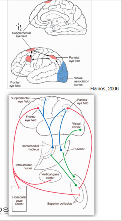
Describe how the frontal eye fields and parietal eye fields contribute to eye movement
A/E
Function
Frontal Eye Fields (BA 8)
A/E:
A:
visual association cortex + thalamus (md)
(regards target location)
E:
brainstem gaze centers + superior colliculus
Function:
Volitional or memory guided saccades
Parietal Eye Fields (posterior IPS)
Indirect influence
A/E:
Reciprocally connected to FEF
E: superior colliculus
Function:
visual selection (attention)
provides "salience map"
***NOTE: Salience map = highlights the most attention‑grabbing or behaviorally relevant parts of the visual field***
Reflexive saccades
Draw out the circuitry of the FEF and PEF
Explain how the brain tracks a moving object
How?
Mech?
Draw it Out
Shifting Gaze:
How?:
Object moving → Saccade brings fovea into alignment → smooth pursuit of eye
Mech:
Extrastriate visual cortex (MT and MST) + FEF + posterior parietal cortex (info about target motion)→ Dorsolateral pontine nucleus (DLPN) (encodes direction/velocity of pursuit) → vestibulocerebellum → brainstem oculomotor system via vestibular nuclei.
Describe Vergence
What is it?
Describe the neural circuitry:
Where are motor error commands created?
Location of Premotor Neurons? Function?
Vergence:
What is it?
Eye movement required to maintain Fovea on target as distance changes
NOTE: Gaze shifts often involve both version and vergence movements
Version = both eye same movement; vergence = opposite
Neural Circuitry:
Motor error commands
by neurons of visual cortex w/ binocular visual fields in response to retinal disparity.
Binocular = area where both retina share
Retinal disparity = difference in images in L/R Retina
Premotor Neurons:
found in supraoculomotor area in midbrain
drives vergence/accomodation
Describe antisaccade task
Procedure
Neural Circuitry
Diagnosis
Consequence of FEF stroke
Antisaccade Task:
Procedure:
Pt shown a dot that moves rapidly → instructured to resist the reflexive saccade + shift gaze in opposite direction
Neural Circuitry invovled:
Dorsolateral prefrontal cortex (DLPFC): saccade inhibition
FEF: triggering (anti)saccade motion
Diagnosis:
Increasing errors = frontal lobe dysfunction (b/c stroke/dementia)
b/c Difficult to ignore or override parietal cortex saliency signal
FEF Stroke:
Transient gaze deviation to side of lesion
difficulty directing gaze to contralateral side.
[REVIEW] Horizontal eye movement control
Describe what happens when these areas area damaged:
Flocculus
Oculomotor Vermis and fastigial nuclei
Nodulus and ventral uvula
Vestibulocerebellum
Flocculus:
ipsilateral smooth pursuit impairment
inability to hold eccentric eye positions
oculomotor vermis and fastigial nuclei:
saccade dysmetria
nodulus & ventral uvula:
produce periodic alternating nystagmus (PAN)
spontaneous horizontal nystagmus (ipsilesional)
Vestibulocerebellum:
VOR disturbances