1.3. Development of Nervous System
 Call Kai
 Learn
Learn Practice Test
 Spaced Repetition
 Match
Match Flashcards
 Knowt Play
1/69

There's no tags or description
Looks like no tags are added yet.
Name | Mastery | Learn | Test | Matching | Spaced | Call with Kai |
|---|
No analytics yet
Send a link to your students to track their progress
70 Terms
what are the 3 stages of human development?
1. pre-embryonic
2. embryonic
3. fetal
describe the pre-embryotic stage of development in utero (include timeline)
1. egg fertilized in uterine tube; cells develop
2. cells divide and form sphere (inner/outer cell layers)
3. Cavity in sphere forms (outer cells/inner cells)
4. Implants into endometrium
5. Embryonic disc:(Ectoderm, mesoderm, endoderm)
conception- day 14
the inner cell mass forms the
embryo
the outer cells form the
placent
the inner cell mass becomes the
3 primary germ layers
describe the embryonic stage of development in utero (include timeline)
organ formation starts in etoderm, mesoderm, and endoderm
day 15- week 8
what forms in the ectoderm (outer)?
sensory organs, epidermis, nervous system
what forms in the mesoderm (middle)?
dermis, muscles, skeleton,
excretory & circulatory systems
what forms in the endoderm (inner)?
gut, liver, pancreas, respiratory system
the organs developed in the embryonic stage _______ here
BEGIN (are not fully developed)
describe the fetal stage of development in utero (include timeline)
nervous system specific... 2 things occur
1. NS continues to develop
2. myelination begins
week 9-birth
what stage(s) does the nervous system form? from germ layer?
- embryonic and fetal stages
- ectoderm
what are the 2 phases of nervous system formation?
neural tube formation and brain formation
when does neural tube formation occur? what stage of development?
day 18-day 26
embryonic stage
what is the neural plate?
long, flat thickening of the ectoderm on the surface of the
embryo from head to tail
the edges of the neural plate fold in forming the
neural groove
the neural tube is formed when the
edges of the neural plate meet (edges curl towards each other)
the edges first close in the
cervical region
the edges continue to close from cervical region and on in both directions and form openings called....
what are the directions called?
neuropores (will eventually close)
rostral and caudal
when the neural tube is developing it begins to look like the
spinal cord (early foundation)
what is also formed when the edges meet and neuropores are formed?
neural crest
the neural tube and crest are surrounded by what layer? what does this layer become?
ectoderm
eventually becomes epidermis
two rings are layers are formed during the formation of the neural tube. the inner layer is ______ matter and the outer layer is ______ matter
gray; white
what structure has an inner gray matter and outer white matter?
spinal cord
the adjacent germ layer of the neural tube is the
mesoderm (skeletal system, muscular system, and dermis layer develop here)
the mesoderm layer divides to form
somites
what are somites?
clusters of cell bulges
(foundation blocks that build dermatomes and myotomes)
what are the 3 parts to the somite and what do they form?
1. sclerotome: vertebrae + skull
2. myotome: skeletal muscle
3. dermatom: dermis
what do myotomes and dermatomes represent?
single spinal nerve contribution to patch of skin or group of muscles with common action
the ventral part of a neural tube is the
motor plate
the dorsal part of a neural tube is the
association plate
motor neurons are typically
multipolar
sensory nerves are typically
pseudounipolar
what comes immediately off of the spinal cord?
dorsal and ventral roots
Somas from motor plate extend axons to their respective ________ region of the somite
myotome
the nerves from the motor plate become
lower motor neurons
what does the neural crest separate into during dermatome development?
two columns on each side of neural tube
the neural crest forms segments during dermatome development. what do these areas correspond to?
dermal areas of somites (appear as bulges that represent spinal levels of SC)
Crest cells at each segment form
peripheral sensory neurons
peripheral sensory neurons are what type of neuron? soma location?
pseudounipolar; sit in dorsal root ganglion
what do the ends of peripheral sensory neurons connect to?
One end connects to spinal cord and the other end connects to somite (dermis)
what is the definition of a dermatome and myotome during development?
The area of a somite that will become skeletal muscle / dermis
what is the definition of a dermatome and myotome in a fully developed adult?
A group of muscles / area of dermis innervated by a single spinal nerve
the entine PNS develops from tissue of the
neural crest (exept LMN)
what does the CNS/LMN develop from?
cells from neural tube
Spinal cord segments are _________ to corresponding vertebrae until the
_____ fetal month
adjacent; 3rd
the _______ _________ grows faster than the ________ __________
vertebral column; spinal cord
what is the significance of the vertebral column growing faster than the spinal cord?
mainly due to efficiency purposes. want to continue to develop skeletal system whereas spinal cord fully develops earlier in development. would rather conserve that "energy" to focus on developing the function of nervous tissue cells
the spinal cord ends at what level vertebrae?
L1/2 (conus medullaris)
what is the cauda equina? where do nerves do?
spinal nerves that come off spinal cord. nerves go to their respective vertebral level/opening
when does the brain begin forming?
around day 28 (end of 4th week)
what structure closing tiggers the neural tube to form 3 enlargements? what are those enlargements?
superior neuropore
• Hindbrain
• Midbrain
• Forebrain
what does the hindbrain consist of?
medulla, pons, cerebellum
(medulla/pons are anterior, cerebellum is posterior)
what is the central canal?
inner part of neural tube where CSF is found. sits between the brainstem and cerebellum
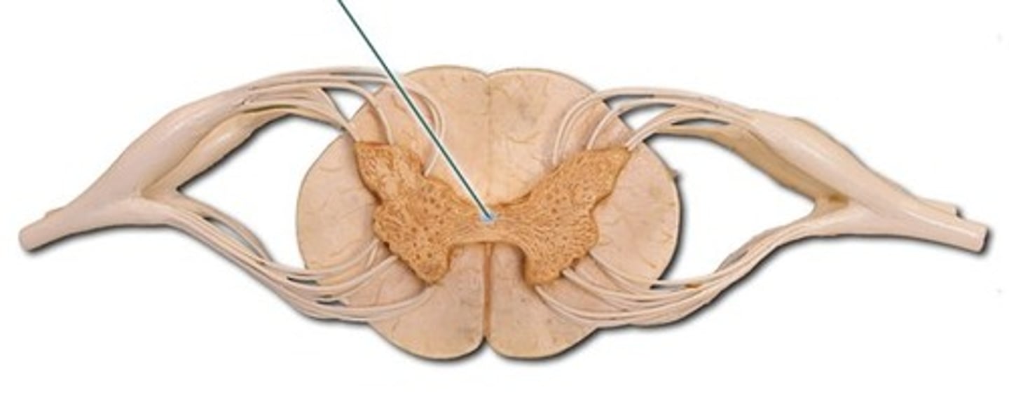
the central canal of the neural tube becomes what in the hindbrain?
4th ventricle
the central canal of the midbrain becomes
cerebral aqueduct
role of cerebral aqueduct?
connects the 4th ventricle to the 3rd ventricle
what does the forebrain consist of?
diencephalon, cerebrum (telencephalon)
what structure forms in the diencephalon of the forebrain?
3rd ventricle
where do the lateral ventricles form in the forebrain?
telencephalon (cerebrum)
as the brain develops through the fetal stage it fold in on itself. what are these folded sheets of tissue called?
sulci and gyrus
what makes neurons and glia?
epithelial cells in the neural tube (migrate to final destination)
nervous system tissue develops up to a certain point. what happens after it develops?
the rest of the time is used to develop function of nerve tissue
when do neurons differentiate?
after they reach final destination
what does neuron function depend on?
which region of the brain they migrate to. as neurons put out to their respective regions, this is where nervous tissue figures out its function
what helps "link" neurons with target cells?
growth cones
where are growth cones found? how do they work?
end of neurons. they communicate with different structures and tell nervous tissues where to/not to go (either attracted to or repelled by tissue)
when does myelination begin? completed?
4th fetal month (4 months after fertilization; week 9)
completed in 3rd year of life
what are critical periods?
- times when neuronal projections compete for synaptic sites (period of nervous system optimization)
- expected level of exposure to stimuli that allow neurons to develop properly
what must be adequate for adequate development? exmaples?
Peripheral and central nervous system experiences
- Retinal cells, optic nerves, visual cortex & proper vision
- Cerebral cortex hearing centers, speech sounds & language development
must receive proper exposure during critical period for proper function to develop