Microbiology - Practical 1
1/53
There's no tags or description
Looks like no tags are added yet.
Name | Mastery | Learn | Test | Matching | Spaced | Call with Kai |
|---|
No analytics yet
Send a link to your students to track their progress
54 Terms
contract transmission (direct-contact transmission)
In the fluid exchange lab, did this simulation represent a disease spread via contact, vehicle, or vector transmission?
propagated epidemic
In the fluid echange lab, did this simulation represent a common source epidemic or a propagated epidemic?
propagated epidemic
an outbreak of disease that occurs through direct or indirect person-to-person contact
common source epidemic
a disease outbreak where a group of people become infected after being exposed to the same single, shared source of an infectious agent or toxin
incidence and prevalence graphs
Incidence Line/Bars: Tracks only the newly diagnosed cases per year. For a rapidly spreading or new outbreak, this line will spike.
Prevalence Line/Bars: Tracks the total number of people currently living with the disease.
The Relationship: Prevalence = Incidence × Duration. Therefore, for chronic conditions like HIV, prevalence is consistently much higher than incidence because it accumulates cases over many years, minus patients who are cured or have died
incidence
the number of new cases (either illness or death) of a disease in a specific population over a defined period. It is a crucial measure used by epidemiologists to track disease risk and understand how fast an illness is spreading. [1, 2]
prevalence
refers to the total number of individuals infected with a specific disease in a given population at a particular point in time. It provides a "snapshot" of the disease's overall burden on a community
prevalence and incidence for acute vs chronic disease
Why they are more similar for acute diseases:
Because an acute disease develops rapidly and resolves in a relatively short period of time (days or weeks), a person is only considered a "case" for a very brief window. By the time a specified time period (like a month or a year) passes, those individuals have either fully recovered or died. Therefore, the total number of people currently sick at any given moment is almost entirely made up of the newly reported cases. [1, 2, 4]
Why they are different for chronic diseases:
In a chronic disease, patients may remain ill and active cases for months, years, or an entire lifetime. Because the disease persists, the total number of existing cases (prevalence) vastly outnumbers the amount of new cases (incidence) reported in any single time period. [1, 2, 4]
BSL-1 organism
well-characterized agents that pose minimal hazards to the environment and do not consistently cause disease in healthy, immunocompetent adults. These microbes are the focus of entry-level and undergraduate educational training and do not require specialized containment or isolation facilities
ex: E.coli
BSL-2
moderate-risk microbes commonly found in geographic areas (indigenous) that pose health hazards if exposed to the skin, inhaled, or ingested. According to OpenStax Microbiology, these agents cause human diseases of varying severity, but effective treatments or preventative measures are generally available
ex: Staphylococcus aureus (common cause of skin infections), Salmonella spp. (foodborne illness), and pathogenic strains of E. coli.
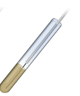
performing an asceptic transfer
In the asceptic transfer lab with s. marcescens and e. coli. The following are the steps to perform an asceptic transfer:
1. Sterilize the Loop
Hold the wire loop in your dominant hand at an angle and place it in the hottest part of a Bunsen burner flame (the inner blue cone). Heat the entire wire until it glows red-hot to incinerate any microbes. Let it cool for 10-20 seconds before touching the culture. [1, 2, 3, 4, 5]
2. Retrieve the Inoculum
From a Tube: Hold the culture tube in your non-dominant hand. Remove the cap with the pinky finger of your dominant hand (never place the cap on the table). Briefly pass the lip of the tube through the flame. Insert the loop to collect a small loopful of the culture, then flame the tube lip again and replace the cap. [1, 2, 3, 4]
From a Plate: Keep the plate right-side up, slightly lift the lid at an angle with your non-dominant hand (using the lid as a shield). Touch the cooled loop to a single, isolated colony. Close the lid immediately. [1, 2, 3]
3. Inoculate the New Media
To a Broth: Uncap, flame the tube, and dip the loop into the liquid medium. Twirl the loop gently to dislodge the bacteria, flame the tube again, and recap. [1, 2]
To an Agar Slant: Uncap, flame, and gently drag the loop across the agar surface in a zigzag "fishtail" motion starting from the bottom, flame, and recap. [1, 2, 3]
To a Petri Plate: Lift the lid slightly, and gently streak the loop over the agar surface to isolate colonies without gouging the agar. [1, 2]
Final Sterilization
Before placing the loop down, immediately re-sterilize it in the Bunsen burner flame from the base to the tip to kill any remaining bacteria
culture media
A liquid or solid substance containing nutrients and other requirements needed to support and sustain the growth of microorganisms.
asceptic
Free of contamination; a condition or process designed to prevent the introduction of unwanted microorganisms. [1, 2, 3, 5]
broth cultures
Physical State: Liquid medium containing essential nutrients without any solidifying agents.
Primary Purpose: Cultivating massive populations of microorganisms in a small volume and assessing metabolic characteristics and growth patterns (such as turbidity, pellicle formation, or sedimentation).
Best Used For: Rapid reproduction of cells, fermentation studies, and biochemical assays. [1, 2, 3, 4, 5]
agar slants
Physical State: Solid or semi-solid medium solidified at an angle in a test tube to provide a larger surface area for growth.
Primary Purpose: Maintaining stock cultures and storing pure strains of microbes for extended periods.
Best Used For: Creating space-saving, easily stored culture stocks. The tube can be tightly capped to prevent the media from drying out
agar deeps
Physical State: Solid or semi-solid medium solidified in a vertical, upright column.
Primary Purpose: Evaluating bacterial motility and testing how bacteria react to varying levels of oxygen (aerotolerance).
Best Used For: Differential metabolic tests. They are typically inoculated using an "inoculating needle". Microbes that grow at the top require oxygen, while those growing only at the bottom require an oxygen-free (anaerobic) environment. [1, 2, 3, 4]
Asceptic Transfer Steps
Sterilize innoculating loop
Cool the loop
Uncap the tubes
Flame the tube mouth
Insert loop into parent culture
Inoculate the fresh media
resterilize tubes and loop
Also remember:
Never lay tube caps flat on the benchtop.
Work quickly and deliberately to minimize the time tubes are open to the air.
Flame tubes from the base to the tip of the opening to avoid splattering.
Always flame the loop before and after the transfer. [1, 2, 3]
dangers of patient contamination
Patient contamination in a healthcare setting introduces, spreads, and breeds pathogens, primarily resulting in Healthcare-Associated Infections (HAIs). These nosocomial infections severely threaten patient safety, leading to extended hospital stays, high healthcare costs, and increased mortality rates, especially among vulnerable or immunocompromised individuals
asepsis in the clinical environment
Asepsis is the state of being free from disease-causing microorganisms. In the clinical environment, it encompasses practices used to prevent the transmission of pathogens to patients, protect healthcare workers, and maintain sterile fields during medical procedures. [1, 2, 3]
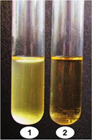
oxygen
The picture is used to help classify the species based on its growth in ___________
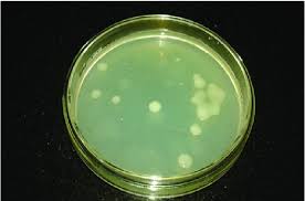
purpose of streak plate isolation
to dilute a high-density, mixed population of microorganisms into distinct, individual colonies on a solid agar surface. Each colony represents a pure, clonal culture originating from a single microbial cel
streak plate isolation steps
The goal of a streak plate isolation is to mechanically dilute a heavy microbial sample across an agar plate, depositing individual cells far enough apart to grow into distinct, genetically pure colonies. The standard four-quadrant method achieves this dilution
Label the plate
sterile loop
inoculate quadrant 1
sterilize and steak quadrant 2
repeat for quadrant 3
final streak (quadrant 4)
sterilize loop and incubate
colony morphology
refers to the visual characteristics of a bacterial or fungal colony growing on a solid culture medium, like agar. Because genetically identical cells clump together to form a single visible mass, observing these distinct features is the first step in identifying microbial species
used in microbiology lab protocols as an initial visual tool to identify microorganisms. By observing colony traits like size, shape, color/pigmentation, texture, and margin, lab technicians can efficiently differentiate between bacterial species
bacterial growth
The tube on the left (the cloudy one) has: _____________
left
Move the stage slowly to the right. In what direction does the image appear to
move?
the working distance is shorter than the 10X objective
Center the specimen and rotate to the 40X objective. Use the fine focus if needed. The distance between the specimen on the slide and the objective is called the working distance. Is the working distance greater or lesser than it was with the 10X objective?
calculating total magnification
Most standard microscopes feature a \(10\text{x}\) eyepiece. To find the total magnification, multiply it by the objective lens you are using: [1, 2]
Scanning Power: 10x (eyepiece) X 4x (objective) = 40x
Low Power: 10x (eyepiece) X 10x (objective) = 100x total
High Power: 10x (eyepiece) X 40x (objective) = 400x total
Oil Immersion: 10x eyepiece) X 100x (objective) = 1000x total [1, 2, 3]
field of view
Objective | Diameter of the Field of View (mm) |
4X | 5 mm |
10X | about 2 mm |
40X | less than 1 mm |
The field of view decreasees
What statement can you make about the field of view as you increase the objective magnification?
the depth of field decreases
What statement can you make about the depth of field as you increase the objective
magnification?
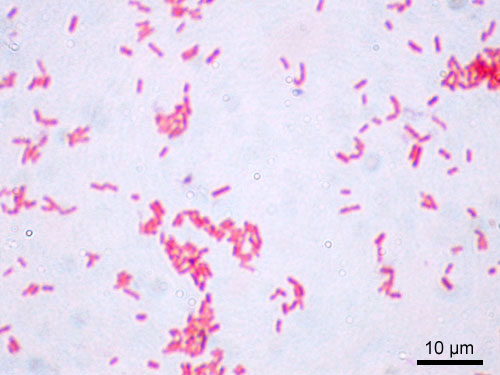
Serratia marcescens
Gram-negative, rod-shaped bacterium and opportunistic pathogen. Belonging to the Yersiniaceae family within the Enterobacterales order, it is widely ubiquitous in soil, water, and plant surfaces. It is well-known for producing a distinct, bright-red pigment called prodigiosin. [1, 2, 3, 4]
Serratia marcescens morphological characteristics
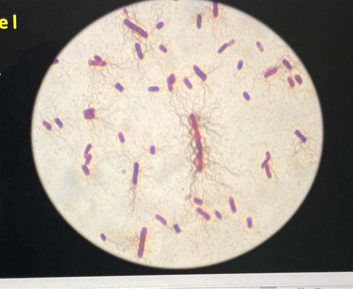
Shape & Size: Short bacilli (rods) typically measuring \(0.5 - 0.8 \,\mu\text{m}\) in width and \(0.9 - 2.0 \,\mu\text{m}\) in length.
Gram Staining: Gram-negative; due to a thin peptidoglycan layer, they do not retain crystal violet and stain pink/red with safranin.
Flagella & Motility: Motile using peritrichous flagella (flagella distributed over the entire cell surface).
Spore Formation: Non-spore-forming.
Capsule: Generally non-encapsulated (or has a very poorly defined capsule), though this can vary across specific clinical isolates
Serratia marcescens physiological characteristics
Oxygen Requirements: Facultative anaerobe. It can generate ATP through aerobic respiration in the presence of oxygen but is also capable of switching to fermentation. [1, 2]
Temperature & pH: Optimal growth occurs at temperatures between 25C and 37C and at a pH of around 9. Prodigiosin (red pigment) production is strictly temperature-dependent and is most robust between 25C and 30C it is significantly reduced or inhibited at 37C
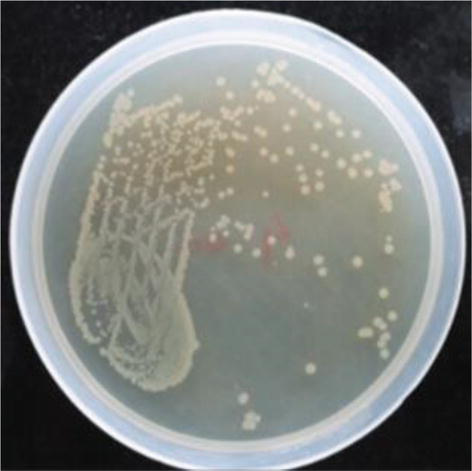
Serratia marcescens cultural characteristics
Colony Morphology: On standard nutrient agar, colonies typically appear circular, convex, smooth, moist, and glistening. [1, 2, 3, 4]
Pigmentation: Depending on the strain and culture age, colonies vary from pale pink to a distinct dark, brick-red due to prodigiosin. S. marcescens grown in clinical settings is often non-pigmented (white or beige
![<ul><li><p><span><strong>Colony Morphology:</strong> On standard nutrient agar, colonies typically appear circular, convex, smooth, moist, and glistening.</span> [1, 2, 3, 4]</p></li><li><p><span><strong>Pigmentation:</strong> Depending on the strain and culture age, colonies vary from pale pink to a distinct dark, brick-red due to prodigiosin. <em>S. marcescens</em> grown in clinical settings is often non-pigmented (white or beige</span></p></li></ul><p></p>](https://assets.knowt.com/user-attachments/900e7e77-8abf-4311-ac00-29d403da7fab.jpg)
Escherichia coli
Gram-negative, rod-shaped bacterium and a primary facultative anaerobe of the human gut. It is highly versatile, non-spore forming, and utilizes both respiratory and fermentative metabolism
Escherichia coli morphological characteristics
Shape: Straight rods (bacilli) with rounded ends, typically \(1.0 - 2.0\:\mu\text{m}\) in length and \(0.5\:\mu\text{m}\) in width.
Arrangement: Occur singly or in pairs.
Motility: Most strains are motile via peritrichous flagella, though some non-motile variants exist.
Structures: Possesses pili (fimbriae) for adhesion and may have a capsule. [1, 2, 3, 4, 5]
![<ul><li><p><span><strong>Shape:</strong> Straight rods (bacilli) with rounded ends, typically \(1.0 - 2.0\:\mu\text{m}\) in length and \(0.5\:\mu\text{m}\) in width.</span></p></li><li><p><span><strong>Arrangement:</strong> Occur singly or in pairs.</span></p></li><li><p><span><strong>Motility:</strong> Most strains are motile via peritrichous flagella, though some non-motile variants exist.</span></p></li><li><p><span><strong>Structures:</strong> Possesses pili (fimbriae) for adhesion and may have a capsule.</span> [1, 2, 3, 4, 5]</p></li></ul><p></p>](https://assets.knowt.com/user-attachments/c156e9c9-81a3-49ba-ac2f-48037febfd18.webp)
Escherichia coli physiological characteristics
Oxygen Requirements: Facultative anaerobe. [1, 2]
Temperature: mesophile
Metabolism: Ferments glucose and lactose with the production of acid and gas. [1]
Escherichia coli culture characteristics
Grows as large, circular, convex, smooth, grayish-white, and opaque/translucent colonies
Bacilllus subtilis
is a Gram-positive, rod-shaped bacterium renowned for its resilience and industrial applications. It naturally occurs in the soil and gastrointestinal tracts, growing optimally under aerobic conditions. Its defining feature is the ability to form stress-resistant endospores
Bacilllus subtilis morphological characteristics
Cell Shape and Size: Vegetative cells are typically rod-shaped
Arrangement: Cells can occur singly, in pairs, or occasionally in short chains.
Spore Morphology: Endospores are ellipsoidal and centrally or sub-terminally located. They do not cause the cell to swell (non-distending) and enable the bacteria to survive harsh conditions like radiation, desiccation, and extreme temperatures.
Staining: Gram-positive (retains crystal violet and stains purple).
Motility: They are motile by means of peritrichous flagella
Bacilllus subtilis physiological characteristics
Oxygen Requirements: Strictly aerobic to facultatively anaerobic.
Temperature: Mesophilic, with an optimal growth temperature range of \(30^{\circ }\text{C}\) to \(37^{\circ }\text{C}\).
pH Range: Grows optimally at a neutral to slightly alkaline pH (\(6.0\)–\(7.5\)).
Enzymes: Produces catalase, oxidase, and hydrolytic enzymes such as amylase (to break down starch) and protease (to break down proteins).
Metabolism: Capable of fermenting various carbohydrates (e.g., glucose, sucrose) to produce acids, but does not produce gas.
Capsule: Can produce a polypeptide capsule made of poly-D-glutamic acid, though this varies depending on the strain and environment
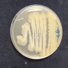
Bacilllus subtilis cultural characteristics
On standard nutrient agar, colonies appear large opaque, and creamy to grayish-white. They often have an irregular or undulate margin with a rough, wrinkled, or dull surface (resembling frosted glass). Soil isolates can display a "swarming" spreading growth across the plate
Staphylococcus epidermidis
Gram-positive, coagulase-negative bacterium that acts as a normal commensal of the human skin flora. While typically harmless, it is an opportunistic pathogen that forms tenacious, antibiotic-resistant biofilms on indwelling medical devices, making it a major cause of hospital-acquired infections
Staphylococcus epidermidis morphological characteristics
Shape and Arrangement: Spherical cells (cocci) that primarily arrange in irregular, grape-like clusters, though they can also appear in pairs or short chains.
Gram Stain: Gram-positive (retains crystal violet, appearing deep purple under a microscope).
Size: Approximately 0.5 to 1.5 μ m in diameter.
Motility and Spores: Non-motile and non-spore-forming.
Capsule: Possesses a polysaccharide/protein capsule that aids in adhering to surfaces and evading the host's immune response. [1, 2, 3, 4, 5]
![<ul><li><p><span><strong>Shape and Arrangement:</strong> Spherical cells (cocci) that primarily arrange in irregular, grape-like clusters, though they can also appear in pairs or short chains.</span></p></li><li><p><span><strong>Gram Stain:</strong> Gram-positive (retains crystal violet, appearing deep purple under a microscope).</span></p></li><li><p><span><strong>Size:</strong> Approximately 0.5 to 1.5 μ m in diameter.</span></p></li><li><p><span><strong>Motility and Spores:</strong> Non-motile and non-spore-forming.</span></p></li><li><p><span><strong>Capsule:</strong> Possesses a polysaccharide/protein capsule that aids in adhering to surfaces and evading the host's immune response.</span> [1, 2, 3, 4, 5]</p></li></ul><p></p>](https://assets.knowt.com/user-attachments/e702493b-5269-407f-9b0f-9c9fdd8ecaa3.jpg)
Staphylococcus epidermidis cultural characteristics
On general media, colonies are small (1–2 mm in diameter), circular, convex, smooth, and glistening.
Pigmentation: Colonies are typically non-pigmented, appearing white, grey, or beige (distinguishing it from the golden-yellow colonies of S. aureus).
Proteus hauseri
a Gram-negative, rod-shaped, facultative anaerobic bacterium historically classified within the Proteus vulgaris group. It is known for its distinct polymorphic cell structure, unique swarming motility across agar surfaces, and metabolic capabilities like strong urease production and hydrogen sulfide (H₂S) release.
Proteus hauseri morphogical characteristics
Cell Shape: Straight, Gram-negative bacilli ranging from 0.4 to 0.8 μm in diameter and 1.0 to 3.0 μm in length.
Motility: Highly motile via numerous peritrichous flagella, which give it a unique ability to undergo morphological phase variation. It can alter between a standard "swimmer" cell and an elongated, hyper-flagellated "swarmer" cell.
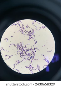
simple stain
simple stain is a rapid microbiology technique that uses a single, positively charged dye (basic stain) to colorize bacterial cells. Because the dye is positively charged and the bacterial cell wall is naturally negative, the stain is attracted to and bonds with the cells, making them clearly visible against a transparent background
simple stain steps
1) Prepare the Smear
2) heat fix the smear
3) stain the slide with methylene blue or crystal violet
4) rinse with water
5) blot dry with bibulous paper
6) view under microscoe
Kocuria rhizophila
Gram-positive, strictly aerobic bacterium in the family Micrococcaceae. Originally isolated from plant roots (rhizosphere), it is a non-motile, non-spore-forming coccus. Ecologically, it is noted for its high halotolerance, capacity to survive in harsh environments, and potential biotechnological applications.
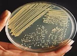
Kocuria rhizophila culture characteristics
Colony Appearance: On initial isolation (e.g., on nutrient agar or blood agar), colonies are typically small (\(2 \text{ to } 3\ mm\)), whitish, round, convex, and raised. [1, 2]
Pigmentation: Upon prolonged incubation or under strict aerobic conditions, colonies may develop a dull, creamy appearance with a non-diffusible yellow tinge or yellowish pigmentation. [1, 2]
![<ul><li><p><span><strong>Colony Appearance:</strong> On initial isolation (e.g., on nutrient agar or blood agar), colonies are typically small (\(2 \text{ to } 3\ mm\)), whitish, round, convex, and raised.</span> [1, 2]</p></li><li><p><span><strong>Pigmentation:</strong> Upon prolonged incubation or under strict aerobic conditions, colonies may develop a dull, creamy appearance with a non-diffusible yellow tinge or yellowish pigmentation.</span> [1, 2]</p></li></ul><p></p>](https://assets.knowt.com/user-attachments/726a500e-da79-460f-9f17-4aa95ec56627.jpg)
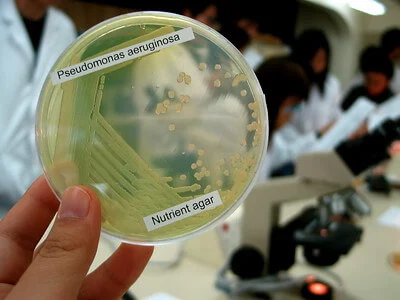
Pseudomonas aeruginosa culture characteristics
large, flat, opaque colonies with irregular margins and a spreading edge. The colonies typically emit a sweet, fruity, or grape-like odor. They frequently produce diffusible pigments that color the agar, most notably the blue-green pyocyanin and the yellow-green pyoverdine
Staphylococcus aureus culture characteristics
forms smooth, round, convex colonies that range from 1 to 4 mm in diameter. The colonies have a glistening, buttery consistency, a well-defined edge, and are most famously known for their distinct golden-yellow pigmentatio
Klebsiella pneumoniae culture characteristics
forms large, raised, and highly mucoid (slimy or sticky) colonies. Because of their abundant polysaccharide capsule, the colonies are typically greyish-white in color, glistening, and circular with a smooth, convex or dome-shaped elevation