BIo 161 - Cell Structure
1/124
There's no tags or description
Looks like no tags are added yet.
Name | Mastery | Learn | Test | Matching | Spaced | Call with Kai |
|---|
No analytics yet
Send a link to your students to track their progress
125 Terms
Which cells are larger, eukaryotic or prokaryotic?
Eukaryotic !!
Why are eukaryotic cells large?
have nucleus
many organelles & membranous structures
Purpose: keep reactions separate & localize reactions (enzymes, reactants) —> increase speed/efficiency
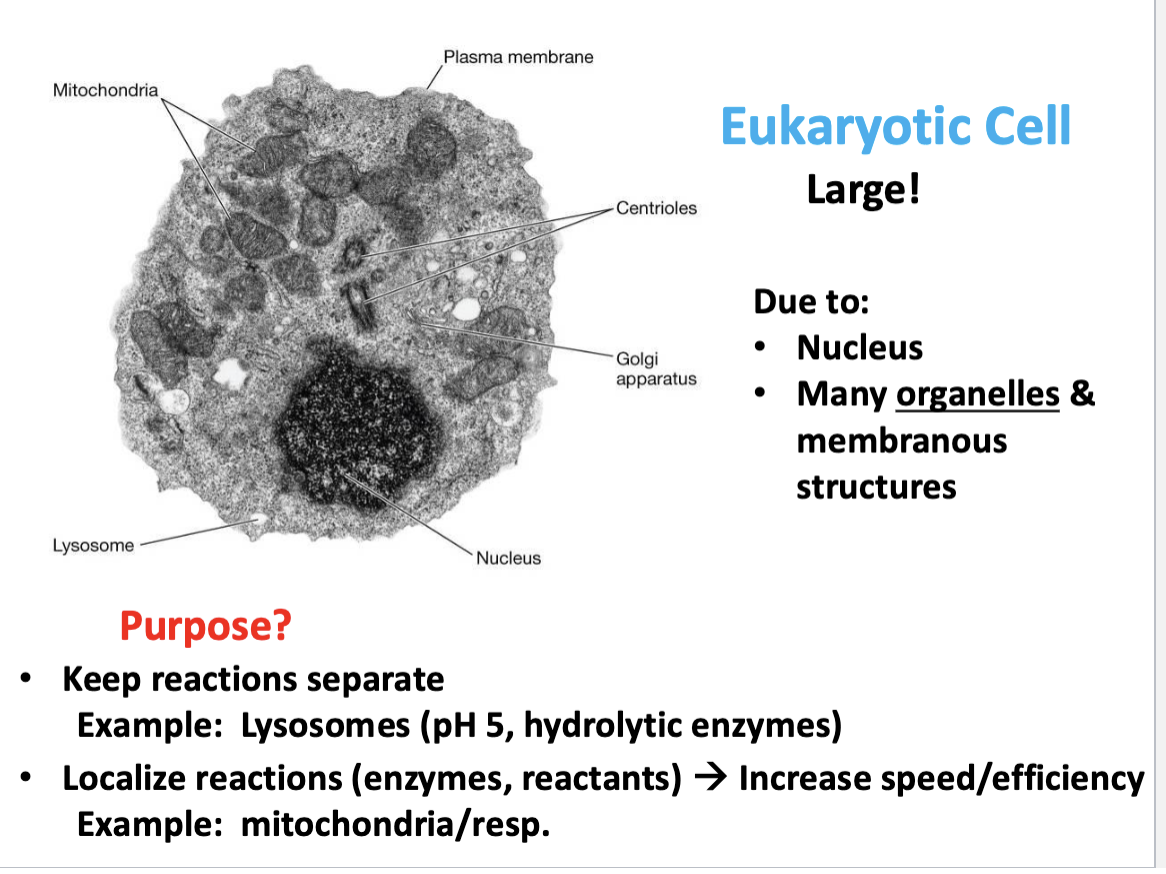
Compartmentalization in eukaryotic cells offers two primary, distinct advantages for cellular function:
Keeps reactions separate:
Specialized compartments, such as lysosomes, maintain unique internal environments—like a low pH and specific hydrolytic enzymes—to perform distinct functions (e.g., waste digestion) without damaging the rest of the cell

Compartmentalization in eukaryotic cells offers two primary, distinct advantages for cellular function: Localizes reactions
By confining specific enzymes and their substrates together, compartments like mitochondria create high local concentrations of reactants, which significantly increases the efficiency and speed of metabolic processes like aerobic respiration

Organelles are what keep things separate…
compartmentalize using lipids to separate functions

The bilayer phospholipid membrane of a eukaryotic cell….
encircles/encloses cytoplasm, regulates transport

What features do all cells have in common?
All cells have:
Plasma membrane → phospholipid bilayer; regulates transport
Cytoplasm → fluid interior (cytosol + structures)
DNA → genetic information
Ribosomes → protein synthesis
Both prokaryotes and eukaryotes have these.
All cells have: Plasma membrane
Plasma membrane → phospholipid bilayer; regulates transport
All Cells have: Cytoplasm
Cytoplasm → fluid interior (cytosol + structures)
All cells have: DNA
DNA → genetic information
All cells have: ribosomes
Ribosomes → protein synthesis
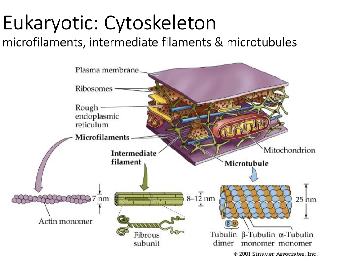
Eukaryotic Cells: Cytoskeleton
microfilaments, intermediate filaments & microtubules
any movement in cell takes place along some component of cytoskeleton
Protein fibers for:
Shape
Transport
Movement
Division
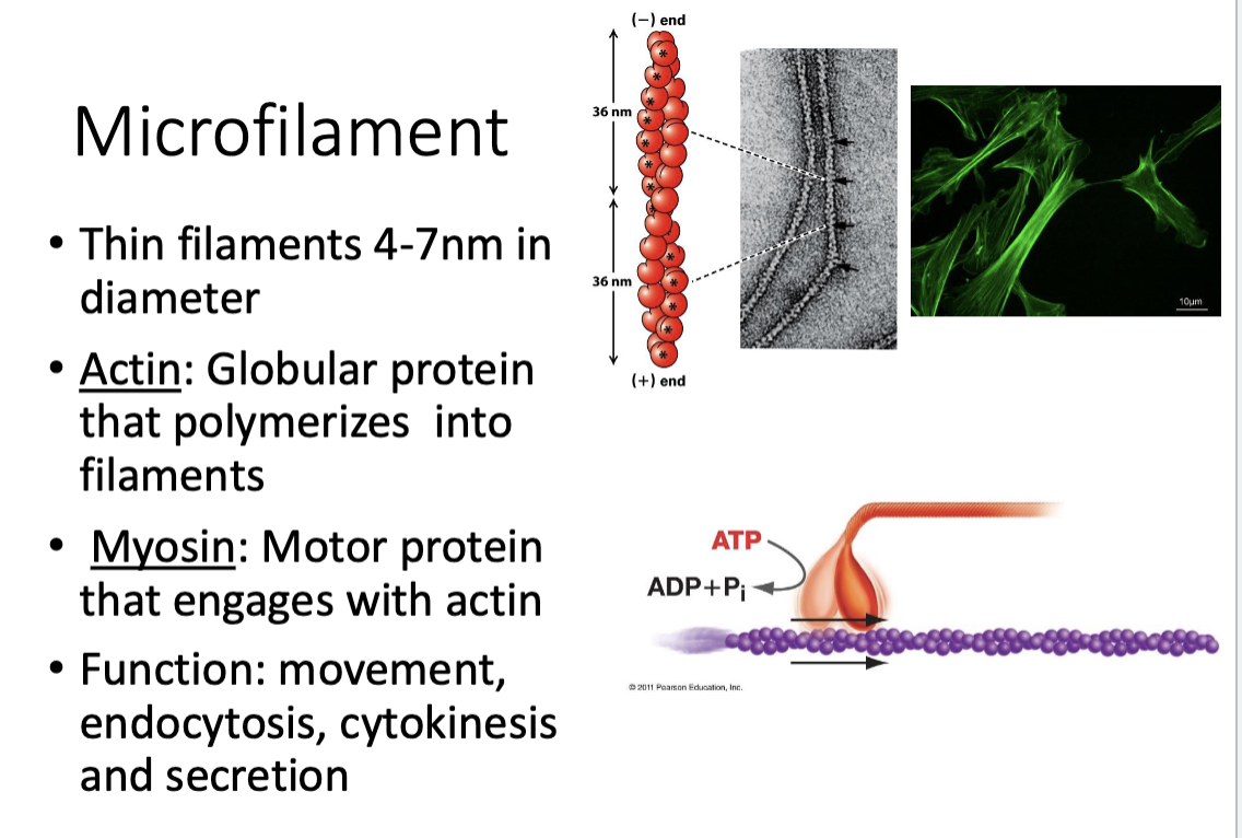
Component of cytoskeleton: What are microfilaments?
very thin filaments (4-7 nm in diameter)
2 types: actin & myosin
function: movement, endocytosis (bringing things into cell), cytokinesis (breaking cell apart), & secretion (vesicles that may be released from cell)

Brief description: endocytosis
bringing things into cell
brief description: cytokinesis
breaking cell apart
brief description: secretion
vesicles that may be released from cell
Pathogens can take advantage of microfilaments in that ….
there are certain bacteria that can usr filaments to move through cell
Type of microfilament: Actin
globular protein that polymerizes into filaments
think “road”

Type of microfilament: Myosin
motor protein that engages with actin
think “motor that drives on that road”
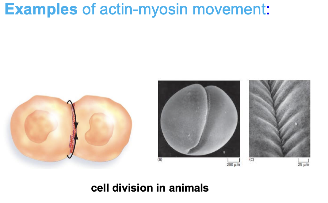
Example of actin-myosin movement: Cell division in animals
cytokinesis —> formation & contraction of a contractile ring at the cell equator
—> ring, made of actin filaments & myosin-II motor proteins, shrinks, pinching the cytoplasm to divide one cell into two daughter cells
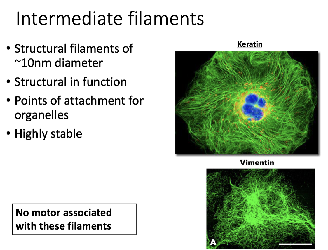
Component of Cytoskeleton: Intermediate filaments
structural filaments (~10 nm diamerer)
STRUCTURAL in function (NO motor proteins associated w/these filaments)

Intermediate filaments are…
true cytoskeleton filaments
points of attachment for organelles
HIGHLY STABLE (pretty static, good for binding)

Examples of intermediate filaments
Keratin
Vimentin
NO MOTOR associated w/intermediate filaments
Component of cytoskeleton: Microtubules
helical shaped polar cylinders (~25nm diameter)
alternating tubulin subunits (protein) forms filaments
—> alpha & beta tubulin
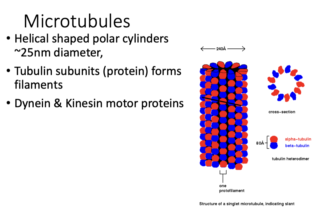
Think of microtubules as…
“freeways” of filaments, largest!!
good for movememnt, dynamic

Associated motor proteins of microtubules
Dynein & Kinesin motor proteins
ATP-driven motor proteins —> transport intracellular cargo by walking along microtubule tracks, convert chemical energy from ATP hydrolysis into mechanical work, typically moving in opposite direction
—> kinesin moves toward + end (cell periphery), while dynein moves toward the - end (cell center)

Components of cytoskeleton: microfilaments, intermediate filaments, & microtubules: Which have no motor associated with them?
Intermediate filaments!
Why none for intermediate filaments?: They are stable support cables, not directional transport tracks.
How did an archea change to develop into a eukaryote?
Key events in eukaryotic evolution!
Key event in Eukaryotic Evolution: 1
Acquisition of a Nucleus !!
—> protect genetic material
—> taking genetic material & compartmentalizing it (prokaryotes don”t)
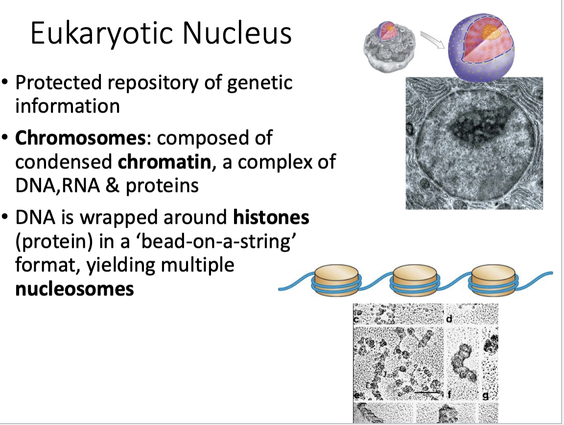
Eukaryotic Nucleus
Protected repository of genetic information
Chromosomes within nucleus

Eukaryotic Nucleus: Chromosomes
Chromosomes: composed of condensed chromatin
Chromatin = complex of DNA, RNA, & proteins

Chromatin that make up chromosomes are
a complex of DNA, RNA, & proteins

Basic repeating structural units that make up chromatin (which make up chromosomes..)
multiple nucleosomes: DNA wrapped around histones (protein) in a ‘bead-on-a-string’ format
Eukaryotic Nuclear Envelope
two membranes (Inner & outer) —> each membrane are phospholipid bilayers
separates genetic material of cell from cytoplasm
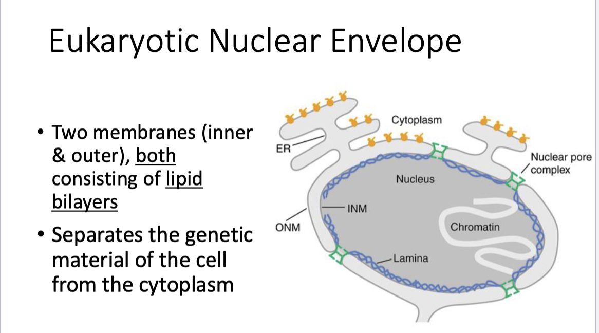
The eukaryotic nuclear envelope has…
nuclear pore complexes!! —> highly regulated process that allows thing into nucleus

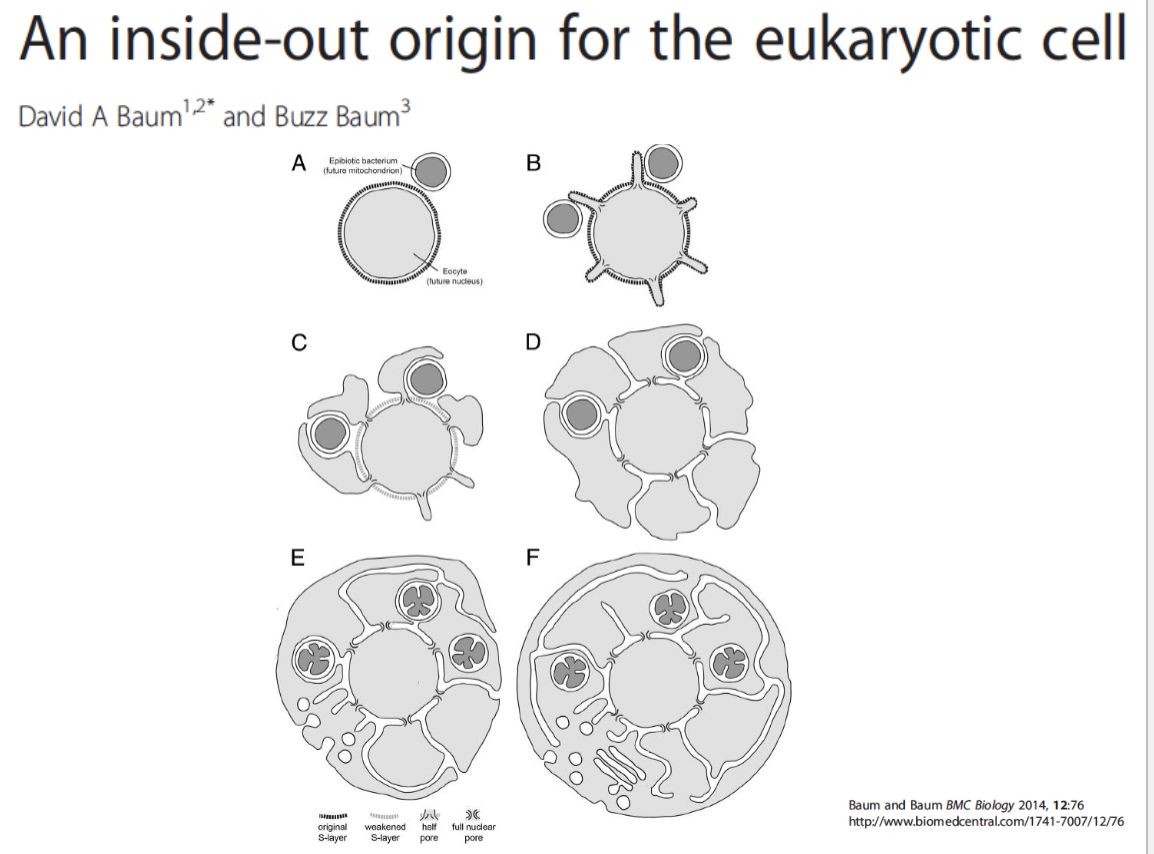
An inside out origin for the eukaryotic cell
blebs extend out —> engulf other organisms
perhaps how nuclear pore complex developed…still unknown
The Nuclear Pore Complex….
regulates entry & exit out of the nucleus
depends on nuclear localization signals & nuclear export signals
made up of proteins that form gates!!

Nuclear Pore Complex: Nuclear Localization Signals (NLS) =
Into the nucleus

Nuclear Pore Complex: Nuclear Export Signals (NES) =
out of the nucleus

Nuclear Pore Complex: Both NLS and NES are
primarily amino acid/peptide based
short peptide sequences that may get cleaved from the protein upon entry/exit
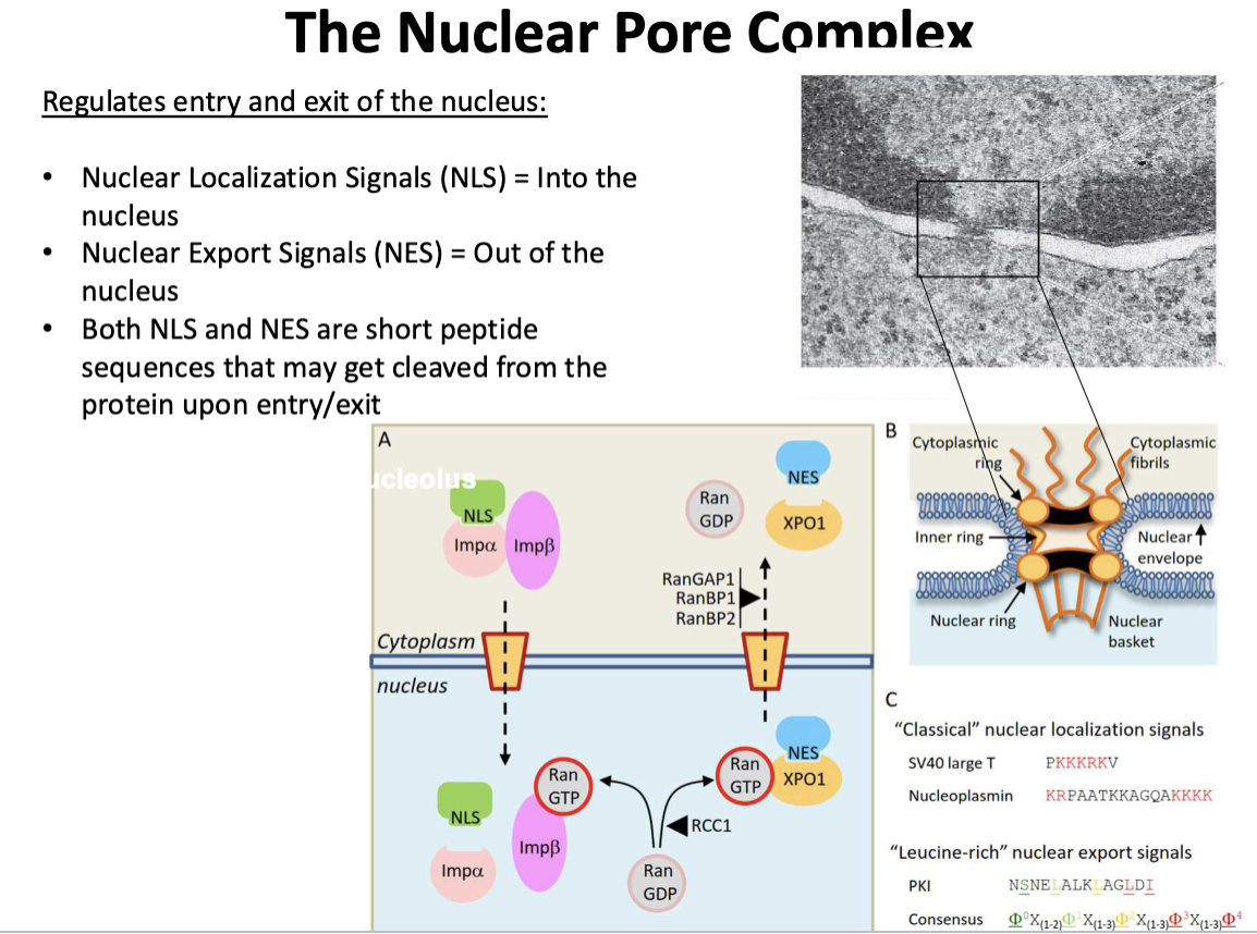

Nuclear Pore Complex “Classical” Nuclear localization Signals
proteins w/lysines will be signal to bind to gates
Key event in Eukaryotic Evolution: 2
Endomembrane system!
membranes within cell
Eukaryotic Plasma Membrane
Plasma membrane
contains high proportion of sphingolipids & sterols (cholesterol) for strength & rigidity
selectively permeable barrier
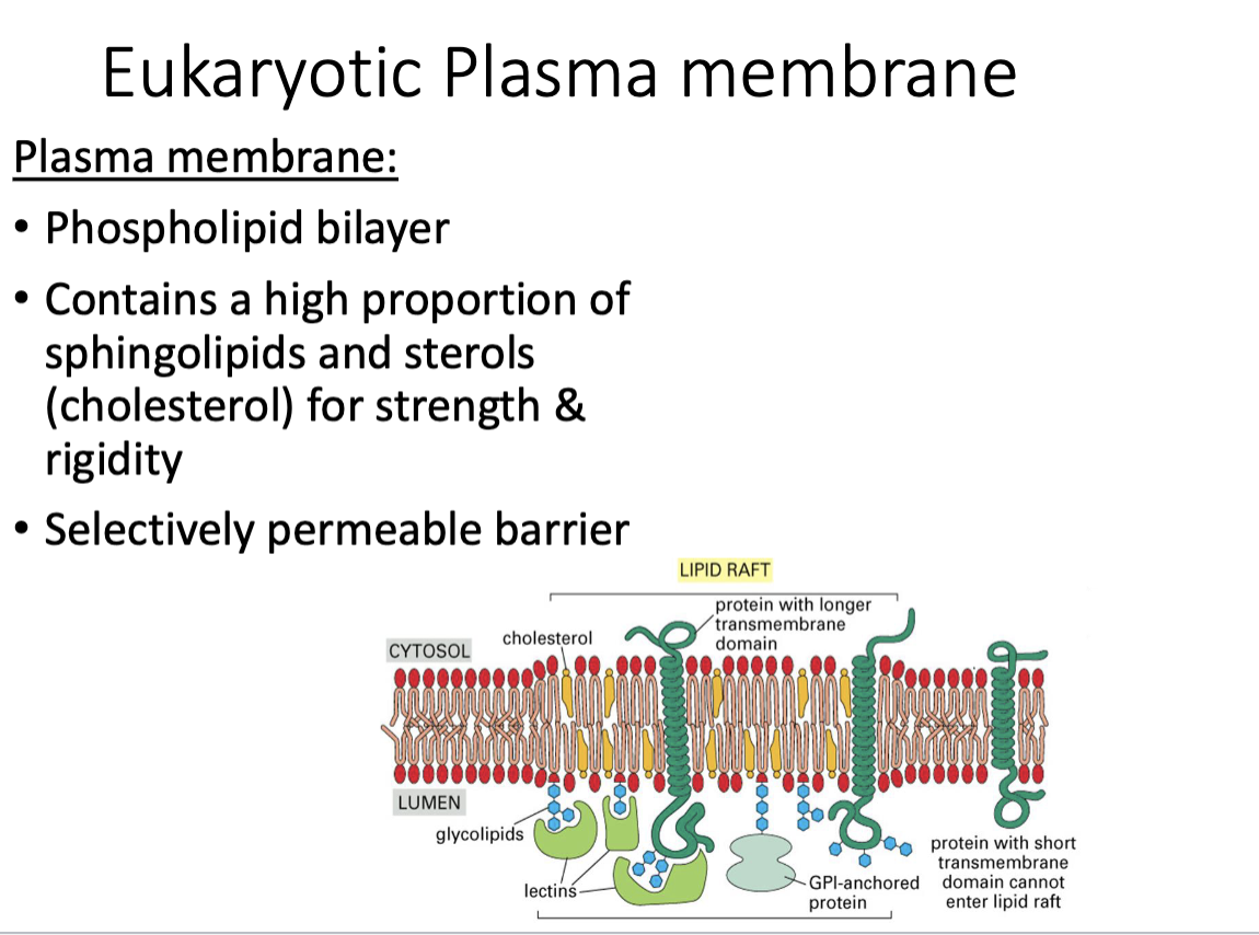
The eukaryotic plasma membrane…
..creates domain
enable cells to organize lipids & proteins into functional, non-uniform regions
membrane domains like lipid rafts = highly dynamic, ordered regions w/cholesterol & sphingolipids —> regulate protein-protein interactions, cell signaling, & membrane protein turnover

Brief Description: Microfilament protein
actin
Brief Description: Microfilament motor?
myosin
Brief description: microfilaments main function?
movement, cytokinesis
Brief Description: Intermediate Filaments Protein
Keratin, Vimentin
Brief Description: Intermediate Filaments Motor?
none!!!
Brief Description: Intermediate Filaments Main Function
structural stability
Brief Description: Microtubules Protein
Tubulin subunits
Brief Description: Microtubules Motor?
Dynein & Kinesin
Brief Description: Microtubules Main Function
intracellular transport & movement
Brief Description: What is the function of the endocytic pathway?
Brings materials into the cell from outside.
Brief Description: What is the function of the secretory pathway?
Moves materials through cell and to plasma membrane/exterior.
Functions of microfilaments?
Movement, endocytosis, cytokinesis, secretion.
The endocytic pathway…
..is observed in all eukaryotic cells
function = to bring materials into the cell (from outside)

Endocytic Pathway: Phagocytosis
generally mediated by receptor binding (trigger) —> blebs extend out —> engulf/eat whole thing/larger components
(not constitutive) —> particle fuses with lysosome

Endocytic Pathway: Macropinocytosis
drinking small fluids, constant, constitutive
Non-specific uptake of extracellular fluid and dissolved materials into large vesicles.
—> go to early endosome & transition to late endosome —> (compartment becomes acidified, breaks components brought in) fuses w/lysosome which degrades components

Endocytic Pathway: Endocytosis
regulated! receptor mediated, smaller components
whatever is brought in go to early endosome, transition to late endosome—> (compartment becomes acidified, breaks components brought in) fuse w/lysosome which degrades components

Endocytic Pathway: Function of the early endosome?
First sorting station for material brought into cell

Endocytic Pathway: Function of the late endosome?
Further processes and transports materials toward lysosomes for digestion.

Endocytic Pathway: Function of lysosome
Digests macromolecules using hydrolytic enzymes at low pH.
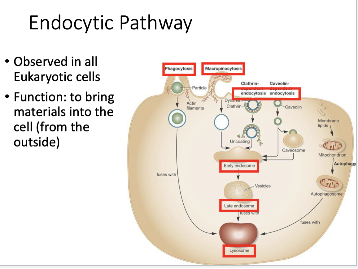
Order of the endocytic pathway?
Endocytosis → Early endosome → Late endosome → Lysosome
Macropinocytosis —> early endosome —> late endosome —> lysosome
Phagocytosis —> particle —> lysosome

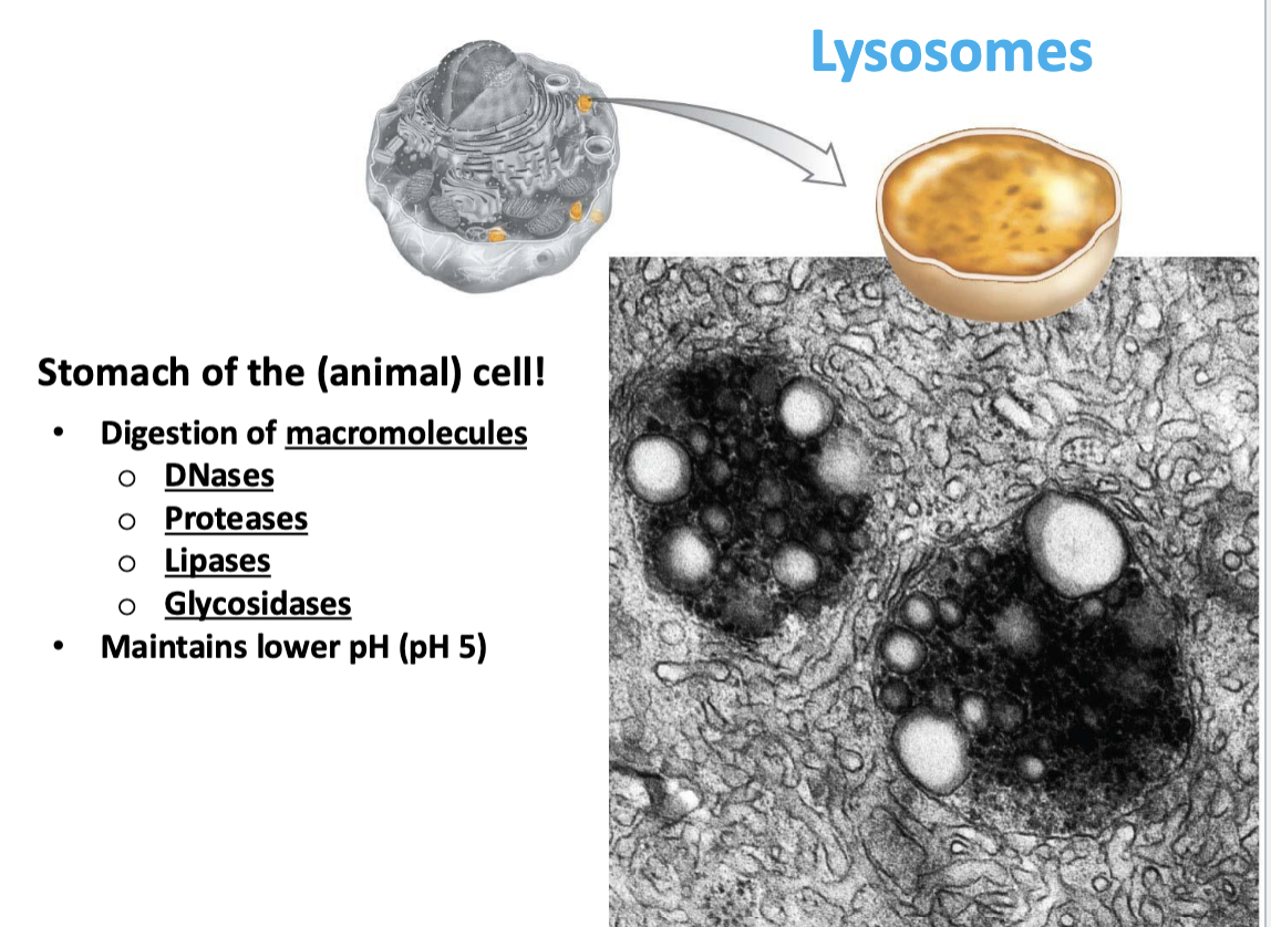
Lysosomes
stomach of the (animal) cell!!
digestion of macromolecules
maintains lower pH (pH 5)
Lysosomes contain
Enzymes that break down macromolecules
DNases (DNA)
Proteases (Protein)
Lipases (Lipids)
Glycosidases (sugars)
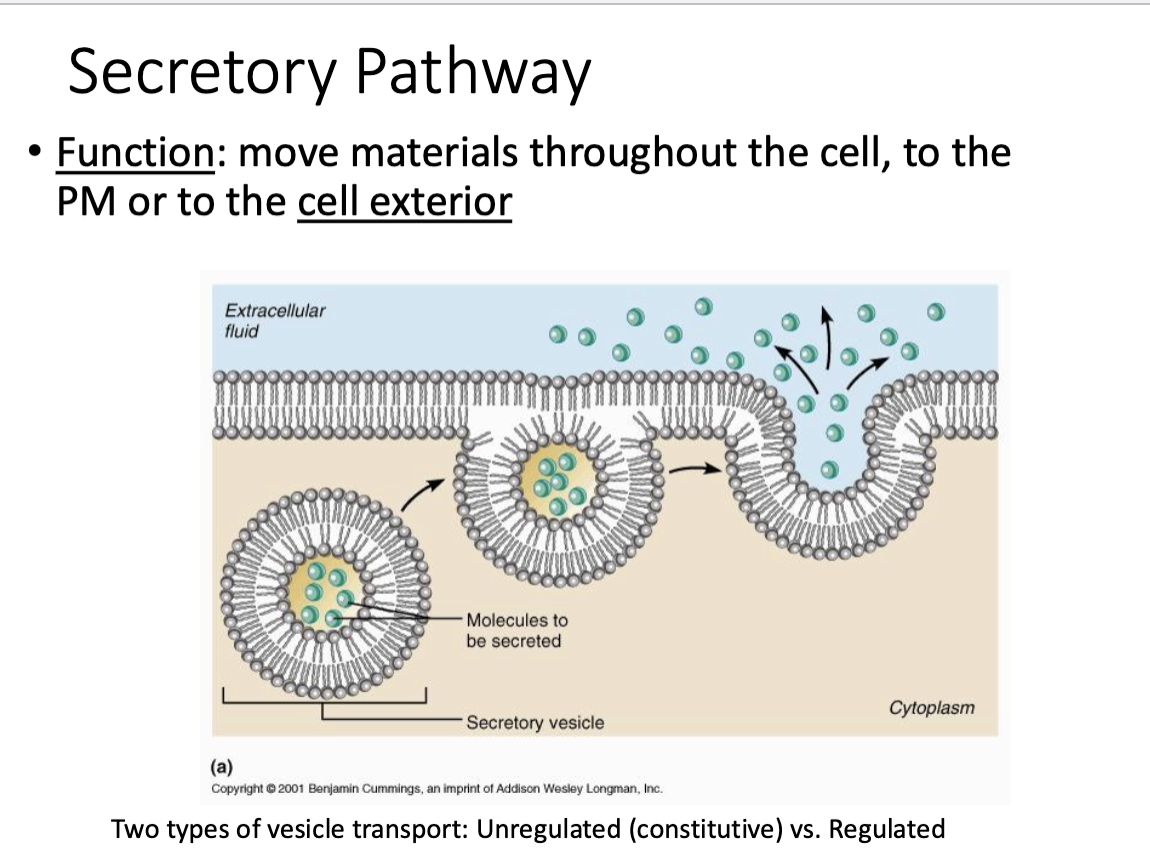
Secretory Pathway
Function: move materials throughout cell to the plasma membrane or to the cell exterior

Secretory Pathway: Two Types of Vesicle Transport
unregulated (constitutive) vs. regulated
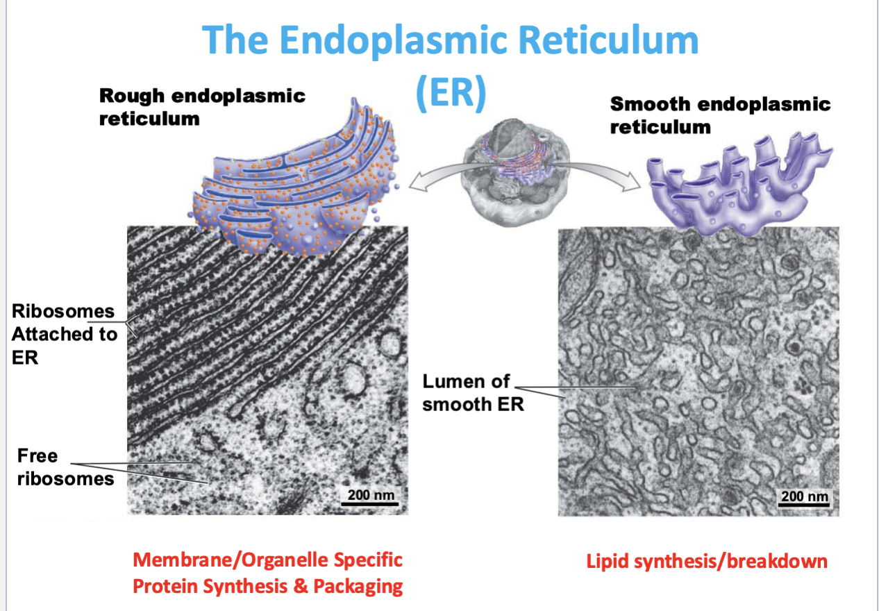
The Endoplasmic Reticulum
complex of membranes derived from outer membrane of nucleus
where “building blocks” are made

Rough Endoplasmic Reticulum
studded w/ribosomes
membrane/organelle specific

Function of rough ER
protein synthesis & packaging for secretion/membranes

Smooth endoplasmic reticulum
no studding w/ribosomes
lipid synthesis/breakdown/modification
Function of Ribosomes
Protein synthesis; forms peptide bonds between amino acids
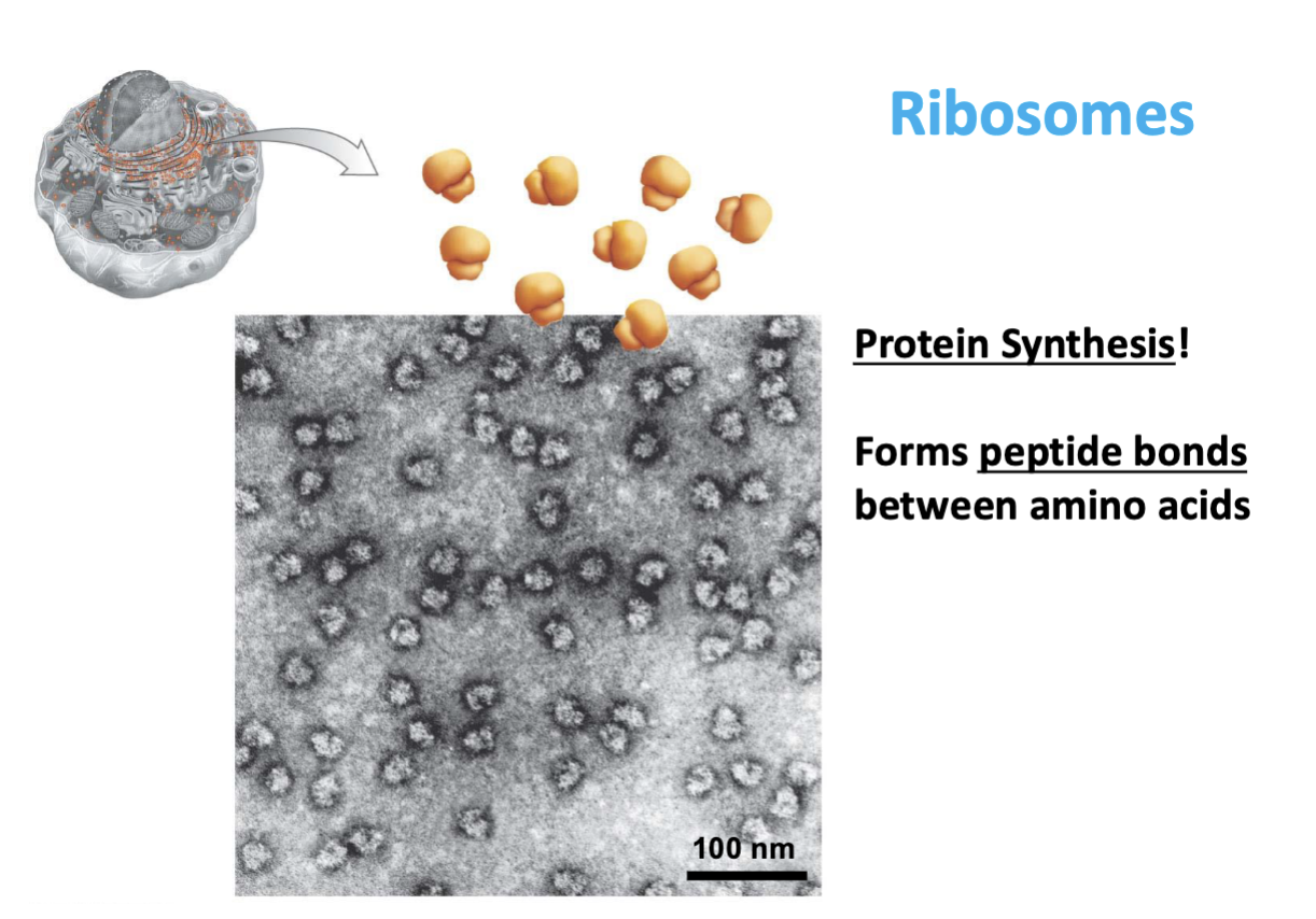
Ribosomes can be..
bound or unbound
free in cytoplasm or on rough ER

What are ribosomes made of?
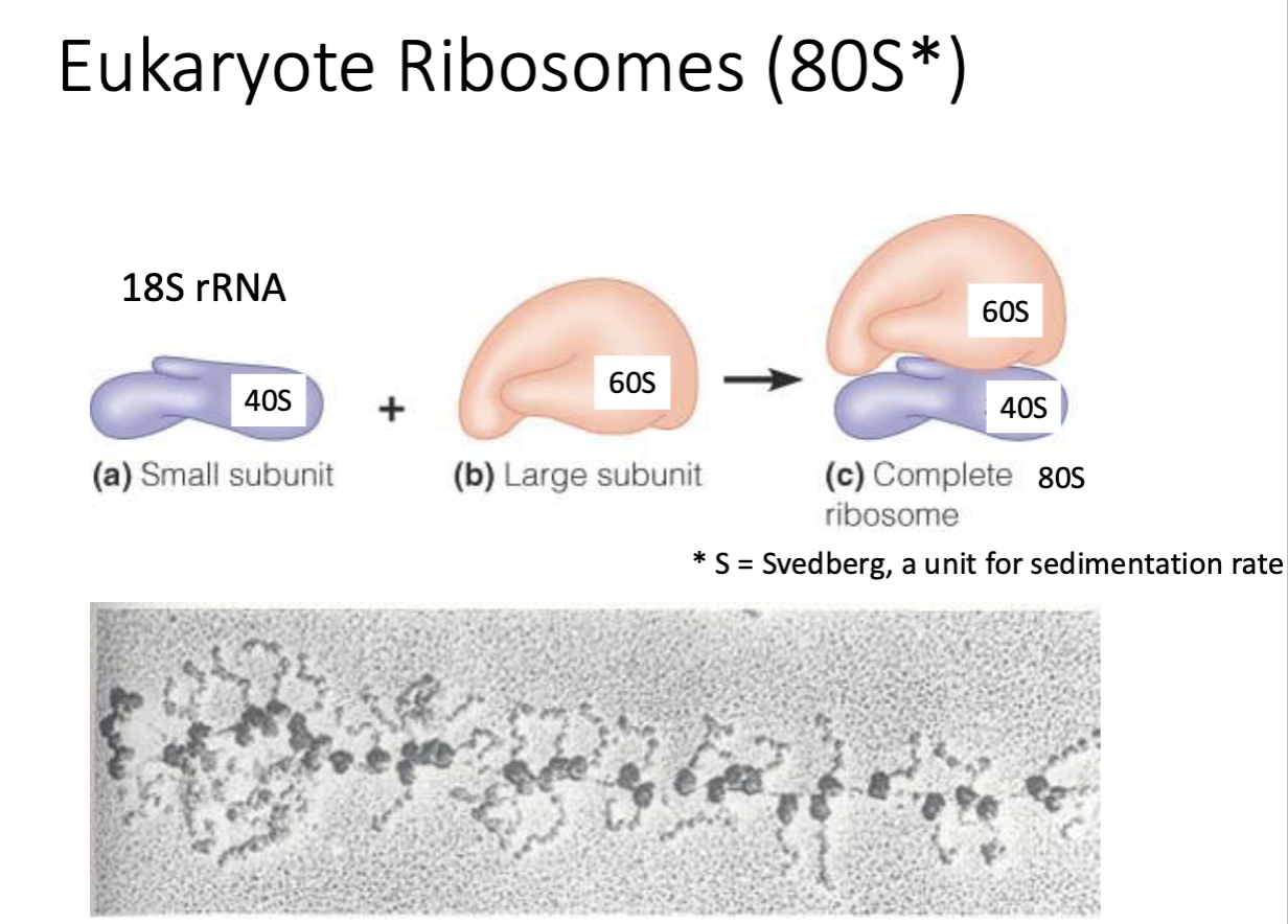
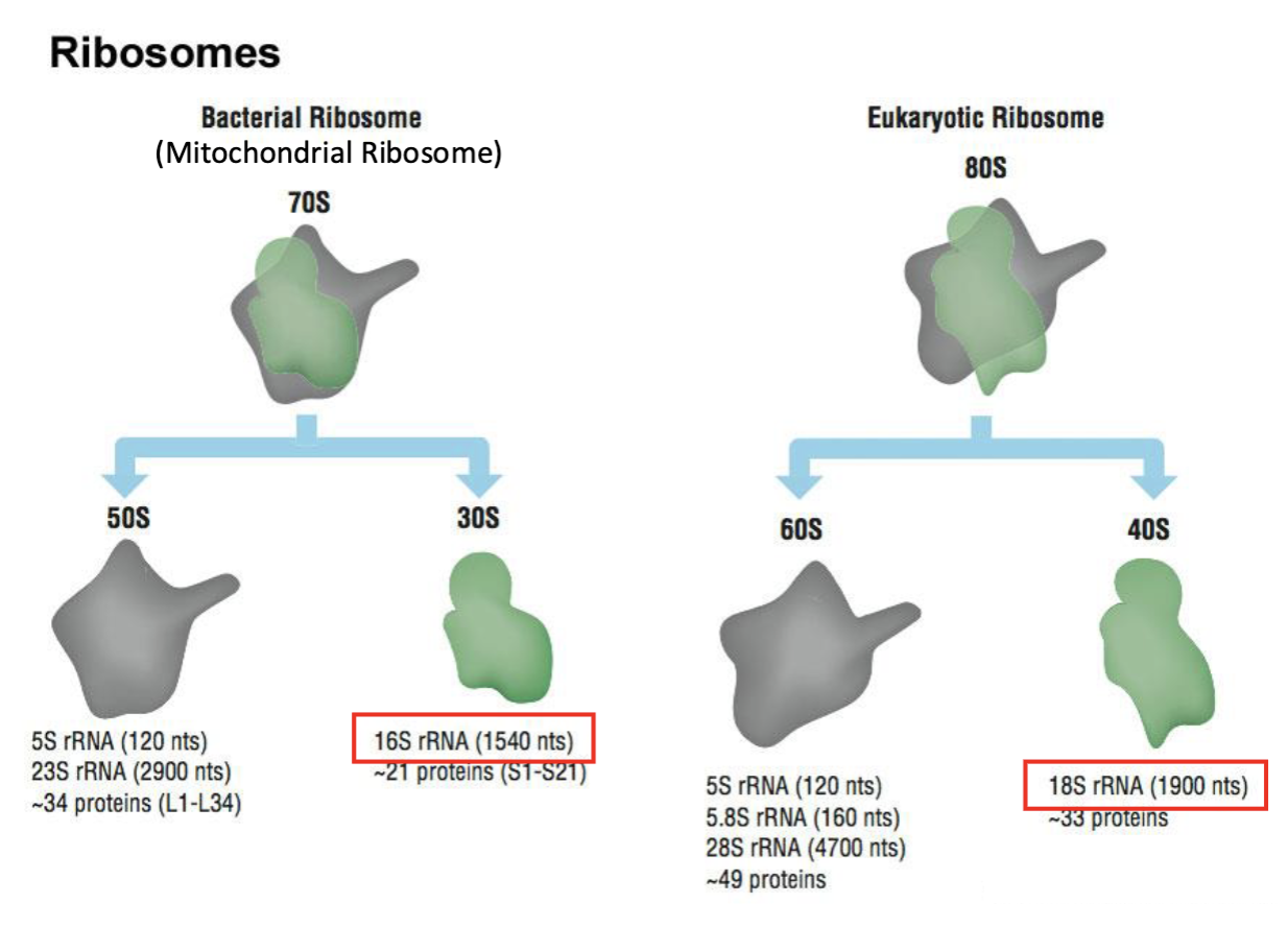
ribosomal RNA (rRNA) (60%) & proteins (40%)

What is the size of eukaryotic ribosomes?
80S (40S + 60S subunits)
(small subunit + large subunit)
* subunits are not additive
Difference between free and ER-bound ribosomes?
Free ribosomes make proteins for cytosol; bound ribosomes make proteins for secretion/membranes.
Ribosomes can be used for
for looking at evolutionary changes of chromosomes (bacterial ribosomes vs eukaryotic ribosome)
The secretory pathway: Step 1
nascent protein (nascent protein = protein currently being synthesized by a ribosome, emerging from its exit tunnel…) —> ENTERS lumen of ER
in lumen, protein spontaneously folds (chaperone proteins allow for proper folding in ER)
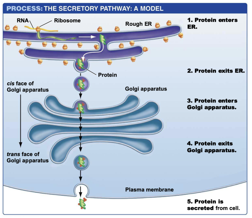
The secretory pathway: Step 2
protein may be tagged w/post translational modification (red dots), exits ER

the secretory pathway: step 3
protein enters golgi apparatus (enters cis face, faces nucleus)
goes through maturation process/sorting

the secretory pathway: step 4
protein exits golgi apparatus (exits from trans face)
(could have been tethered to membrane)
OR enclosed in vesicle

the secretory pathway: step 5 (if in vesicle)
protein is secreted from cell

Cis face of golgi apparatus
interior portion (faces nucleus)

Trans face of golgi apparatus
outer portion

Free vs ER-bound ribosomes
Determined by signal sequence on growing protein.
If signal sequence present:
→ ribosome binds ER
If absent:
→ ribosome stays free
Free ribosomes make proteins for:
Cytosol
Nucleus
Mitochondria
Peroxisomes
Chloroplasts
Why organelle abundance varies by cell type
Cells specialize.
Examples:
Muscle cells → many mitochondria (need ATP)
Pancreas cells → lots rough ER/Golgi (protein secretion)
Liver cells → lots smooth ER (detox)
White blood cells → many lysosomes (digestion)
Structure matches function.
Why protein sorting matters
Proteins must reach the correct location.
Wrong location = nonfunctional or harmful.
Secretory pathway:
Nucleus → Rough ER → Vesicles → Golgi → Vesicles → membrane / lysosome / secretion
Golgi Apparatus Main Functions
Protein/lipid processing, packaging, and delivery.

What enters the cis face of the Golgi?
Materials from the ER.
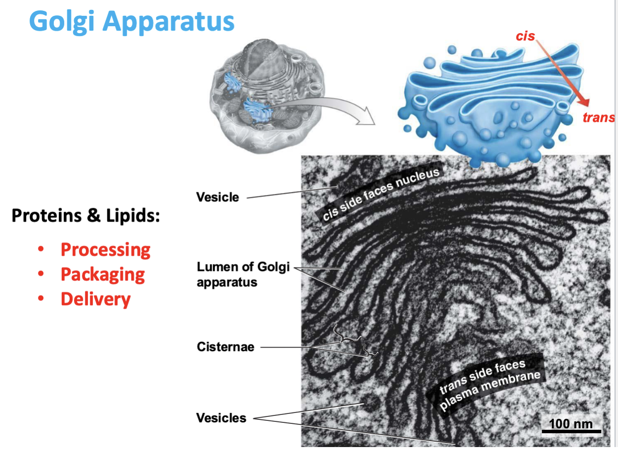
What exits the trans face of the Golgi?
Processed materials in vesicles.

Key Event in Eukaryotic Evolution 3
Acquisition of Mitochondria
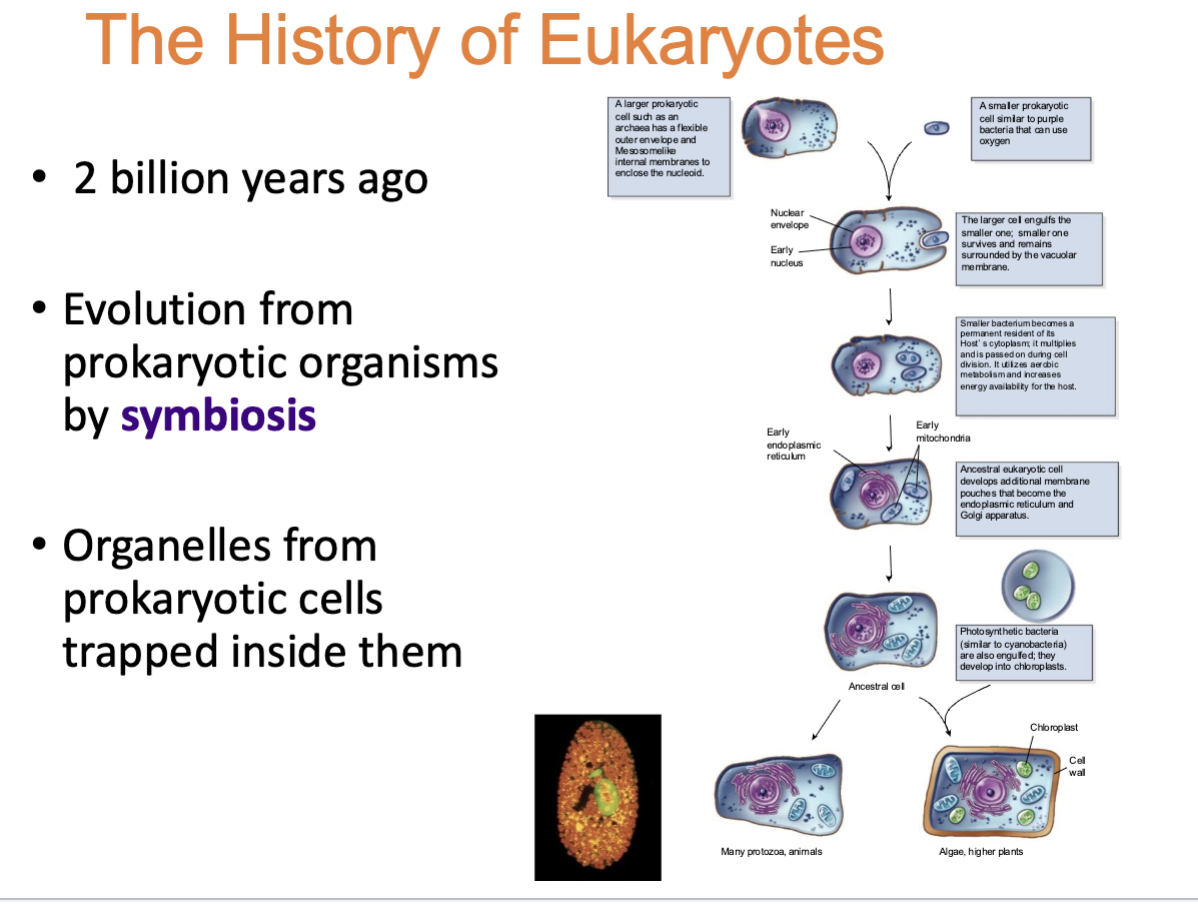
The history of Eukaryotes
2 billion years ago
evolution from prokaryotic organisms by symbiosis
Organelles from prokaryotic cells trapped inside them
Mitochondria
semi-independent organelle
Matrix: DNA (circular) & ribosomes 70s
reproduce by binary fission (asexual reproduction)

Mitochondria Function
energy production & synthesis
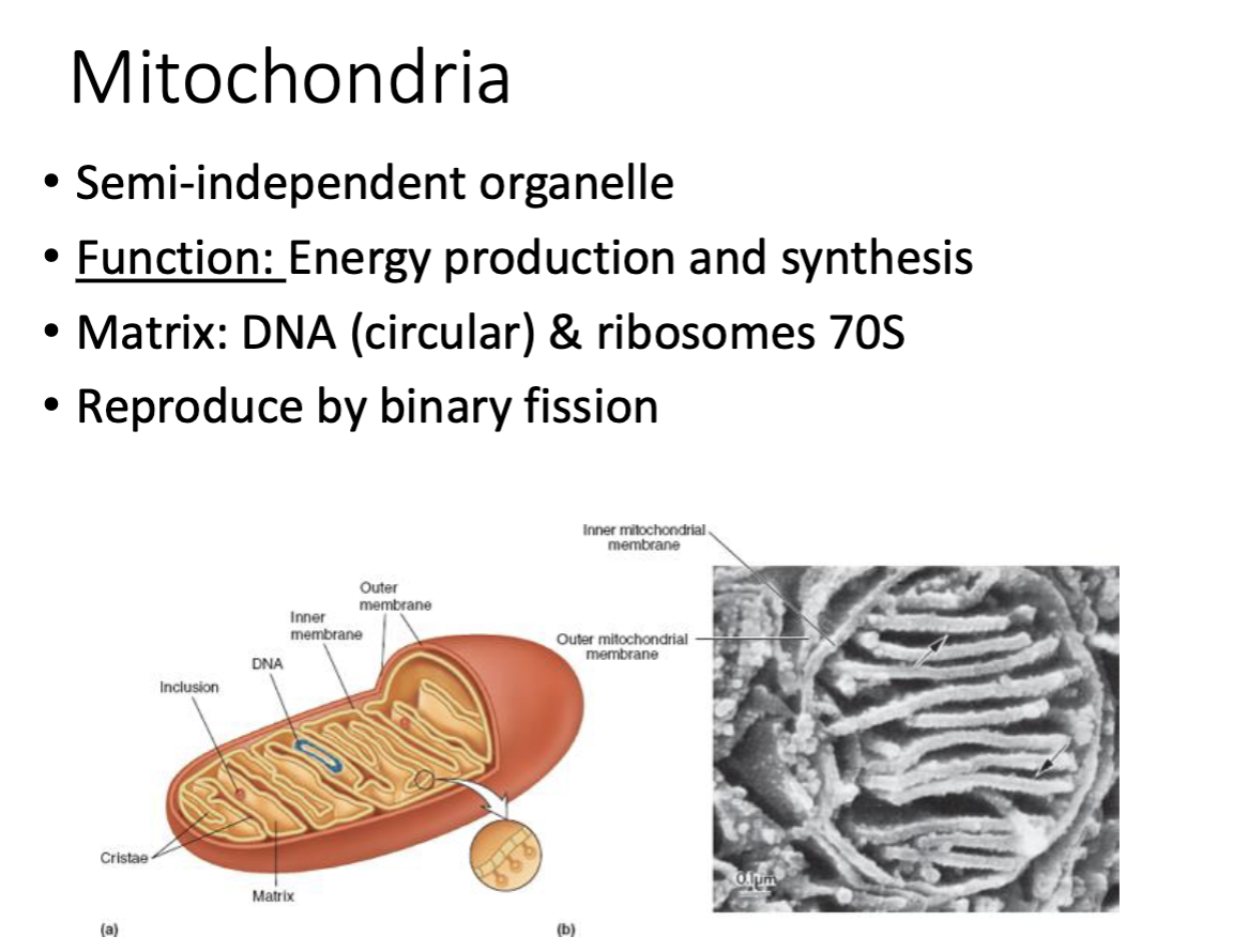
Why are mitochondria considered semi-independent?
Have their own circular DNA, 70S ribosomes, and reproduce by binary fission (asexual reproduction).
Evidence mitochondria evolved from bacteria?
Own DNA, own ribosomes, binary fission (asexual reproduction), double membrane.
Function of chloroplasts?
Photosynthesis and energy transformation.
What features do chloroplasts share with mitochondria?
Own DNA, own ribosomes, high membrane surface area, semi-independent.
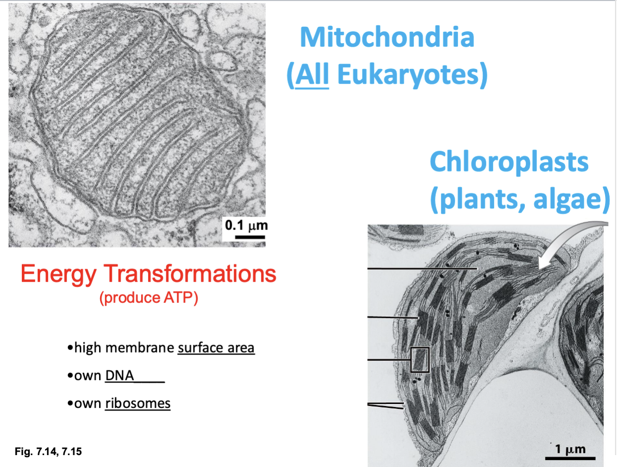
Mitochondria & Chloroplasts function for
energy transformation (produce ATP)
Mitochondria in ALL eukaryotes
Chloroplasts in plants & algae
