ANSC 251 - Garbage Brain
1/164
Earn XP
Description and Tags
I am going to kill myself, not even tomodachi life 2 can save me.
Name | Mastery | Learn | Test | Matching | Spaced | Call with Kai |
|---|
No analytics yet
Send a link to your students to track their progress
165 Terms
Name the facial bones
Frontal, nasal, maxilla, incisive, orbit
Orbital margin
Zygomatic arch
Pterygopalatine fossa
Fossa for lacrimal sac
infraorbital foramen
Facial bone formed by frontal, lacrimal & zygomatic bones
Orbital margin
Facial bone formed by maxilla, zygomatic process of temporal bone & zygomatic bones
Zygomatic arch
Origin of masseter muscle
Zygomatic Arch
Muscle that arises from pterygopalatine fossa
Pterygoid muscle
What foramen are found in pterygopalatine fossa?
Caudal palatine foramen
Sphenopalatine foramen
Maxillary foramen
Fossa containing nasolacrimal canal for the nasolacrimal duct
Fossa for lacrimal sac
Rostral opening for infraorbital canal, contains infraorbital arteries and veins
Infraorbital foramen
Bones of the braincase
Basioccipital
Tympanic
Petrosal part of temporal bone
Basiphenoid
Presphenoid
Name some of the ventral surface of the skull components
Paracondylar process
Tympanic bone
Mastoid process
Oval foramen
Tympano-occipital fissue
Mandibular fossa
What is found in the tympanic bone?
Tympanic bulla + external acoustic meatus
Annular cartilage of external ear attaches to its periphery
External acoustic meatus
Ventral skull component that is the origin for digastricus muscle
Paracondylar process
Foramen in which the manidbular nerve exits from
Oval foramen
Mandibular fossa components
Temporomandibular joint
What forms the temporomandibular joint?
Zygomatic process of temporal bone articulating with condyles of mandible
Termination of _____ parts of celidocephalicus and sternocephalicus
Mastoid process
What does the tympano-occipital fissure allow passage to?
Glossopharyngeal
Vagus
Accessory nerves
Internal carotid artery
Internal jugular vein
Postganglionic axons from cranial cervical ganglion
Occipital bones of the skull
Occipital condyles
Nuchal crest
External occipital protuberance
Foramen magnum
Mastoid foramen
Passageway of spinal cord (from occipital bones)
Foramen magnum
Term that is also called sagittal crest of the skull
External occipital protuberance
Area where dorsal portion of parietal bone meets with caudal portion of occipital bone
Nuchal crest
Passage for meningeal arteries and veins
Mastoid foramen
Genetic disorder of occipital bone mesoderm
Caudal occipital malformation syndrome
Function of hyoid apparatus
Stabilize tongue and larynx by suspending them from skull
Names of hyoid apparatus bones (prob won’t need to know names)
Stylohyoid
Epihyoid
Ceratohyoid
Basihyoid
Thyrohyoid
Components of mandible
Lower jaw
Lower and upper jaw
Masseteric fossa
Coronoid process
Mandibular foramen
Condylar process
Mandibular notch
Angular process
What does the lower jaw articulate with
Mandibular fossa of zygomatic process of temporal bone
Where do the upper and lower jaw meet?
At the symphisis
Origin and insertion of masseter muscle
O: zygomatic arch
I: masseteric fossa
True or false: coronoid process is the ventral half of ramus
False: dorsal
Process that contains depression for insertion of temporal muscle
Coronoid process
Forms temporomandibular joint and is part of the manible bones
Condyle process
U shaped depression between condylar and coronoid processes
Mandibular notch
Caudal opening of mandibular canal and transmits inferior alveoli a. v. and n., opens up at the 3 mental foramina to supply sensory innervation to lower lip and chin
Mandibular foramen
Process that hooked eminence ventral to condylar process + attachment of pterygoid muscle medially and masseter laterally
Angular process
Name the 4 types of teeth
Incisors
Canine
Premolars
Molars
Teeth placement
upper: embedded in maxilla bone lower: embedded in mandible
1 on left and right side
Canine
Teeth placement
upper: embedded in maxilla bone lower: embedded in mandible
4 on left and right side
Premolars
Teeth placement
upper: embedded in incisive bone lower: embedded in mandible
3 on left and right side
Incisors
Teeth placement
upper: embedded in maxilla bone
2 on left and right side
lower: embedded in mandible
3 on left and right side
Molars
Total number of canine teeth
a) 24
b) 20
c) 32
d) 42
d)
Which teeth are known as sectorial of shearing teeth?
Upper 4th premolar and lower 1st molar
Term given to baby teeth
Deciduous teeth
When do deciduous teeth begina and end erupting?
3 to 6 weeks
At what age do permanent teeth erupt and when do they end
4 to 6 months
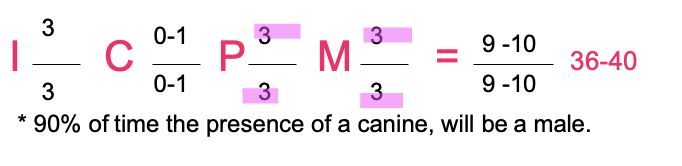
Which animal does this correspond to?
a) dog
b) pig
c) horse
d) ox
c)
True or false, dogs have 3 upper molars (left and right) and 3 lower molars (left and right)
False: 2 upper per side and 3 lower per side
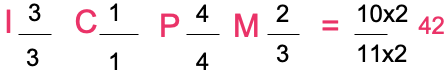
How many premolars do horses have per side?
3 upper and 3 lower

Which animal does this correspond to?
a) dog
b) pig
c) horse
d) ox
d)

Which animal does this correspond to?
a) dog
b) pig
c) horse
d) ox
b)
Which animal does this correspond to?
a) dog
b) pig
c) horse
d) ox
a)
Cavities of the skull: what does the cranial cavity contain?
Brain and its coverings
BLood vessels
Name of roof of braincase + what it’s formed by
Calvaria formed by parietal and frontal bones
What is the nasal aperture composed of?
2 symmetrical halves separated by a median nasal septum
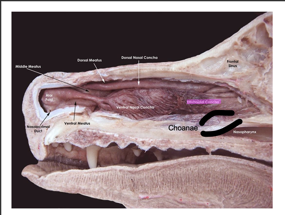
Components of nasal cavity
Nasal aperture
Choanae
Conchae
Caudal end of nasal septum where 2 nasal cavities open into nasopharynx
Choanae
Function of conchae
Act as baffles to warm and cleanse inspired end + contain olfactory neurons
What are the dorsal and ventral conchans divided into?
4 primary passages (meatuses):
Dorsal nasal meatus
Middle nasal meatus
Ventral nasal meatus
Common nasal meatus
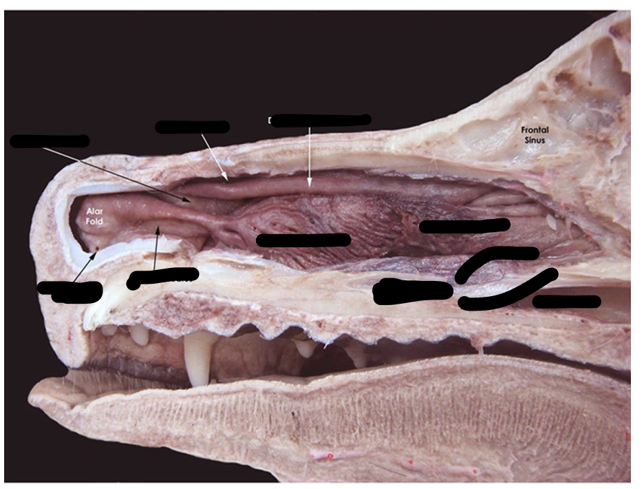
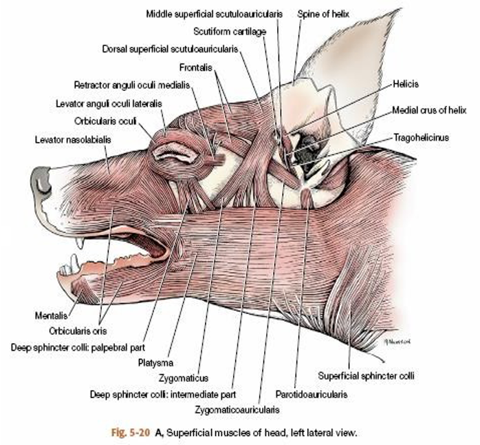
Name those parts
answers are here
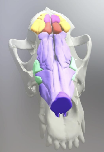
Can you name these parts?
Purple: nasal cavity airways
Orange: L & R rostral frontal sinus
Yellow: L & R lateral frontal sinus
Pink: L & R medial frontal sinus
Light blue: L & R maxillary recess
Component that communicates with ventral nasal meatus
L & R maxillary recess
Name the muscles of the face
Platysma
Orbicularis oris
Buccinator
Levator nasolabialis
Innervation of face muscles
All: facial nerve
Exception: Levator palpebrae superioris innervated by oculomotor nerve superior division
Face muscle action: returns food from vestibule to occlusal surface of teeth
Buccinator
Face muscle action of Levator nasolabialis
Dilates nostrils and raises upper lip
Face muscle action of platysma
Draws commissure of lips caudally
Face muscle action: controls shape and size of mouth opening
Orbicularis oris
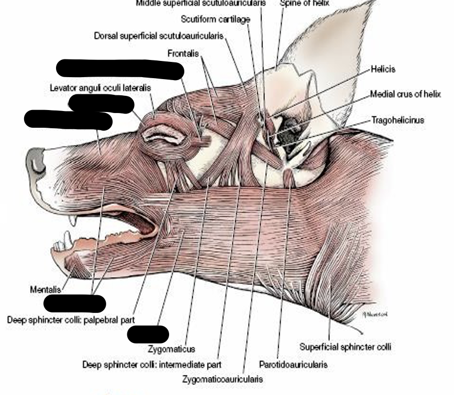
Name missing muscles
Name the eyelid components
Palpebrae
Lacrimal gland
Plica semilunaris/nictitating membrane
Muscles + eyelids
Junction of upper and lower eyelid
Commisure
What attaches the commisure of eyelids?
Medial and lateral palebral ligaments
Which eyelids bear the cillia?
Only upper eyelids
What does the joining of palpebrae form and where do they meet?
Form medial and lateral palpebral comisure at the end of fissure
Mucous membrane covering inner eyelid
Palpebral conjunctiva
Both upper and lower palpebrae border the _______
Palpebral fissure
Eyelid component that is ventral to zygomatic process of frontal bone
Lacrimal gland
Where does the lacrimal gland secrete into?
Conjunctival sac
Describe destination of lacrimal flow
Serous fluid passes across cornea to be collected by puncta and passes through lacrimal duct of each lid to lacrimal sac —> nasolacrimal duct —> nasal meatus of nasal cavity where evaporation takes place
Opening of lacrimal duct
Puncta
Function of plica semilunaris
Lubricates the cornea
Concave fold of palpebral conjunctiva and cartilage that moves horizontally across the eyebal
Plica semilunaris
What causes the cherry eye condition?
Tear gland of nictating membrane prolapsing
Name some muscles of the eyelids
Orbicularis oculi
Retractor anguli oculi lateralis
Levator palperae superioris
Elevates upper lid, eye muscle innervated by oculomotor nerve
a) orbicularis oculi
b) retractor anguli oculi lateralis
c) levator palperae superioris
c)
Action: closes palpebral fissure
a) orbicularis oculi
b) retractor anguli oculi lateralis
c) levator palperae superioris
a)
Attached to medial palpebral ligament and closes eyelids
a) orbicularis oculi
b) retractor anguli oculi lateralis
c) levator palperae superioris
b)
Cavity lying outside teeth and gums + inside the lip and cheeks
Vestibule
What ducts can be found within the vestibule?
Parotid duct
Zygomatic gland
Duct found through cheek on small papilla opposite the caudal end of upper shearing tooth
Parotid duct
Duct that opens into vestibule lateral to last upper molar tooth
Ducts of zygomatic gland
Bounded dorsally by hard palate + part of soft palate, laterally & rostrally by dental arches and ventrally by tongue
Oral cavity proper
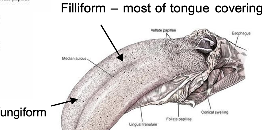
What are the 5 types of papillae in the tongue?
Filiform
Conical
Fungiform
Foliate
Vallate
Which papillae of the tongue are known as the taste buds?
Fungiform
Foliate
Vallate
Associate the taste buds with the correct taste:
a) Fungiform
b) Foliate
c) Vallate
Sweet
Salt
Bitter
a) 2
b) 3
c) 1
Papillae that is nongustatory
Filiform
Papillae that is mechanical & tactile (not very gustatory)
Conical
Elastic limb of free portion of tongue
Lyssa
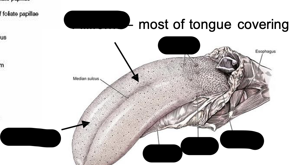
Identify the structures