Therex test 3*
1/143
There's no tags or description
Looks like no tags are added yet.
Name | Mastery | Learn | Test | Matching | Spaced | Call with Kai |
|---|
No analytics yet
Send a link to your students to track their progress
144 Terms
Elbow open-packed position
70° FLX and 10° supination
Elbow closed-packed position
Extension
Radiohumeral open packed-position
full extension and supination
Radiohumeral closed-packed position
elbow flexed to 90° and forearm supinated 5°
Proximal radioulnar open-packed
70° flexion and 35° supination
Proximal and distal radioulnar closed packed position
5 ° supination
Distal radioulnar open packed
10° supination
Lateral Epicondylitis
overuse injury of the common wrist extensor origin
the extensor carpi radialis most commonly affected
inc pain with extension and radial deviation
What is the most important exercise for management of overuse injuries
eccentric contractions
Medial Epicondylitis
An overuse injury that affects the wrist flexors’ origin
mainly flexor carpi radialis
inc pain with resisted wrist flexion and full passive wrist extension
Medial Valgus Stress Overload
effects the MCL aka the UCL
seen in throwing athletes
Cubital Tunnel Syndrome
aka ulnar tunnel syndrome
The ulnar nerve gets entrapped at the elbow
Acute rupture of the medial (ulnar) collateral ligament
longer recovery period needed
An internal brace of collagen tape is used to reinforce the ligament for athletes who don’t need full reconstruction
Lateral collateral ligament injury
2nd most commonly dislocated
via hyperextension or posterior-lateral rotation
can be treated conservatively or with a graft surgery
What type of fracture is this
Supracondylar
What type of fracture is this
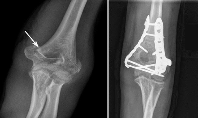
Distal humeral fracture
What complications can occur with a distal humeral fracture
nonunion, malunion, contractures
VASCULAR COMPROMISE
What causes a Volkmann ischemic contracture
a distal humeral fracture that causes brachial artery obstruction due to displaced fragments
What are the 6 symptoms indicating Vascular Obstruction
severe pain in the forearm
limited/pain with finger movement
purple discoloration
paresthesia and loss of sensation
loss of radial pulse
pallor, anesthesia, paralysis
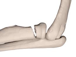
What type of fracture is this

Intercondylar fracture (T or Y)
What treatments for an intercondylar fracture are considered for young patients vs older patients
young= ORIF
old= bag of bones
What type of fracture is this
Radial head fracture
Myositis ossificans
abnormal bone formation in other tissues
Cozens test
for lateral epicondylitis
pronate forearm and extend and radial deviate the wrist
Tinel’s sign
Ulnar nerve compression
poke the medial side of the elbow
Radiocarpal (wrist) open-packed
neutral with slight ulnar deviation
Radiocarpal (wrist) losed-packed position
extension with radial deviation
Anatomical Snuffbox
The depression created by the abductor pollicis longus, extensor pollicis longus, and extensor pollicis brevis muscles
Colles fracture
Most common distal radial fracture
When the fragment is displaced in a dorsal direction (FOOSH)
“dinner fork”
Smith Fracture
fall of the dorsum of the hand
fragment displaced in a palmar direction
aka reverse Colles fracture
What is the point of the 6-pack exercises
to maintain MP and IP ligament length with wrist immobilization
Distal ulnar fractures
usually occur in combination with distal radius fractures
What is the most common carpal fracture
scaphoid fractures
Important scpahoid facts
often dismissed as a sprain
high incidence of nonunion
take much longer to heal
Boxer’s fractures
fractures of the neck of the fourth or fifth metacarpal
occur when the patient strikes a hard object wuith a clenched fist
Bennett’s fracture
palmar base of the proximal first metacarpal
How do most ligament sprains occur
with wrist hyperextension
Triangular Fibrocartilage Complex (TFCC)
formed by ligaments and an articular disk
Injury occurs via an axial force applied to the ulnar side of the wrist
The healing is poor
Skier’s Thumb
An acute sprain of the ulnar collateral ligament of the thumb
Tendinitis
inflammation of the tendon
Tenosynovitis
inflammation of the synovial sheath of the tendon
Tendinosis
degeneration of the tendon
De Quervain’s Disease
not a true clinical disease
abductor pollicis longus and the extensor pollicis brevis tendon and sheaths
radial-sided pain
Trigger Finger
catching of the finger or thumb
via swollen sunovium
Mallet finger
interuption of the extensor tendon mechanism over the DIP joint
flexor digitorium profundus
DIP flexion
Boutonnière deformity
central extensor tendon and triangular ligament interruption
PIP FLX and DIP hyperextension
Swan-neck Deformity
PIP hyperextension and DIP FLX
Dupuytren’s disease
firm nodules under the palm
via overactive fibroblasts/contractures
full FLX
Fasciotomy
cutting the contracted fascia blindly by inserting a small blade
Dermofasciectomy
removal of the skin overlying the diseased tissue, as well as the diseased tissue
Carpal tunnel syndrome
median nerve compression/entrapment
Ulnar tunnel syndrome
compression in the Guyon canal/tunnel
between pisiform and the hamate
Radial sensory nerve compression (wartenberg’s syndrome)
sensation affected only
back of the thumb, radial side of the hand, and radial PIP joint
repetitive pronation
Complex Regional Pain syndrome (CRPS)
abnormall sever of prolonged pain response; can be type one or two
Ulnar collateral ligament instability test
tear of UCL
Allen test
occlusion of radial or ulnar artery
Froment’s sign
ulnar nerve compromise/paralysis
Phalen’s test
tests carpal tunnel syndrome
Tinel’s sign
tests for carpal tunnel syndrome
Finkelstein’s test
tests for tenosynovitis in the thumb (de Quervain’s disease)
What are the two arteries found around the hip joint?
retinacular arteries
femoral arteries
Hip open-packed position
30° FLX , 30° ABD, slight lateral rotation
Hip closed-packed position
full extension and medial rotation
LE D1 FLX PNF pattern
hip: FLX/Add/ER
knee: FLX
ankle: DF/inversion
LE D1 EXT PNF pattern
hip: EXT/ABD/IR
knee: EXT
ankle: PF/eversion
LE D2 FLX PNF pattern
hip: FLX/ABD/IR
knee: FLX
ankle: DF/eversion
LE D2 EXT PNF pattern
hip: EXT/ADD/ER
knee: EXT
ankle: PF/inversion
What is another name for the intertrochanteric fracture
extracapsular
What is another name for intracapsular fracture
femoral neck or subcapital
what is another name for the subtrochanteric fracture
proximal femoral shaft
What are some common clinical complications of hip fracture
malunion, delayed union, nonunion avascular necrosis
What is legg-calve-perthes disease
femoral head flattening that causes a disruption of the blood supply
What kind of orthosis is given to someone to keep the femoral head seated within the acetabulum when they have Legg-Calve-Perthes disease
abduction orthosis
Hemiarthroplasty is performed when a patient has
femoral head osteonecrosis
What is Osteoarthritis (OA)
focal loss of articular cartilage with variable subchondral bone reaction
What are some indications of a THA
RA, OA, fractures, WB pain, deterioration
Total hip replacement complicaations
blood clots
loosened components
nerve injury
dislocations
THA universal precautions
avoid hip ADD
avoid IR
avoid FLX over 90
avoid combining them
Pincer impingement
overhanging rim of the acetabulum; labrum and articular cartilage damage
Cam impingment
head of femur; damage to articular cartilage
Greater Trochanteric Pain Syndrome (GTPS)
pain with abduction
trochanteric bursitis
tears of glute med or min
Thomas test
hip flexion contractures or tightness
90-90 SLR test
hamstring tightness
Trendelenburg test
testing for weak glute med
Knee open-packed position
25° FLX
Knee closed-packed position
full EXT and lateral tibial rotation
what are the rules of rehabilitation according to Gray
Create a safe environment
Do not hurt the patient
Be agressiove w/o breaking first 2 rules
What are the progression steps post knee injury
progress ROM/quad control
establish normal gait
demonstrate ascending normal steps
demonstrate decending normal steps
running
plyometrics
Sould the time frame of a assessment-based guidline incorportate fixed or flexible time
flexible time frame
Anterior Cruciate Ligament (ACL)
resists anterior translation of the tibia on the femur
controls hyperextension of the knee
females tear more than men
What test is used to assess the integrity of the ACL
Lachmans test
Anterior Drawer test
Pivot shift
What is the most specific ACL test
Lachman
How is the Lachman performed?
knee in 25-30° FLX, the clinician attempts to displace the tibia anteriorly
How is the anterior drawer test performed?
The knee is flexed to 90°, and the clinician attempts to provide an anteriorly directed force onto the tibia
Posterior Cruciate Ligament Injuries
The primary restraint to the posterior displacement of the tibia
strongest ligament in the knee, injury occurs less often
What tests can be done to test the PCL
posterior drawer test
Godfrey posterior sag test
active quad drawer test
What is the most common and accurate PCL test
Posterior drawer test
How is the posterior drawer test performed
The knee is flexed to 70° and the clinician stabilises the ankle on the table as the patient attemps to straighten the knee using the quad
Which ligament has a longer rehab/WB percaution time ACL or PCL
PCL
Medial collateral ligament injuries
The most common ligament injury in the knee
via valgus forced applied to the knee while the foot is fixed