exam 3: all sets combined
1/262
There's no tags or description
Looks like no tags are added yet.
Name | Mastery | Learn | Test | Matching | Spaced | Call with Kai |
|---|
No analytics yet
Send a link to your students to track their progress
263 Terms
What internal structures do protozoa have?
Nucleus, nucleolus, ER, ribosomes, Golgi complex, food vacuoles.
-complex metabolic processes
Where can protozoa be located in the body?
Extracellular vs intracellular; tissue vs blood vs gut.
How do protozoa move?
Cilia, flagella, pseudopodia, microtubules.
What life cycles do protozoa have?
Direct and indirect life cycles; motile and non-motile.
How do protozoa reproduce?
Asexual (fission) and sexual (gametogony).
What are the 2 kinds of flagellates?
Mucosoflagellates
intestinal or repro tracts
direct
hemoflagellates
blood and tissues
use vectors as intermediate host
What animals get Trichomonads?
dogs & cats (Trichomonas brixi)
birds (T. gallinae, T. stableri, T. gypaetinii)
humans (T. vaginalis & T. tenax)
What does T. vaginalis cause in humans?
trichomoniasis
Where are trichomonads typically found?
GI or reproductive tracts.
What type of life cycle do trichomonads have?
Direct life cycle.
Where are trichomonads found in cats and cattle?
Cats - intestinal tract; Cattle - reproductive tract.
What life stage do trichomonads have?
Only trophozoite stage, no cysts (~10-25 × 3-15 µm).
How do trichomonads reproduce?
Longitudinal binary fission within the host.
How long do trichomonads survive in the environment?
Short-lived in the environment and the infective stages.
How can pseuocysts occur with Trichomonads?
Formed under adverse conditions; can't survive in the environment.
What Tritrichomonas species are important?
Tritrichomonas foetus (bovine) and Tritrichomonas blagburni (feline).
What organism causes feline intestinal trichomonosis?
Tritrichomonas blagburni
What organism causes bovine genital trichomoniasis?
Tritrichonmonas foetus
How is Tritrichomonas blagburni transmitted?
Fecal-oral route - colonizes distal ileum and colon.
What is the prevalence of Tritrichomonas blagburni?
~4-10% in pet cases.
Which cats are most at risk?
Cats in shelters and catteries.
-infected cats maintain good health, normal appetite and condition
What are clinical signs of feline trichomonosis?
One-eight bowel movements/day
chronic large bowel foul-smelling diarrhea
mucus/fresh blood
straining in litterbox
How is feline trichomonosis diagnosed?
examine fecal samples of trophozites - direct exam or wet mount with stained, thin fecal smear
fecal culture with InPouch-TF-Feline
PCR of the internal transcribed spacer region (ITS1 and ITS2) and the 5.8S rRNA gene
Histo
What can Tritrichomonas blagburn have co-infection with?
Giardia spp.
What sample handling can avoid harming diagnosis?
Fecal flotation and refrigeration is detrimental.
What drug treats Tritrichomonas blagburni?
Ronidazole (30 mg/kg PO QD for 14 days).
T/F: Feline T. blagburni is resistant to all commonly used anti-protozoal drugs.
true
Treatment with ________ gives a false sense of effectiveness as trichomonads feed on colonic bacteria.
Antimicrobials
-trichomonads are protozoa, not bacteria. it would allow the parasite to thrive
How is Tritrichomonas foetus transmitted?
Sexually transmitted.
Where does Tritrichomonas foetus live in cattle?
Cows: vagina and uterus
Bulls: penis and prepuce.
common in herds with natural service
What disease effects does Tritrichomonas foetus cause?
Infected bulls and cows are asymptomatic and look normal, but causes abortion and infertility (reduced calf crop).
Bulls - less than 3 y/o - transient carriers and can clear infection, older - chronic carriers for life
some cows show pyometra, endometritis and adventital placentation
Where is Tritrichomonas foetus endemic to?
West of Mississippi River
How is bovine trichomoniasis diagnosed?
culture w/ Diamond’s medium and InPouch-TF-Bovine
collect preputial smegma for iso of T. foetus
trophs in vaginal secretions of heifers 14-20 days post breeding
what do you take a PCR of in T. blagburni?
internal transcribed spacer region (ITS1 and ITS2) and the 5.8S rRNA gene
what do you take a PCR of in T. foetus?
ITS1 and 5.8S rRNA genes from InPouch-TF culture
control/rx of T. foetus
infected cows’ clear infection within 4-5 months of sexual rest (susceptible to reinfection)
no effective treatments for Bovine T. foetus - culling of bulls
vaccine available for cows only
What are hemoflagellates?
Blood- or tissue-dwelling flagellated protozoan parasites.
what organism causes American trypanosomiasis (Chagas disease)
trypanosomes - Trypanosoma cruzi
Reduviidae - Reduviid - Triatomine "kissing bugs" and vertebrate host
what organism causes canine visceral leishmaniasis
leishmania donovani complex (L. donovani, L. infantum/chagasi)
sand flies and the vertebrae host
What type of life cycle do hemoflagellates have?
Indirect life cycle with mammals and arthropods as vectors.
What is kinetoplastid DNA (kDNA)? What protozoa has it?
Hemoflagellates have it
Organized into an incredible network of interlocked rings
unlike any other type of DNA in nature which resembles chainmail that medieval knights wore
Hemoflagellates transmission- biological
Biological: Vectors with parasite multiplication (Triatomine bugs, Sand flies, Tsetse fly – T. brucei).
Hemoflagellates transmission- mechanical
Mechanical: Biting flies (mouthpiece cross-contamination), Congenital, Blood Transfusion, Venereal,, Oral ingestion of vector (mainly Trypanosoma cruzi)
How is Trypanosoma cruzi transmitted?
Triatomine bug bite, blood transfusion, organ transplant, venereal, oral ingestion.
Asexual reproduction both in the vertebrate (extra- and intra-cellular stages) and the vector (infective stages develop here)
how does Trypanosoma cruzi enter the body after being transmitted
Bug defecates after taking a blood meal, host rubs bite wound spreading the feces and the parasite into wound and enters the body
Where can Trypanosoma cruzi species be found?
In and around beds and bedrooms, especially under or near mattresses or nightstand.
areas of rodent infestation
Trypanosoma cruzi reservoir hosts
woodrats, raccoons, skunks, coyotes, opossums, armadillos, dogs, cats, pigs
~39-45% antibody seroprevalence rates for dogs in SE USA
affecting 6-8 million people worldwide with >10,000 attributed deaths annually
What are the 2 morphological forms of Trypanosoma cruzi in the mammalian host?
Amastigotes; Intracellular, ovoid with internalized flagella, performs binary fission.
Often found in cardiac and smooth muscle cells, neural and glial cells.
Trypomastigotes; Extracellular, motile, blood stage forms, low numbers in blood,
what is acute chagas disease typically found in
young dogs less than 6 months and 1-2 weeks following infection
What are signs of acute Chagas disease?
severe and rapid weakness, loss of appetite, myocarditis, pericardial effusion, meningoencephalitis, sudden death.
what is chronic chagas disease typically found in
adult dogs with slowly progressive infection (~8-12 weeks) increased parasitemia
clinical signs & abnormal ECG are absent at this time
What are signs of chronic Chagas disease?
generalized lymphadenopathy, lethargy, organomegaly, chronic myocarditis, dilation of heart, ECG abnormalities and eventual death
How is Chagas disease diagnosed?
serology (IFA or direct microscopy)
trypomastigotes in blood and CSF, amastigotes found in tissue biopsy
what do you take a PCR of in T. cruzi diagnosis
kDNA or nDNA from blood
what is xenodiagnosis in T. cruzi
unfed, clean bugs are allowed to feed on suspected individual’s blood. after 1-3 weeks, look for metacyclic trypomastigotes in the gut of the bug
What drug treats Trypanosoma cruzi?
Benznidazole 20 mg/kg PO once per week.
who should you screen for control/prevention of T. cruzi
blood donors and breeding dogs
What organism causes canine visceral leishmaniasis?
Leishmania infantum (Leishmania donovani complex).
How is Leishmania transmitted?
Phlebotomine sandflies (Phlebotomus and Lutzomyia)
indirect transmission by Phlebotomus sp. (eastern hemisphere) and Lutzomyia sp (western hemisphere)
Lutzomyia sp are now in at least 21 US states
travel related cases
Leishmania infantum reservoir hosts
dog, fox, human, rodent, wild mammal
outbreaks in foxhounds - vertical transmission over several generations
autochthonous infections in dogs in TX, OH, NY, OK - horiztonal infections - dog fighting
the 2 morphological forms of L. infantum
amastigotes (intracellular in host): reticuloendothelial cells, skin, bone marrow, liver and spleen
promastigotes (in the sandfly): extracellular, motile, and infectious stage
asexual in both
What percentage of infected dogs show disease?
~1 in 5 develop clinical disease.
what is L. infantum typically seen in
dogs < 3 years of age and older than 8 yrs
What skin signs occur with Leishmania?
Alopecia (regional/facial) and non-pruritic ulcerative, nodular, or papular dermatitis.
L. infantum clinical signs
Chronic wasting, epistaxis, diarrhea, conjunctivitis, uveitis, retinitis, severe muscle atrophy, swollen limbs and joints, lameness, lymphadenopathy, polyarthritis, and protein-losing nephropathy, which may lead to renal failure
How is Leishmania diagnosed?
serology (IFA, ELISA, microscopy)
amastigotes found in aspirates and biopsy, touch presentations of affect tissues
PCR of kDNA or kinetoplast minicircle
What drugs treat Leishmania?
Miltefosine and allopurinol.
What are protozoa?
Microscopic unicellular, heterotrophic, eukaryotic organisms.
What are the four organization types of protozoa?
Ciliates, flagellates, amoebae, and parasitic sporozoans.
How many protozoa species have been described?
>50,000 described species.
What internal structures do protozoa have similar to eukaryotes?
Nucleus, nucleolus, ER, ribosomes, Golgi complex, food vacuoles.
Where can protozoa be located in the body?
Extracellular vs intracellular; tissue vs blood vs gut.
What types of locomotion do protozoa use?
Cilia, flagella, pseudopodia, microtubules.
What types of life cycles do protozoa have?
Direct and indirect life cycles.
How do protozoa reproduce?
Asexual (fission) and sexual (gametogony).
What are the general characteristics of protozoa ciliates?
Majority commensals
few parasitic
direct life cycle
extracellular
reproduce asexually by binary fission
What are the morphologic features of ciliates?
2 nuclei (macro- and micro-) and cilia.
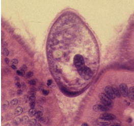
What is Balantidium coli and where is it found?
Extracellular pathogen in large intestine lumen.
Who is the reservoir for Balantidium coli?
Pig is reservoir.
Is Balantidium coli zoonotic
yes
How is Balantidium coli transmitted?
Fecal-oral route.
What are the clinical signs of Balantidium coli?
Usually nonpathogenic, occasional diarrhea.
How is Balantidium coli diagnosed?
Cysts in feces, sometimes trophozoites in direct smears (diarrheal stool)
What are the life cycle stages of Balantidium coli?
Trophozoite and cyst.
What is the treatment for Balantidium coli?
Tetracycline (500 mg/kg, 4x/daily for 10 days).
What happens during the trophozoite stage in Balantidium coli
metabolically active, feeding and reproducing stage
size features of the trophozoites in Balantidium coli
~30-150 × 25-120 um (largest protozoan parasite of humans)
size features of the cysts in Balantidium coli
non-multiplying, non-motile with no cilia found
smaller, spherical cysts (~40-70 × 40-60 um)
What causes 'Ich' in freshwater fish?
Ichthyophthirius multifiliis.
What causes 'Ich' in saltwater fish?
Cryptocaryon irritans.
what is the name of the disease caused by ciliates of fish
white spot disease
What are the three life-cycle stages of Ich?
Trophonts: large ciliated, feeding stages, live in cysts in gills and skin (white spots)
tomonts: encysted repro stage, release tomites
theronts: tomites transform to theronts, free swimming infective stage
What are the clinical signs of Ich?
White spots on skin and gills.
what does penetration by a theront in white spot disease cause
irritation, small blisters, abundant mucus production, skin sloughing/scale loss
white spot disease diagnosis
obtain gill or skin scraping for microscopic evaluation
look for trophonts or tomites
observation of even single organism warrants rx!
white spot disease rx
slowly raising water temp to 28-30 C/82-86 F - speeds up parasite life cycle
combined w copper sulfate, malachite green, or formalin for 10-14 days
vacuum the gravel during water changes to remove cysts
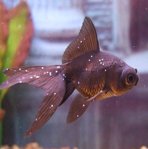
what is this disease
white spot disease
pig reservoir
Balantidium coli
How is Giardia transmitted?
Waterborne (fecal-oral)/food/surfaces.
What is the most significant Giardia species?
Giardia duodenalis. (AKA: G. lamblia or G. intestinalis)
-9 total species